Chapter 16 and 17 cell bio

0.0(0)
studied byStudied by 0 people
call kaiCall Kai
learnLearn
examPractice Test
spaced repetitionSpaced Repetition
heart puzzleMatch
flashcardsFlashcards
GameKnowt Play
Card Sorting

1/102

encourage image

There's no tags or description

Looks like no tags are added yet.

Last updated 5:27 AM on 12/6/24
Name
Mastery
Learn
Test
Matching
Spaced
Call with Kai

No analytics yet

Send a link to your students to track their progress

103 Terms

1
New cards

Chapter 16: Define signal transduction and list the basic components involved in this process in cells.

  • signals that pass between cells

  • the signaling cell produces a particular type of extracellular signal molecule that is detected by the target cell.

  • Target cells possess proteins called receptors that recognize and respond specifically to the signal.

  • signal transduction begins when the receptor on a target cell receives an incoming extracellular signal and then produces intracellular signalling molecules that alter cell behavior.

2
New cards

Distinguish the main types of signal-mediated cell-cell communication and identify the type of extracellular signal molecule involved in each

  • signal molecules can be proteins, peptides, amino acids, nucleotides, steroids, fatty acids, derivatives or even dissolved gases.

  • the most "public" style cell-cell communication: endocrine signaling

  • some less public signaling is paracrine signaling

3
New cards

Endocrine signaling

secretes the signal into the animals bloodstream, made by endocrine cells in the pancreas

4
New cards

Explain how the same signal molecule can induce different responses in different target cells

  • a cells response depends on the set of intracellular signaling molecules each receptor produces or how these molecules alter the activity of the effector proteins

5
New cards
Recall how a combination of signals can evoke a response that is different from the sum of the effects that each signal can trigger on its own
this is called "tailoring" of cells response occurs in part because the intracellular relays systems activated by the different cell signals interact. thus the presence of one signal might enable a cell to survive another might drive to differentiate in some specialized way and another might cause it to divide. in the absence of proper signals, most animal cells are programmed to kill themselves
6
New cards

Differentiate the types of cell responses that occur rapidly with those that take minutes or hours to execute

  • response times corresponds to the complexity of the response

  • for ex: cell growth and division can take a long time to execute since the response requires changes in gene expression and production of new proteins

7
New cards

transmembrane receptors

detect a signal on the outside and relay the message, in a new form across the membrane into the interior cell

8
New cards

steps of an intracellular signaling pathway

  • signal is relayed onward

  • the signal received is amplify

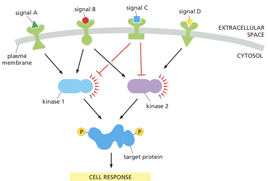
  • detect signals from more than one pathway and integrate them before relaying a signal onward

  • signal is distributed to more than one effector protein

  • response to the signal is regulated through feedback

9
New cards

Compare positive and negative feedback and contrast the types of responses produced by each

  • Positive: a component that lies downstream in the pathway acts on an earlier component in the pathway to enhance the response to the initial signal

  • Negative: a downstream component acts to inhibit an earlier component in the pathway to diminish the response to the initial signal

10
New cards

Summarize how phosphorylation can act as a molecular switch, and identify the types of proteins that add and remove this chemical modification

  • protein kinase covalently attaches to a phosphate group onto the switch protein

  • protein phosphatase takes the phosphate off again

11
New cards

Distinguish the two main types of GTP-binding proteins

  1. trimeric GTP-binding proteins: AKA G-proteins that relay messages from G-protein coupled receptors

  2. monomeric GTPases: help to hydrolyze GTP

    • Guanine nucleotide exchange factors (GEFs) activate the protein by exchange of GDP for GTP

    • GTPases-activating proteins (GAPs) turn them off by promoting GTP hydrolysis

12
New cards

Describe how monomeric GTPases toggle between active and inactive form

  • depends on whether they have GTP or GDP bound to them respectively

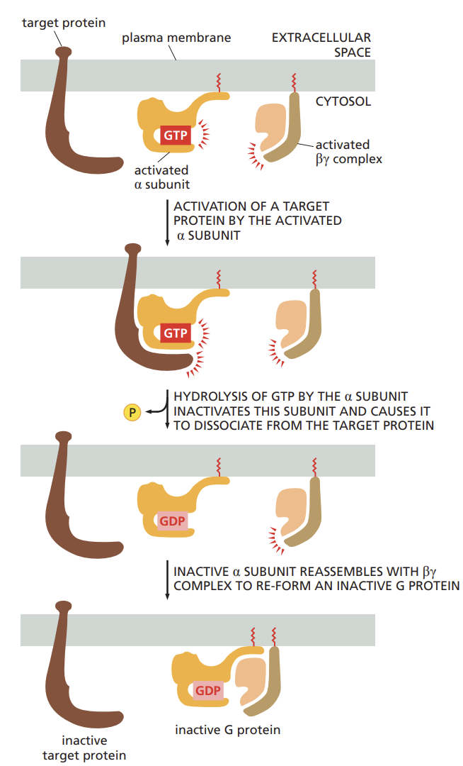
  • once activated by GTP binding many of these proteins have intrinsic GTP hydrolyzing (GTPase) activity, and they shut themselves off by hydrolyzing their bound GTP to GDP

13
New cards

Differentiate the three main classes of cell-surface receptors and provide an example of each.

-all are coupled receptors

1) ion-channel: change the permeability of the plasma membrane to select ions thereby altering the membrane potential and if the conditions are right producing an electrical current

2) g-protein: activate membrane-bound trimeric GTP-binding protein, which then activate/inhibit an enzyme or an ion channel in the plasma membrane, initiating an intracellular cascade

3) enzyme: either act as enzymes or associate with enzymes inside the cell. when stimulated the enzymes can activate a wide variety of intracellular signaling pathways

14
New cards

List some foreign substances that alter physiology by interacting with cell-surface receptors.

  • heroin, nicotine, tranqs and chili peppers

  • these substances either block or overstimulate the receptor's natural activity

15
New cards

Describe the type of signal transduction carried out by ion-channel-coupled receptors.

  • rapid transmissions of signals across the synapses in the nervous system

  • convert a chemical signal, a neurotransmitter, into an electrical chemical signal, voltage

  • when the neurotransmitter binds to ion channel-coupled receptors on the surface of a target cell, the receptor alters its conformation so as to open a channel in the target cell's membrane, making it permeable.

ex. Na+, K+, Ca 2+

16
New cards

structure of G-protein-coupled receptors (GPCRs)

  • each is made of a single polypeptide chain that threads back and forth across the lipid bilayer seven times

  • specific for a particular set of receptors and for a particular set of target enzymes or channels in the plasma membrane

17
New cards

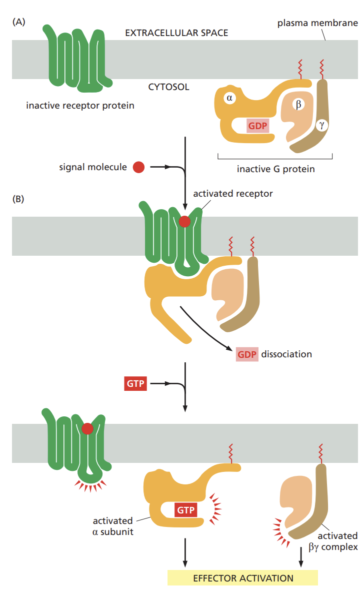
the general structure of a G protein and how the protein responds when activated by a GPCR

  • composed of three subunits (alpha beta and Y) two of which are tethered to the plasma membrane by short lipid tails.

  • in the unstimulated state, the alpha subunit is GDP bound and the g-protein is idle.

  • when an extracellular signal binds to its receptor the altered receptor activates a G-protein by causing the alpha subunit to decrease its affinity for GDP which is exchanged for GTP.

  • alpha subunit and the beta(y) can each interact directly with target proteins in the plasma membrane, which in turn may relay the signal to other destinations in the cell.

<ul><li><p>composed of three subunits (alpha beta and Y) two of which are tethered to the plasma membrane by short lipid tails. </p></li><li><p>in the unstimulated state, the alpha subunit is GDP bound and the g-protein is idle. </p></li><li><p>when an extracellular signal binds to its receptor the altered receptor activates a G-protein by causing the alpha subunit to decrease its affinity for GDP which is exchanged for GTP.</p></li><li><p>alpha subunit and the beta(y) can each interact directly with target proteins in the plasma membrane, which in turn may relay the signal to other destinations in the cell.</p></li></ul><p></p>
18
New cards

Summarize the factors that determine the duration of a GPCR-stimulated response

  • the longer the target proteins remain bound to an alpha subunit or beta(Y) complex the more prolonged the relayed signal will be.

<ul><li><p>the longer the target proteins remain bound to an alpha subunit or beta(Y) complex the more prolonged the relayed signal will be.</p></li></ul><p></p>
19
New cards

cholera toxin:

  • the protein cholera toxin enters the cell that line the intestine and modifies the alpha subunit of g-protein called Gs

  • this modification prevents Gs from hydrolyzing when its bound to GTP, thus locking the g-protein in an active state

  • results in diarrhea and dehydration

20
New cards

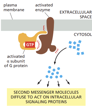
Relate the speeds of the responses produced by G proteins activating an ion channel versus activating a membrane-bound enzyme.

  • when g-protein interact with ion channels they cause an immediate change in the state and behavior of the cell

  • the interaction of activated g-proteins with enzymes are less rapid and more complex as they lead to the production of additional intracellular signalling molecules

21
New cards

Name the classes of enzymes that are the most frequent targets of G proteins, and list the second messenger molecules they produce

  • adenylyl cyclase and phospholipase C (PLC)

  • adenylyl cyclase produces a small molecule called cyclic AMP

  • PLC produces inositol trisphosphate and diacylglycerol.

<ul><li><p>adenylyl cyclase and phospholipase C (PLC)</p></li><li><p>adenylyl cyclase produces a small molecule called cyclic AMP</p></li><li><p>PLC produces inositol trisphosphate and diacylglycerol.</p></li></ul><p></p>
22
New cards

Outline how cyclic AMP is produced in response to G protein activation, and recall how caffeine can potentiate this response.

  • activated G protein alpha subunit switches on the adenylyl cyclase, causing an increase in the synthesis of cyclic AMP from ATP

  • to help terminate the signal, a second enzyme called cyclic AMP phosphodiesterase, rapidly converts cyclic AMP to ordinary AMP

  • caffeine: inhibits phosphodiesterase in the nervous system which blocks cyclic AMP degradation and keeps the concentration of this second messenger high.

23
New cards

Compare a signaling pathway in which cyclic AMP produces a response within seconds to one in which the response takes minutes or hours to develop.

  • cyclic AMP phosphodiesterase is continuously active inside the cell

  • eliminates cyclic AMP quickly

  • so the cytosolic concentration of this second messenger can change rapidly in response to extracellular signals rising or falling

  • Cyclic AMP is water soluble so it can carry the signal throughout the cell traveling from the site on the membrane where it is synthesized to interact with proteins located in the cytosol, nucleus, or on other organelles

24
New cards

Recall the location and action of the second messenger molecules produced by activated phospholipase C.

  • phospholipase C (PLC) activated by GCPRs or RTKs

  • once activated PLC propagates the signal by cleaving a lipid molecule that is a component of the plasma membrane

  • this cleavage produces two second messenger molecules: inositol 1,4,5-trisphosphate (IP3) and diacylglycerol (DAG)

<ul><li><p>phospholipase C (PLC) activated by GCPRs or RTKs</p></li><li><p>once activated PLC propagates the signal by cleaving a lipid molecule that is a component of the plasma membrane</p></li><li><p>this cleavage produces two second messenger molecules: inositol 1,4,5-trisphosphate (IP3) and diacylglycerol (DAG)</p><p></p></li></ul><p></p>
25
New cards

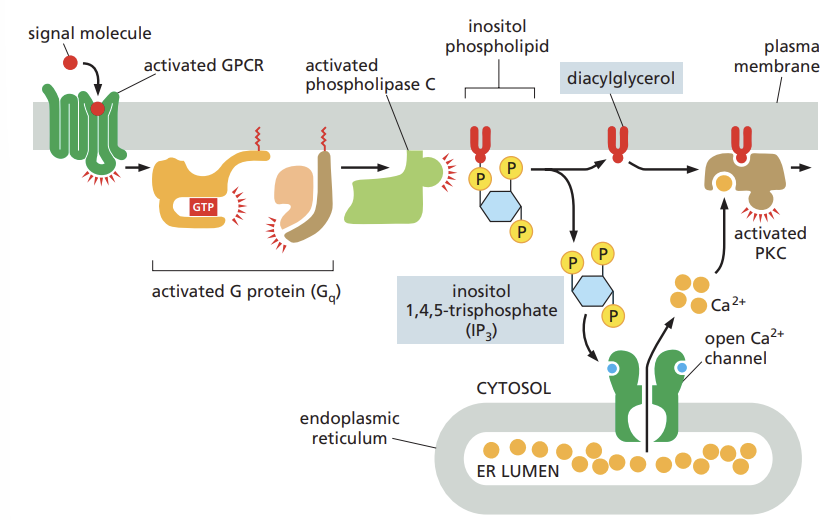
List several biological processes triggered by calcium ions.

  • when a sperm fertilizes an egg , Ca2+ channels open

  • a rise in cytosolic Ca 2+ triggers the egg to start development for muscles cells, a signal from nerve triggers a rise in cytosolic Ca 2+ that initiates muscle contraction.

  • in many secretory cell including nerve cells Ca 2+ triggers secretion

26
New cards

Explain how cells keep the concentration of calcium ions in the cytosol low and how they terminate a calcium ion signal

  • membrane-embedded Ca pumps actively remove Ca from the cytosol sending it either into the ER or across the plasma membrane and out of the cell

  • As a result a steep electrochemical gradient of Ca exist across both ER membrane and the plasma membrane.

27
New cards

Review how calcium-responsive proteins such as calmodulin propagate a calcium ion signal.

  • calmodulin is present in the cytosol

  • When Ca binds to calmodulin the protein undergoes a conformational change that enables it to interact with a wide range of target proteins in the cell altering their activity

  • target is the Ca/calmodulin-dependent protein kinases (CaM-kinases)

  • when these kinases are activated by binding to calmodulin complexes with Ca, they influence other processes in the cell by phosphorylating selected proteins

28
New cards

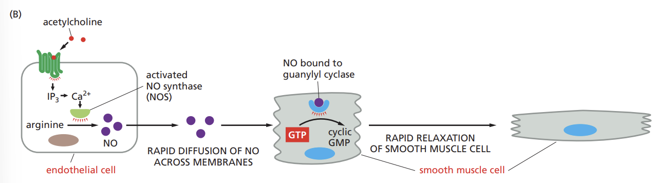
Outline how the gas nitric oxide (NO) can act as a signaling molecule to trigger the relaxation of smooth muscle cells.

  • nitric oxide diffuses readily from its site of synthesis and slips into neighboring cells,

  • the distance the gas diffuses is limited by its reaction with oxygen and water in the extracellular environment, which converts NO into nitrates and nitrites within seconds.

29
New cards

Recall why nitric oxide acts as a paracrine signal only on cells near its site of synthesis.

  • highly reactive gas molecule with a very short half-life, so it quickly breaks down, limiting its ability to reach distant cells

<ul><li><p>highly reactive gas molecule with a very short half-life, so it quickly breaks down, limiting its ability to reach distant cells</p></li></ul><p></p>
30
New cards

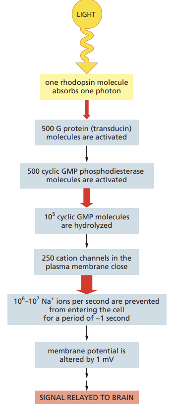
Outline how GPCRs in the photoreceptors of the retina transmit an extremely rapid signal in response to stimulation by light.

  • But photoreceptors also provide a beautiful illustration of the advantage of intracellular signaling cascades

  • in particular, such cascades allow spectacular amplification of the incoming signal and allow cells to adapt so as to be able to detect signals of widely varying intensity.

31
New cards

Summarize how adaptation in the intracellular signaling cascade of photoreceptors allows the eye to respond to dim or bright light.

  • adaptation occurs in intracellular signaling pathways that respond to extracellular signal molecules

  • allows cells to respond to fluctuations in the concentrations

  • positive and negative feedback mechanisms allow cells to respond equally well to the signaling equivalents of shouts and whispers.

<ul><li><p>adaptation occurs in intracellular signaling pathways that respond to extracellular signal molecules</p></li><li><p>allows cells to respond to fluctuations in the concentrations</p></li><li><p>positive and negative feedback mechanisms allow cells to respond equally well to the signaling equivalents of shouts and whispers.</p></li></ul><p></p>
32
New cards

enzyme-coupled receptors

  • transmembrane proteins that display their ligand-binding domains on the outer surface of the plasma membrane

  • instead of associating with a G-protein however the cytoplasmic domain of the receptor either acts as an enzyme itself or forms a complex with another protein that acts as an enzyme.

33
New cards

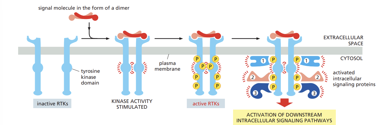
Review how the binding of a signal molecule activates RTKs to trigger the assembly of an intracellular signaling complex

  • phosphorylation of tyrosines on the receptor tails triggers the assembly of an intracellular signaling complex on the tails

  • The newly phosphorylated tyrosines serve as binding sites for a variety of signaling proteins that then pass the message on to yet other proteins

  • the binding of a signaling molecule with an RTK activates tyrosine kinase in the cytoplasmic tail of the receptor

<ul><li><p>phosphorylation of tyrosines on the receptor tails triggers the assembly of an intracellular signaling complex on the tails</p></li><li><p>The newly phosphorylated tyrosines serve as binding sites for a variety of signaling proteins that then pass the message on to yet other proteins</p></li><li><p>the binding of a signaling molecule with an RTK activates tyrosine kinase in the cytoplasmic tail of the receptor</p></li></ul><p></p>
34
New cards

Recall how signals transmitted by RTKs can be terminated.

  • tyrosine phosphorylation is reversed by tyrosine phosphatases which remove the phosphates that were added to the tyrosine of both the RTKs and other intracellular signaling proteins in response to the extracellular signal

  • in some cases activated RTKs are inactivated in a more brutal way: they are dragged into the interior of the cell by endocytosis and then destroyed by digestion in the lysosomes.

35
New cards

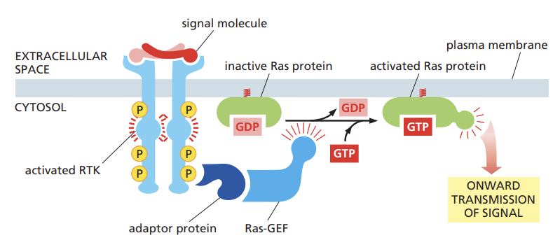
outline how intracellular signaling proteins activate RTKs and travel

  • A signal molecule activates the RTK, which then phosphorylates itself

  • Adaptor proteins bind to the phosphorylated RTK and recruit SOS, a Ras-GEF, which activates Ras by exchanging GDP for GTP

  • Active Ras then transmits the signal downstream, leading to cellular responses.

<ul><li><p>A signal molecule activates the RTK, which then phosphorylates itself</p></li><li><p>Adaptor proteins bind to the phosphorylated RTK and recruit SOS, a Ras-GEF, which activates Ras by exchanging GDP for GTP</p></li><li><p>Active Ras then transmits the signal downstream, leading to cellular responses.</p></li></ul><p></p>
36
New cards

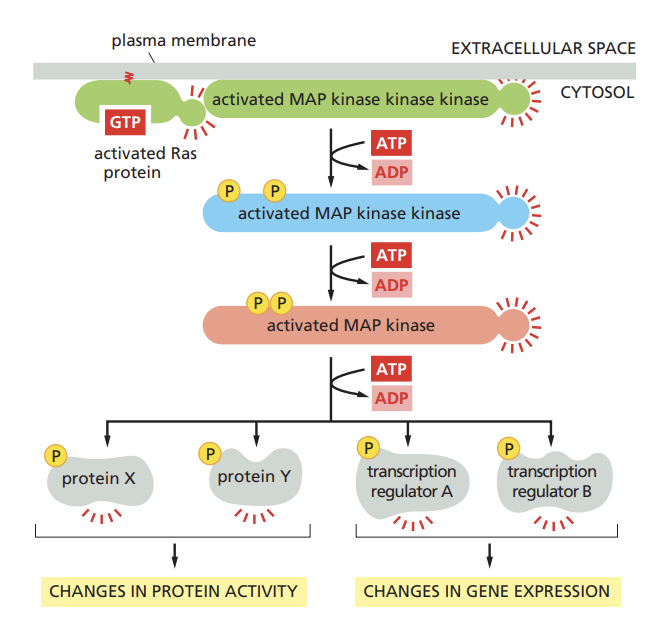
Outline how RTKs activate the MAP kinase signaling module.

  • When Ras is activated (by binding GTP), it initiates a phosphorylation cascade

  • This cascade involves a series of serine/theronine kinases being activated in sequence

  • Phosphorylation can change the activity, localization, or interactions of proteins, leading to cellular responses like gene expression or changes in cellular behavio

<ul><li><p>When Ras is activated (by binding GTP), it initiates a phosphorylation cascade</p></li><li><p>This cascade involves a series of serine/theronine kinases being activated in sequence</p></li><li><p>Phosphorylation can change the activity, localization, or interactions of proteins, leading to cellular responses like gene expression or changes in cellular behavio</p></li></ul><p></p>
37
New cards

Indicate how Ras can fuel uncontrolled proliferation in cancer.

  • a mutant form of Ras inactivates the GTPase activity of Ras

  • so that the protein cannot shut itself off

  • promotes uncontrolled cell proliferation and the development of cancer

38
New cards

Review how extracellular signals that promote cell growth and survival activate PI-3-kinase signaling pathways.

  • one important signaling pathway that these RTKs activate to promote cell growth and survival involves the enzymes phosphoinositide 3-kinase(PI 3-Kinase) which phosphorylates inositol phospholipids in the plasma membrane

  • these phosphorylated lipids serve as docking sites for specific intracellular signaling proteins, which relocate from the cytosol to the plasma membrane, where they can activate one another.

<ul><li><p>one important signaling pathway that these RTKs activate to promote cell growth and survival involves the enzymes phosphoinositide 3-kinase(PI 3-Kinase) which phosphorylates inositol phospholipids in the plasma membrane</p></li><li><p> these phosphorylated lipids serve as docking sites for specific intracellular signaling proteins, which relocate from the cytosol to the plasma membrane, where they can activate one another.</p></li></ul><p></p>
39
New cards

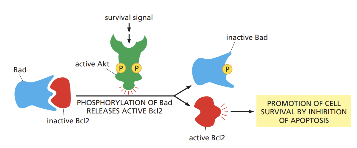
Compare how Akt promotes cell survival via Bad and stimulates cell growth via Tor.

  • akt (AKA serine/threonine kinase or protien kinase b) promotes cell growth and survival of many cell types often by inactivating the signaling proteins it phosphorylates

  • Akt phosphorylates and inactivates a cytosolic protein called bad

  • in its active state, Bad encourages the cell to kill itself by indirectly activating a cell-suicide programed called apoptosis

  • phosphorylation by Akt thus promotes cell survival by inactivating a protein that otherwise promotes cell death.

<ul><li><p>akt (AKA serine/threonine kinase or protien kinase b) promotes cell growth and survival of many cell types often by inactivating the signaling proteins it phosphorylates</p></li><li><p>Akt phosphorylates and inactivates a cytosolic protein called bad </p></li><li><p>in its active state, Bad encourages the cell to kill itself by indirectly activating a cell-suicide programed called apoptosis</p></li><li><p>phosphorylation by Akt thus promotes cell survival by inactivating a protein that otherwise promotes cell death.</p></li></ul><p></p>
40
New cards

Review how different types of receptors can trigger a rise in the cytosolic concentration of calcium ions.

  • intracellular Ca2+ stores and external Ca2+ entering across the plasma membrane

  • intracellular Ca2+ release mechanisms involves the phosphoinositide specific phospholipase C (PI-PLC)-derived second messenger IP3, which acts by binding to a specific receptor on the endoplasmic reticulum and releases Ca 2+

41
New cards

Describe a method to identify proteins that interact in response to stimulation by an extracellular signal.

  • one involves using a protein as "bait" isolating the receptor that binds to insulin, one could attach insulin a chromatography column

  • cells that respond to the hormone are broken open with detergents that disrupt their membrane, releasing the transmembrane receptor proteins

  • protein-protein interactions in a signal pathway can also be identified by co-immunoprecipitation.

42
New cards

Outline how a set of mutant RTKs can be used to determine which tyrosines serve as docking sites for the intracellular signaling proteins that propagate the signal.

  • a series of mutant receptors can be constructed, each missing a different from its cytoplasmic domain

  • in this way, the specific tyrosine required for binding can be determined

  • similarly one can determine whether this phosphotyrosine docking site is required for the receptor to transmit a signal to the cell

43
New cards

Review how a technology such as RNA interference or CRISPR can be used to assess the importance of a particular protein in a signaling pathway.

  • the activity of a specific signaling protein can be inhibited or eliminated

  • in this case of Ras, one could shut down the expression of the Ras gene in cells by RNA interference or CRISPER

  • such cells do not proliferate in response to extracellular mitogens, indicating the importance of normal Ras signaling in the proliferative response

44
New cards

Explain how mutant proteins can be used to determine the order in which proteins participate in a signaling pathway.

  • by examining other mutant animals, many of the genes that encode the protein involved in a signaling pathway can be identified

  • to determine whether these proteins lie upstream or downstream of Ras one could create cells that express an inactive mutant to form of each protein and then ask whether these mutant cells can be "rescued" by the addition of a continuously active form of Ras.

45
New cards

46
New cards

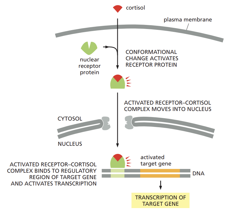
Outline how steroid hormones trigger the transcription of different sets of target genes.

  • cortisol, estradiol, and testosterone and the thyroid hormones such as thyroxine are hydrophobic molecules that pass through the plasma membrane of the target cell and bind to receptor proteins located in either the cytosol or the nucleus

  • regardless of their initial location, these intracellular receptor proteins are referred to as nuclear receptors because when activated by hormone binding they enter the nucleus, where they regulate transcription.

<ul><li><p>cortisol, estradiol, and testosterone and the thyroid hormones such as thyroxine are hydrophobic molecules that pass through the plasma membrane of the target cell and bind to receptor proteins located in either the cytosol or the nucleus</p></li><li><p>regardless of their initial location, these intracellular receptor proteins are referred to as nuclear receptors because when activated by hormone binding they enter the nucleus, where they regulate transcription.</p></li></ul><p></p>
47
New cards

Contrast the cell signaling systems used by plants and animals.

  • plants have hundreds of genes encoding receptor serine and threonine kinases

  • but they are structurally different then the receptors found in animals

  • the plant receptors are thought to play an important part in a large variety of cell signaling processes including those governing plant growth, development and disease resistant

  • in contrast to animal cells, plant cells seem not to use RTKs, steroid hormones type nuclear receptors or cyclic AMP they seem to use few GPCRs.

48
New cards

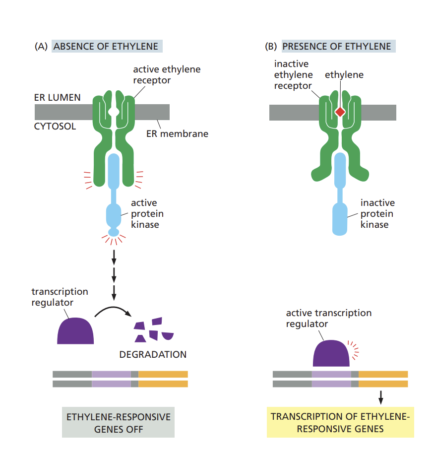
Describe how the ethylene signaling pathway regulates ripening of fruits.

  • ethylene receptor is located in ER

  • it is active in the absence of ethylene, and inactive when ethylene is present

presence of ethylene:

  • ethylene binds to the receptor and causes a conformational change in the receptor, which activates the signaling pathway

  • the protein kinase is deactivated and the the transcription regulator is activated

absence of ethylene:

  • the receptor interacts with and activates a protein kinase which inhibits downstream signaling

<ul><li><p>ethylene receptor is located in ER</p></li><li><p>it is active in the absence of ethylene, and inactive when ethylene is present </p></li></ul><p>presence of ethylene: </p><ul><li><p>ethylene binds to the receptor and causes a conformational change in the receptor, which activates the signaling pathway</p></li><li><p>the protein kinase is deactivated and the the transcription regulator is activated </p></li></ul><p>absence of ethylene: </p><ul><li><p> the receptor interacts with and activates a protein kinase which inhibits downstream signaling</p></li></ul><p></p>
49
New cards

Explain how multiple signaling pathways can integrate information to produce a coordinated cell response.

connection and interactions that occur between different signaling pathways

<p>connection and interactions that occur between different signaling pathways</p><p></p>
50
New cards
Chapter 17: Contrast the structures of the subunits that form intermediate filaments, actin filaments, and microtubules.
cytoskeleton is built on a framework of three types of protein filaments: intermediate filament, microtubules, and actin filament. Each type of filament has distinct mechanical properties and is formed from a different protein subunit. a family of fibrous proteins forms the intermediate filaments; globular tubulin subunits form microtubules; and globular actin subunits form actin filaments
51
New cards
Describe the location and main function of intermediate filaments.
intermediate filaments main function is to enable cells to withstand the mechanical stress that occurs when cells are stretched. in the smooth muscle cells where they were first discovered, their diameter is about 10 nm is between that of the thinner actin filaments and the thicker myosin filaments, are the toughest and most durable of the cytoskeleton filaments. found in the cytoplasm of most animal cells typically form a network throughout the cytoplasm surrounding the nucleus and extending out to the cell periphery
52
New cards
Recall how the structure of intermediate filaments relates to their strength and durability.
is like a rope in which many long strands are twisted together to provide tensile strength- an ability to withstand tension without breaking. fibrous subunits each containing central elongated rod domain with distinct unstructured domains at either end.
53
New cards
Summarize how intermediate filaments are assembled and describe their polarity.
the rod domain consists of an extended alpha helical region that enables pairs of intermediate filament proteins to form stable dimers wrapping around each other in a coiled-coil configuration. two of these coiled-coil dimers running in opposite directions associate to form a staggered tetramer. these tetramers are soluble subunits of intermediate filaments. because the two dimers point in the opposite directions both ends of the tetramer are the same as the two ends of assembled intermediate filaments. depend on noncovalent bonding; it is the combined strength of the overlapping lateral interactions along the length of the proteins that gives intermediate filaments their great tensil strength.
54
New cards
Review how intermediate filament proteins can differ from one another and how these differences relate to the function of the intermediate filament.

the central rod domains of different intermediate filaments proteins are all similar in size and amino acid sequence so that when they pack together they always form filaments of similar diameter and internal structure. by contrast, the terminal head and tail domains vary greatly in both size and amino acid sequence from one type of intermediate filament protein to another. these unstructured domains are exposed on the surface of the filaments, where they allow it to interact with specific components in the cytoplasm

1) keratin filaments in epithelial cells

2) vimentin and vimentin related filaments in connective tissue cells, muscle cells, and supporting cells of the nervous system

3) neurofilaments in nerve cells

4) nuclear lamins which strengthen the nuclear envelope

55
New cards
Describe three disorders that involve defects in intermediate filaments.

keratin genes mutation can form a rare human disease called epidermolysis bullosa simplex where the skin is highly vulnerable to mechanical injury, and even gentle pressure can rupture its cells, causing the skin to blister.

neurofilaments can form neurodegenerative disease amyotrophic lateral sclerosis. axon degeneration and muscle weakness and muscle weakness seen in patients

56
New cards
Compare the structure of the nuclear lamina with that of cytoplasmic intermediate filaments.
whereas cytoplasmic intermediate filaments form ropelike structures the intermediate filaments lining and strengthening inside surface of inner nuclear membrane are organized as two dimensional meshwork. the intermediate filaments that form this tough nuclear lamina are constructed from a class of intermediate filament proteins called lamins. the nuclear lamina disassembles and re-forms at each cell division when the nuclear envelope breaks down during mitosis and then reforms in the daughter cells.
57
New cards
Review how the nuclear lamina disassembles and re-forms during each cell division
collapse and reassembly of the nuclear lamina is controlled by the phosphorylation and dephosphorylation of the lamins. phosphorylation of lamins by protein kinases weakens the interactions between the lamin tetramers and causes the filaments to fall apart. dephosphorylation by protein phosphatases at the end of mitosis allows the lamins to reassemble
58
New cards
Recall how intermediate filaments are stabilized by cross-linking accessory proteins, and explain how these proteins help to position the nucleus within the cell interior.
intermediate filaments are further stabilized and reinforced by accessory proteins such as plectin that cross link the filaments into the bundles and connect them to microtubules to actin filaments and to adhesive structures in desmosomes. plectin and other proteins also interact with protein complexes that link the cytoplasmic cytoskeleton to structures in the nuclear interior, including chromosomes and nuclear lamina. these bridges which span the cytoskeleton and they are involved in many processes, including the movement and positioning of the nucleus within the cell interior and the overall organization of the cytoskeleton.
59
New cards
Review the general location and functions of microtubules
these long and relatively stiff, hollow tubes of proteins can rapidly disassemble in one location and reassemble one another. in a typical animal cell microtubules grow from a small structure near the center of the cell called the centromere. extending out towards the cell periphery, they create a system of tracks within the cell along with vesicles, organelles and other cell components can be transported. the cytoplasmic microtubules are the part of the cytoskeleton mainly responsible for transporting and positioning membrane-enclosed organelles within the cell for guiding the intracellular transport of various cytosolic macromolecules
60
New cards
List several examples of organizing centers from which microtubules grow
extending out towards the cell periphery, they create a system of tracks within the cell along with vesicles, organelles and other cell components can be transported. the cytoplasmic microtubules are the part of the cytoskeleton mainly responsible for transporting and positioning membrane-enclosed organelles within the cell for guiding the intracellular transport of various cytosolic macromolecules
61
New cards
Describe the structure of microtubules and recall how microtubules are assembled from tubulin dimers
molecules of tubulin each of which is a dimer composed of two very similar globular proteins called alpha-tubulin and beta-tubulin, bound tightly together again by noncovalent interactions. the tubulin dimers stack together again by noncovalent bonding to form the wall of hollow, cylindrical microtubule.
62
New cards
State the polarity of a microtubule and summarize how this polarity affects its assembly and function.
tubelike structure is made of 13 parallel protofilaments, each linear chain of tubulin dimers with alpha and beta tubulin alternating along its length. each protofilament has a structural polarity with alpha tubulin exposed at one end and beta tubulin at the other and this polarity is the same for all the protofilaments in the microfilaments. thus the microtubule as a whole has a structural polarity the end with beta tubulin showing is called plus end and the opposite end which contains exposed alpha tubulin is called minus end
63
New cards
Describe the structure of a centrosome and review how the centrosome nucleates the growth of microtubules
the centromere--which is typically close to the cell nucleus when the cell is not in mitosis--organizes and array of microtubules that radiates outward through the cytoplasm. the centrosome consists of a pair of centrioles surrounded by a matrix of proteins. the centrosome matrix includes hundreds of ring shaped structures formed from a special type of tubulin called y-tubulin and each y-tubulin ring complex serves as the starting point or nucleation site for the growth of one microtubule.
64
New cards
Explain why microtubules require organizing centers such as centrosomes to nucleate their growth.
it is much harder to start a new microtubule from scratch, by first assembling a ring of alpha/beta tubulin dimers, than it is to add such dimers to a preexisting y-tubulin ring complex. although purified alpha/bet-tubulin dimers at a high concentration can polymerize into microtubules spontaneously in vitro, the concentration of free alpha/beta in a living cell is too low to drive the difficult first step of assembling the initial ring of a new microtubule. by providing organizing centers at specific sites and keeping the concentration of free alpha/beta-tubulin dimers low cells can control precisely where microtubules form
65
New cards
Describe dynamic instability and indicate how this behavior relates to microtubule function.
behavior of switching back and forth between polymerization and depolymerization. it allows microtubules to undergo rapid remodeling and is crucial for their function. the microtubule may shrink partially and then to no less suddenly start growing again or it may disappear completely to be replaced by new microtubule that grows from the same y-tubulin ring complex.
66
New cards
Summarize how dynamic instability is controlled by GTP hydrolysis.
the dynamically instability of microtubules stems from the intrinsic capacity of tubulin dimers to hydrolyze GTP. this energetically favorable reaction which generates GDP and inorganic phosphate is similar to the hydrolysis of ATP. each free tubulin dimer contains one GTP molecule tightly bound to a beta-tubulin, which hydrolyzes the GTP to GDP shortly after the dimer is added to a growing microtubule. the GDP produced by this hydrolysis remains tightly bound to the beta-tubulin.
67
New cards
Contrast the actions of colchicine and Taxol and explain why both are used to treat human cancers

colchicine: which binds tightly to free tubulin dimers and prevents their polymerizations into microtubules, the mitotic spindle rapidly disappears the cell stalls in the middle of mitosis unable to partition the chromosomes into two groups. the mitotic spindle is normally maintained by a balanced addition and loss of tubulin subunits; when tubulin addition is blocked by colchicine tubulin loss continues until the spindle disappears

taxol: its binds tightly to microtubules and prevents them from losing subunits. because new subunits can still be added the microtubules can grow but cannot shrink.

68
New cards
Recall how and why cells modify the dynamic instability of their microtubules.
cells are able to modify the dynamic instability of their microtubules for particular purposes. microtubules become more dynamic, switching between growing and shrinking much more frequently than cytoplasmic microtubules normally do. this change enables microtubules to disassemble rapidly and then reassembles into the mitotic spindle. when a cell has differentiated into a specialized cell type, the dynamic instability of its microtubules is often suppressed by proteins that bind to the ends or sides of the microtubules and protect them against disassembly. the stabilized microtubules then serve to maintain the organization of the differentiated cell
69
New cards
Compare the movement of cell components—including organelles, membrane vesicles, and macromolecules—by free diffusion and by microtubule-guided transporting
it is cytoplasm is seen to be continual motion, the mitochondria and the smaller membrane enclosed organelles and vesicles travel in small jerky steps- moving for a short period stopping then moving. This saltatory movement is much more sustained and directional than the continual small, brownian movements caused by random thermal movements. these movements can occur along either microtubules or actin filaments. in both cases the movements are driven by motor proteins, which use the energy derived from repeated cycles of ATP hydrolysis to travel steadily along the microtubules or actin filament in a single direction. because motor proteins also attach to other cell components they can transport this cargo along the filaments
70
New cards
Outline how microtubules participate in cell polarization
most differentiated animal cells are polarized; that is one of the cell is structurally or functionally different from the other. cells specialized for secretion have their golgi apparatus positioned toward the site of secretion. the cells polarity is a reflection of the polarized system of microtubules in its interior, which help to position organelles in their required location within the cell and to guide the streams of vesicular and macromolecular traffic moving between one part of the cell to another.
71
New cards
Compare kinesins and cytoplasmic dyneins in terms of their structure, their movement along the microtubule, and how they interact with cargo.
the motor proteins that move along cytoplasmic microtubules, such as those in the axon of a nerve cell, belong to two families; the kinesins generally move toward the plus end of a microtubule (outward from the cell body), the dyeins move toward the minus end (toward the cell body). kinesins and cytoplasmic dyneins are generally dimers that have two globular ATP-binding heads and a single tail members of second class of dyneins. they both interact with microtubules in a stereospecific manner so that the motor protein will attach to a microtubules in only one direction. the tail of motor protein binds stably to some cell component such vesicle or organelle and there by determines the type of cargo that the motor protein transports.
72
New cards
Compare the roles that kinesins and cytoplasmic dyneins have in positioning the organelles in a eukaryotic cell, and describe the effect that colchicine treatment has on organelle placement
kinesins attach to the outside of the ER membrane pull the ER outward along the microtubule stretching it like a net. cytoplasmic dyneins attach to the golgi membranes pull the golgi apparatus along microtubules in the opposite direction, inward toward the nucleus. but when treated with colchicine a drug that promotes microtubule disassembly both the ER and Golgi apparatus change their location dramatically. the Er which is physically connected to the nuclear envelope collapses around the nucleus and the golgi which is not attached to any other organelle fragments into small pieces small vesicles which then disperse throughout the cytoplasm.
73
New cards
Summarize how fluorescent marker proteins and non-hydrolyzable ATP analogs can be used to study the activity of motor proteins such as kinesin or myosin.
observation of kinesin molecules labeled with a fluorescent marker protein revealed that this motor protein marches along microtubules processively-that is each molecule takes multiple steps along the filaments before falling off. combingine of these observations with assays of ATP hydrolysis, researchers have confirmed that one molecule of ATP is hydrolyzed per step. kinesin can move in a processive manner because it have two heads. this enables it to walk toward the plus end of the microtubules in a "hand over hand fashion"
74
New cards
Compare the functions and movements of cilia and flagella

-cilia are hairlike structures covered in plasma membrane, that extend from the surface of many kinds of eukaryotic cells. each cilium contains a core stable microtubules arranged in a bundle, that grow from a cytoplasmic basal body which serves as an organization center. motile cilia beat in a whiplike fashion either to move fluid over the surface of a cell or to propel single cells through a fluid.

-flagella that propel sperm and many protozoa are usually much longer than cilia are. they are designed to move the entire cell rather than moving fluid across the cell surface. flagella propagate regular waves along their length, propelling the attached cell along.

75
New cards
Describe the arrangement of microtubules inside a cilium or flagellum.

each cilium contains a core stable microtubules arranged in a bundle, that grow from a cytoplasmic basal body which serves as an organization center.

the movement of a cilium or flagellum is produced by bending that takes place as its microtubules slide against each other. the microtubules are associated with numerous accessory proteins which project a regular positions along the length of the microtubule bundle.

76
New cards
Outline how ciliary dynein allows a cilium to bend.
ciliary dynein is attached by its tail to one microtubules in each other doubtlet while its globular heads interact with the adjacent microtubules to generate a sliding force between the two microtubules. because of the multiple links the adjacent microtubules is converted to a bending motion.
77
New cards
List several cell structures formed by actin filaments.

depending on which of these proteins they associate with actin filaments can form stiff and stable structures such as:

microvilli on the epithelial cells lining the intestine

small contractile bundles that contract and act like tiny muscles in most animal cells. they also can form temporary structures such as dynamic protrusions formed at the leading edge of a crawling cell or the contractile ring that pinches the cytoplasm in two when an animal cell divides

78
New cards
Compare actin filaments and microtubules in terms of width, length, polarity, and cross-linking.
each filament is a twisted chain of identical globular actin monomers of all which point in the same direction along the axis of the chain. like a microtubule therefore an actin filament has a structural polarity with a plus end and minus end. actin filaments are thinner, more flexible and usually shorter than microtubules, there are however many more of them so the total length of all actin filaments in a cell is generally many times greater than the total length of all the microtubules. they are generally found in cross-linked bundles and networks which are much stronger than individual filaments
79
New cards
Compare the polymerization of actin filaments to that of microtubules.
like microtubules, actin filaments can grow by the addition of monomers at either end but like their rate of growth is faster at the plus end than at the minus end. a naked actin filament like a microtubule without associated proteins is inherently unstable and it can disassemble from both ends. in living cells free actin monomers carry a tightly bound nucleoside triphosphate in this case ATP.
80
New cards
Explain treadmilling and identify the conditions under which this behavior takes place.
at the minus end in contrast ATP is hydrolyzed faster than a new monomers can be added; because ADP actin destabilizes the structure the filaments loses subunits from its minus end at the same time as it adds them to the plus end. individual monomers this move through the filament from the minus end a behavior called treadmilling. when the rates of addition and loses are equal the filament remains the same size
81
New cards
Compare the actions of cytochalasin and phalloidin and describe their effects on cell behavior.
actin filament function can be perturbed experimentally by certain toxins such as cytochalasin to prevent actin polymerization or others such as phalloidin stabilize actin filaments against depolymerization. addition of these toxins to cells or tissues even in low concentration instantaneously freezes cell movements such as cell locomotion. thus as with microtubules many of the functions of actin filaments depend on the ability of the filament to assemble and disassemble. the rates of which depend on the dynamic equilibrium between the actin filaments, the pool actin monomers and various actin-binding proteins
82
New cards
Outline the functions of common actin-binding proteins, including thymosin, profilin, formins, actin-related proteins (ARPs), and myosin

cells contain small proteins such thymosin and profilin that bind to actin monomers in the cytosol, preventing them from adding to the ends of the actin filaments. by keeping actin monomers in reserve until required these proteins play crucial role in regulating actin polymerization. when actin filaments are needed other actin binding proteins such as forminins and actin related proteins to promote actin polymerization.

-Myosins are a large super-family of motor proteins that move along actin filaments, while hydrolyzing ATP to forms of mechanical energy that can be used for a variety of functions such as muscle movement and contraction

83
New cards
Describe the structure and function of the cell cortex.
highly concentrated in a layer just beneath the plasma membrane called the central cortex. actin filaments are linked by actin binding proteins into a meshwork that supports the plasma membrane and gives it mechanical strength. the rearrangement of actin filaments within the cortex provide much of the molecular basis for changes in both cell shape and cell locomotion
84
New cards
Differentiate between lamellipodia and filopodia.
a crawling fibroblast in culture regularly extends thin, flattened lamellipodia at its leading edge. these extensions contain a dense meshwork of actin filaments oriented so that most of the filaments have their plus end close to the plasma membrane. many cells also extend thin, stiff protrusions called filopodia both at the leading edge and elsewhere on the surface. are both exploratory motile structures that form and retract with great speed, moving at around 1 um per second. these protrusions are thought to be generated by the rapid local growth of actin filaments, which assemble close to the plasma membrane and elongated by the additions of actin monomers at their plus end.
85
New cards
Distinguish the roles played by actin in the protrusion, attachment, and contraction involved in cell movement
the molecular mechanisms of these and other forms of cell crawling entail coordinated changes among many molecules in different regions of the cell; there is no single easily identifiable locomotory organelle such as flagellum, three interrelated processes are known as essential (1) the cell sends out protrusions at its "front" or leading edge. (2) these protrusions adhere to the surface over which the cell is crawling and (3) the rest of the cell drags itself forward by traction on these points of anchorage
86
New cards
Explain how ARPs and formins aid in the assembly and extension of protrusions at the cell's leading edge.
promote the formation of a web of branched actin filaments in a lamellipodia. ARPs form complexes that bind to the sides of existing actin filaments and nucleate the formation of new filaments, which grow out at an angle to produce side branches. with the aid of additional actin binding proteins. this web undergoes continual assembly at the leading edge and disassembly further back, pushing the lamellipodium forward.
87
New cards
Summarize how different members of the Rho family of GTPases alter the organization of actin filaments
these extracellular signals act through a variety of cell-surface receptor proteins, which activate various intracellular pathways. many of these pathways converge on a group of closely related monomeric GTPases that are part of the Rho protein family. monomeric GTPases behave as molecular switches that control intracellular processes by cycling between an active GTP-bound site and an inactive GDP-bound state. different RHO family members alter the organizations of actin filaments in different ways
88
New cards
Compare the structure, binding properties, and general function of myosins I and II.

myosin-1 molecules which are present in all cell types, have a head domain and a tail. the head domain binds to an actin filament and has the ATP hydrolyzing motor activity that enables it to move along the filament in a repetitive cycle of binding, detachment and rebinding. the tail varies among the different types of myosin-1 and determines what type of cargo the myosin will carry.

myosin 2: structurally and mechanistically more complex, muscle cells use of its specialized form to drive muscle contraction. these are proteins who are dimers with two globular ATPases heads at one end and a single coiled-tail at the other. they bind to each other to form a bipolar myosin filament, which smaller contractile muscle contractions

89
New cards

List tissues in which actin and myosin filaments are organized in contractile bundles

  • myosin filament: double headed arrow, with two sets of myosin heads pointing outwards

  • one set binds to actin filaments in one orientation and moves the filaments one way; the other set binds to other actin filaments in the opposite orientation and moves the filaments in the opposite direction

  • the myosin filaments slides sets of oppositely orientated actin filaments past one another

  • if actin filament and myosin filaments are organized together in a bundle, the bundle can generate a strong contractile force

  • clearly seen in muscle contraction, but it also occurs in the much smaller contractile bundles of actin filaments and myosin-2 filaments that assemble transiently in nonmuscle cells, and in the contractile ring that pinches dividing cells into two.

90
New cards
Distinguish the structures of a skeletal muscle fiber and a myofibril.

skeletal muscles are huge, multinucleated individual cells formed by the fusion of many smaller cells. the nuclei of the contributing cells are retained in the muscle fiber and lie just beneath the plasma membrane.

-the bulk of the cytoplasm is made up of myofibrils, the contractile elements of the muscle cell. these cylindrical structures are 1-2 um in diameter and maybe as long as a muscle cell itself. each consists of a chain of identical tiny contractile units or sarcomeres.

91
New cards
Describe the structure of a sarcomere.
each sarcomere is about 2.5 um long and the repeating pattern of sarcomeres gives the vertebrate myofibril a striped appearance. sarcomeres are highly organized assemblies of two types of filaments--actin filaments and myosin filaments composed of a muscle specific form for myosin-2
92
New cards
Outline the sliding-filament mechanism of muscle contraction and relaxation, and describe how ATP binding and hydrolysis drive the conformational changes that underlie this movement.
when a muscle is stimulated to contract, the myosin heads start to walk along the actin filament in repeated cycles of attachment and detachment. during each cycle a myosin head binds and is hydrolyzes one molecule of ATP. this causes a series of conformational changes that move the tip of the head by about 5nm along the actin filament toward the plus end. this movement, repeated with each round of ATP hydrolysis propels the myosin molecule unidirectionally along the actin filament.
93
New cards
Explain how excitation of the muscle cell membrane triggers a rise in the cytosolic concentration of calcium ions.
the neurotransmitter released from the nerve terminal triggers an action potential in the muscle cell plasma membrane. this electrical excitation spreads in a matter of millisecond into a series of membranous tubes, called transverse ( or T) tubules that extend inward from the plasma membrane around each myofibril. the concentration of Ca (2+) in the cytosol rises, Ca (2+) binds to tropomyosin and induces the change in shape.
94
New cards
Summarize how calcium ions trigger muscle contraction and how contraction is subsequently reversed
in response to electrical excitation, which passes along the plasma membrane and to the T tubules, much of this Ca (2+) is released in the cytosol through a specialized set of ion channels that open in the sarcoplasmic reticulum membrane. in rise in cytosolic Ca (2+) concentration activates a molecular switch made of specialized accessory proteins closely associated with the actin filaments.
95
New cards
Explain how calcium ions stimulate contraction in nonmuscle cells or in smooth muscle, and compare this mode of activation with that of skeletal muscle
myosin-2 in nonmuscle cells is also activated by a rise in cytosolic Ca (2+) but the mechanism of activation is different from that of the muscle-specific myosin-2. an increase in Ca (2+) leads to the phosphorylation of nonmuscle myosin-2, which alters the myosin conformation and enables it to interact with actin. a similar activation mechanism operates in a smooth muscle that is present in the walls of a lot of different areas. this mode of myosin activation is relatively slow because time is needed for enzyme molecules to diffuse to the myosin heads and carry out the phosphorylation, and subsequent dephosphorylation. However, this mechanism has the advantage that--unlike the mechanism used by skeletal muscle cells.
96
New cards

paracrine signaling

signal diffuses locally through the extracellular fluid, local mediators on to nearby cells

97
New cards

neuronal signaling:

signaling that is delivering messages quickly/ specifically to individual target cells through private lines, extracellular signal is called a neurotransmitter

98
New cards

contact dependent signaling

allows adjacent cells that initially similar to become specialized to form different cell types

99
New cards

receptor protein

  • performs the primary step in signal transduction

  • recognizes the extracellular signal generates new intracellular signals in response.

100
New cards

RTKs (receptor tyrosine kinases):

  • activated in response to extracellular signals

  • consists of receptors with a cytoplasmic domain that functions as a tyrosine kinase, which phosphorylates tyrosines on specific intracellular signaling proteins

Explore top flashcards

Polyatomic Ions
Updated 892d ago
flashcards Flashcards (25)
Module 31
Updated 912d ago
flashcards Flashcards (25)
science
Updated 805d ago
flashcards Flashcards (85)
MKTG Research Final
Updated 426d ago
flashcards Flashcards (138)
Sat vocab
Updated 136d ago
flashcards Flashcards (110)
Polyatomic Ions
Updated 892d ago
flashcards Flashcards (25)
Module 31
Updated 912d ago
flashcards Flashcards (25)
science
Updated 805d ago
flashcards Flashcards (85)
MKTG Research Final
Updated 426d ago
flashcards Flashcards (138)
Sat vocab
Updated 136d ago
flashcards Flashcards (110)