Section 15 scatterplots
1/27
There's no tags or description
Looks like no tags are added yet.
Name | Mastery | Learn | Test | Matching | Spaced | Call with Kai |
|---|
No analytics yet
Send a link to your students to track their progress
28 Terms
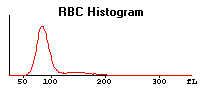
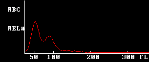
Normal Red Cell Histogram
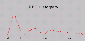
Cold agglutination
Has some agglutinated cells sticking together which accounts for the larger peaks on the far right of the X-axis.
Realoux does not have this affect bc a saline diluent will break up those cells
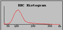
Microcytosis/anisocytosis
Will have increased RDW
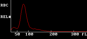
Microcytosis w/ PLT clumping or schistocytes
left peak indicates clumping or schistos
Dimporphic cell poputions
Could be from blood transfusions
Impedance, direct current (Coulter principle)
- passage of each cell through aperture
- creates electrical pulse which appears as a spike on the oscilloscope screen
- the hight of each spike is proportional to the size of the cell
- how the volume of the cell is determined
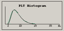
Normal PLT curve
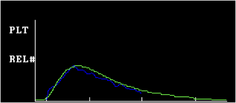
Giant Platelets
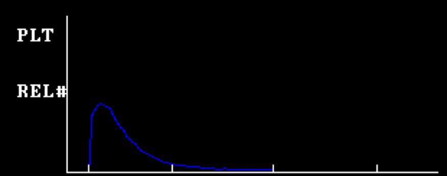
Small Platelets
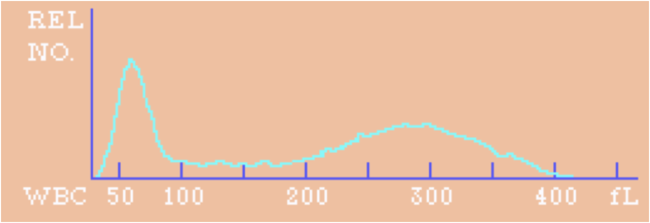
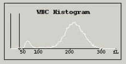
Normal White Blood Cell Histogram
Love Bombing Makes Emo Girls Sick
Lymphocytes
Basophils
Monocytes
Eosinophils
Segmented PMN
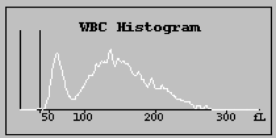
Immature Neutrophils 1 & 22
Immature neutrophils 1 - More blasts. Wider range of immature PMN
Immature Neutrophils 2- Increased in later stage immature cells ie. bands, metas, etc
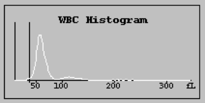
Lymphocytosis
Basically, just lymphocytes in the WBC population. \
Crowding of lymphocytes in BM disrupts production of other WBC
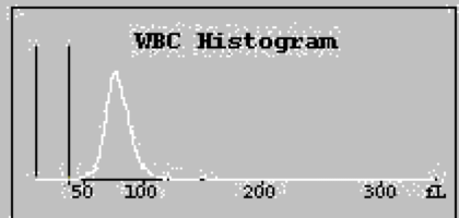
Variant Lymphs.
Think Reactive lymphs.
They’re larger than usual.
Immature Neutrophils 2
Immature Neutrophils 2- Increased in later stage immature cells ie. bands, metas, etc
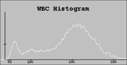
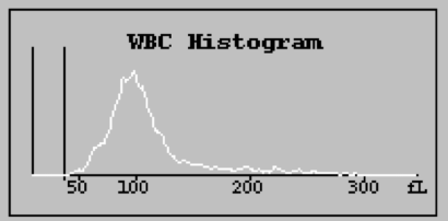
Eosinophilia
Center of the distribution has a huge peak
Blasts
Not sure of mechanism.
Laser Light
- light scatter measures cell surface granularity using broad range of angles
- over 60 angles of light scatter are analyzed
- flow principle
Radio/other high frequency waves
- Conductivity (VCS)- Related to conductivity
- measures internal cell structures such as nucleus & granules using radiographic imaging similar to ultrasound
- conductivity is proprietary technology
Hydrodynamic Focusing
- laminar glow ensures single file cell passage
- coincidence effects minimized
- principle of flow cell cytometry
- cells combined w/ sheath fluid forces them to go 1 by 1
Sweepflow
- eliminates recirculation of cells
- cells pushed away from critical (counting) zone by diluent
- coincidence correction (if it gives off an odd signal it doesn't get included in the count)
- provides more accurate histograms & cell sizing for reliable RBC and PLT indices
- triplicate counting
Fluorescent Flow Cytometry
- Sysmex
- fluorescent stain: nucleic acid and cytoplasmic organelles
- measures fluorescence and side angle light scatter to differentiate
- side fluorescence light: RNA/DNA
- side scatter light: internal cell structures
- forward scattered light: cell size information
VCS Technology
Volume- Total Cell volume
Measures Total Cell Volume using the Reference Method of Direct Current Impedance
Conductivity- Nuclear Volume/ internal composition
Measures Internal Cell Structures such as nucleus and granules using Radiographic Imaging Similar to Ultrasound
Light Scatter- cell surface characteristics/cell shape
Light Scatter Measures Cell Surface Granularity Using a Broad Range of Angles. Over 60 angles of light scatter are analyzed.
Thresholds in Cell Counting
- threshold: electronically set size limit used to choose a pulse size
- choose a limit
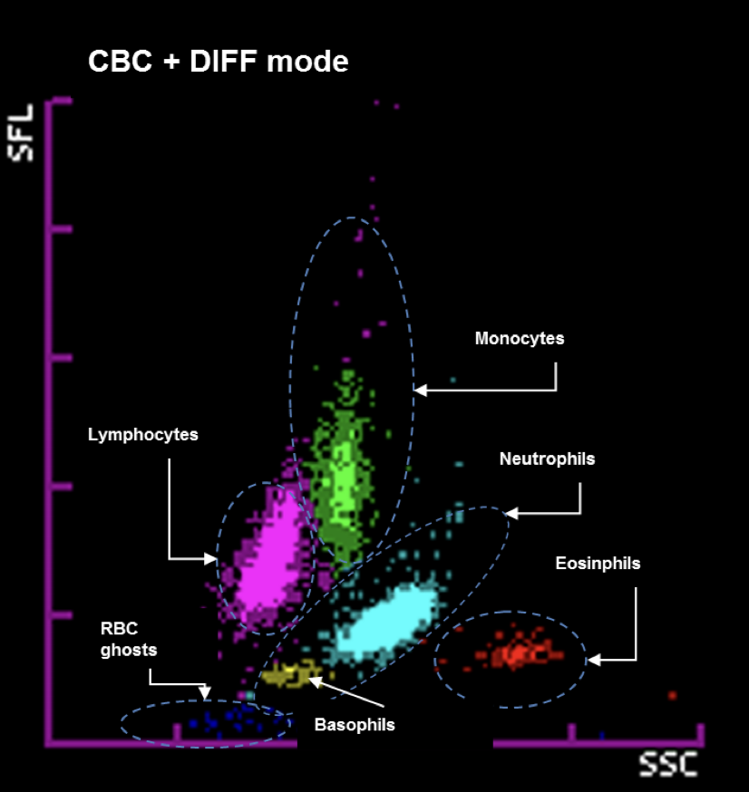
Normal WBC scattergrams
Go Lick My Nose Bitch, Ewwwwwww
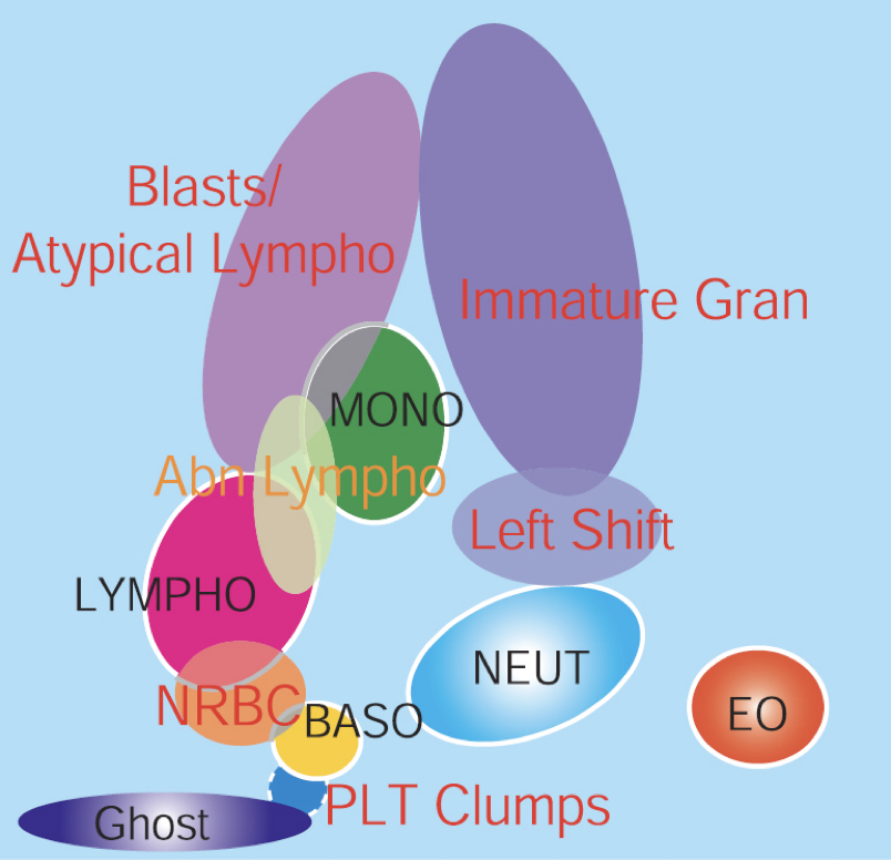
Abnormal WBC scattergram
Left shit is on the right of the plot
The higher up on the Y-axis, the younger the cell pops are, ie blasts
Abnormal lymphs are going to be reactive lymphs, which makes sense based on the size relative to normal lymphs
Channels in Cell Counting
- the area between thresholds
- we can use a series of thresholds to sort cells of a population into many subgroups by size
Histograms
Plot of size vs. the number of cells
Scatterplots/Scattergrams
- every cell treated in the same manner
- each cell given an X, Y, Z coordinate on the data plot
- all cell populations are directly measured