MICRB 410 Exam 3
0.0(0)
Card Sorting
1/168
Earn XP
Description and Tags
Last updated 4:02 AM on 11/8/22
Name | Mastery | Learn | Test | Matching | Spaced | Call with Kai |
|---|
No analytics yet
Send a link to your students to track their progress
169 Terms
1
New cards
Three phases of cell signaling
1. reception
2. transduction
3. response
2. transduction
3. response
2
New cards
Second messengers
small molecules that relay signals from cell-surface receptors to effector proteins in signal transduction
ex: Ca2+
ex: Ca2+
3
New cards
Is recognition of antigen by TCR and BCR enough to transduce a signal?
No, it is insufficient. There are additional signaling molecules that are required to transduce the signal
4
New cards
Do TCR and BCR have ITAMS?
No, other molecules associated with the receptors do
5
New cards
ITAM
immunoreceptor tyrosine-based activation motif
6
New cards
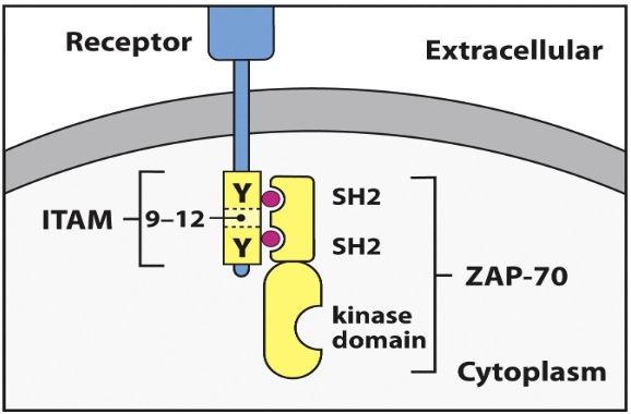
What is the TCR complex made of? Where are the ITAMs located?
TCR
CD3δε--single dimer (2 ITAMs)
CD3γε-single dimer (2 ITAMs)
two ζ chains (3 ITAMS each = 6 ITAMs)
total of 10 ITAMs
TCR + CD3 = TCR complex
CD3δε--single dimer (2 ITAMs)
CD3γε-single dimer (2 ITAMs)
two ζ chains (3 ITAMS each = 6 ITAMs)
total of 10 ITAMs
TCR + CD3 = TCR complex
7
New cards
What is the BCR complex made of?
BCR associated with CD79a(Igα) and CD79b (Igβ) and they each have one ITAM
8
New cards
How do ITAMs activate signaling pathways?
Each ITAM has two tyrosines (Y) that are phosphorylated by ZAP-70 when the ligand binds
9
New cards
ZAP-70 function and structure
A kinase that phosphorylates ITAMs
Has a kinase domain and two SH2 domains
The SH2 domains bind to the Tyrosine domains on the ITAMs and phosphorylates them
Has a kinase domain and two SH2 domains
The SH2 domains bind to the Tyrosine domains on the ITAMs and phosphorylates them
10
New cards
What does signal strength depend on?
1. affinity of receptor for ligand
2. concentration of signaling molecules
3. positive and negative feedback pathways
2. concentration of signaling molecules
3. positive and negative feedback pathways
11
New cards
What are the three amino acids that can be phosphorylated?
tyrosine, serine, and threonine
12
New cards
What are the two ways that receptors are activated by kinases and the signaling pathway is activated?
1. Receptors have kinases as intracellular domain. When the ligand binds, the receptors dimerize and the kinases phosphorylate each other and the activated kinases phosphorylated downstream substrates
2. Kinase is bound noncovalently to intracellular tail of receptor. When the ligand binds, the kinases phosphorylate each other due to close proximity. Activated kinases phosphorylate downstream substrates
2. Kinase is bound noncovalently to intracellular tail of receptor. When the ligand binds, the kinases phosphorylate each other due to close proximity. Activated kinases phosphorylate downstream substrates
13
New cards
Phosphatases
remove phosphate groups, important part of phosphorylation that makes it rapid and reversible
14
New cards
Phosphotyrosines (pY)
binding sites on ITAMs for SH2 domains
15
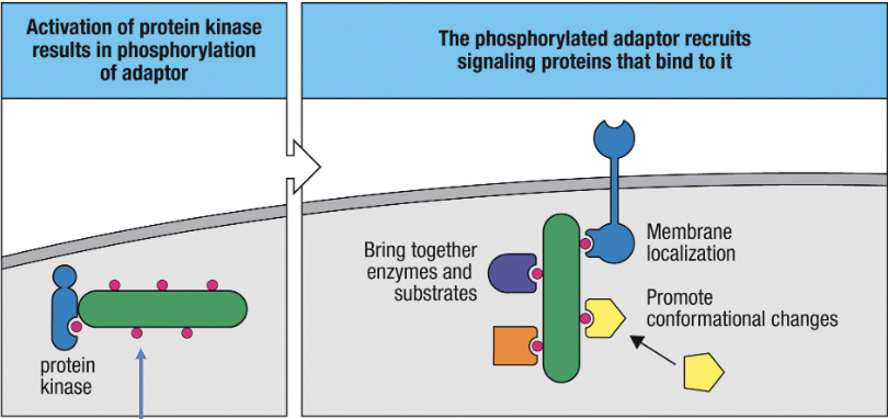
New cards
Adaptor proteins
Help to assemble signaling complexes
Have many sites of phosphorylation (so they can be activated by kinases) and lack enzymatic activity
Function by recruiting other proteins to signaling complex and those proteins bind to the phosphorylated sites of the adaptor protein
Have many sites of phosphorylation (so they can be activated by kinases) and lack enzymatic activity
Function by recruiting other proteins to signaling complex and those proteins bind to the phosphorylated sites of the adaptor protein
16
New cards
Grb2
adaptor protein with an SH2 domain and 2 SH3 domains
Binds to Sos protein with its SH3 domain and binds to phosphorylated kinase domain on receptor to transduce a signal
Binds to Sos protein with its SH3 domain and binds to phosphorylated kinase domain on receptor to transduce a signal
17
New cards
Ras
small GTP binding protein (GTPase)
inactive when bound to GDP and active when bound to GTP
inactive when bound to GDP and active when bound to GTP
18
New cards
GTPase
small GTP binding protein that catalyzes the hydrolysis of GTP to GDP
19
New cards
What activates Ras?
GEF removes GDP so that Ras can bind GTP and become active
20
New cards
What is the GEF for Ras?
Sos
21
New cards
GAPs
GTPase-activating proteins that accelerate the conversion of GTP to GDP
22
New cards
What are the three ways that signaling proteins are recruited to the membrane?
1. Tyrosine phosphorylation of membrane associated adaptor proteins that recruits phosphotyrosine-binding proteins
2. Recognition of activated GTPases
3. PI3K phosphorylates PIP2 to make PIP3 which is recognized by signaling proteins like Akt and Itk
2. Recognition of activated GTPases
3. PI3K phosphorylates PIP2 to make PIP3 which is recognized by signaling proteins like Akt and Itk
23
New cards
Akt and Itk
signaling proteins that recognize PIP3
24
New cards
PI3K
PI3-Kinase that phosphorylates PIP2 to make PIP3
25
New cards
What are the two parts of signal amplification?
1. signal cascade
2. release of second messenger
2. release of second messenger
26
New cards
Calmodulin
Ca2+ binding protein that binds effector proteins
27
New cards
SHP
a phosphatase that plays a role in shutting down signaling pathways
28
New cards
What can a deficiency in phosphatases cause?
autoimmunity and cancer
29
New cards
Ubiquitination. What does it do in signaling pathways?
covalent attachment of one or more ubiquitin
can be used to activate or inhibit signaling responses
can be used to activate or inhibit signaling responses
30
New cards
Polyubiquitination
marks protein for degradation in proteasome
31
New cards
E3 ubiquitin ligase
transfers ubiquitin to protein
32
New cards
K48 linkages and K63 linkages for polyubiquitination
K48 = targets proteins for degradation
K63= serves as scaffold for activation
K63= serves as scaffold for activation
33
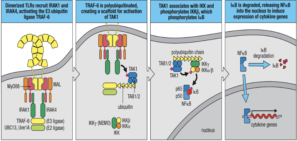
New cards
TRAF-6
E3 ubiquitin ligase
34
New cards
Explain the NFkB activating pathway
1. TLRs dimerase and recruit IRAKs which activate E3 ubiquitin ligase TRAF-6
2. TRAF-6 is polyubiquilated and creates scaffold for TAK1 activation
3. TAK1 associates with IKK and phosphorylates IKKβ which phosphorylates Iκβ
4. Iκβ is degraded and NFκB is released into the nuclease to induce expression of cytokine gene
2. TRAF-6 is polyubiquilated and creates scaffold for TAK1 activation
3. TAK1 associates with IKK and phosphorylates IKKβ which phosphorylates Iκβ
4. Iκβ is degraded and NFκB is released into the nuclease to induce expression of cytokine gene
35
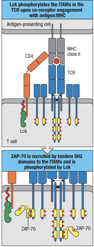
New cards
Lck
a kinase that phosphorylates 3 additional sites to fully activate ZAP-70
always associated with CD4 or CD8
always associated with CD4 or CD8
36
New cards
What are the two TCR co-receptors?
CD4 and CD8
37
New cards
How do T cell Co-receptors help the phosphorylation of ITAMS in the TCR complex?
1. Lck attached to co-receptor
2. co-receptor associates with MHC and Lck is now close to ITAMs
3. Lck phosphorylates ITAM of TCR complex
4. ZAP-70 binds to phosphorylated ITAMs
5. Lck phosphorylates ZAP-70
2. co-receptor associates with MHC and Lck is now close to ITAMs
3. Lck phosphorylates ITAM of TCR complex
4. ZAP-70 binds to phosphorylated ITAMs
5. Lck phosphorylates ZAP-70
38
New cards
How does Lck associate with a co-receptor?
Has a unique amino-terminal motif that has two cysteine residues that bind a Zn ion that is also bound to a similar motif in the cytoplasmic domain of CD4 or CD8
39
New cards
CD45
dephosphorylates the SH2 domain of Lck to make it primed
40
New cards
Csk
phosphorylates SH2 domain of Lck to make it inactive
41
New cards
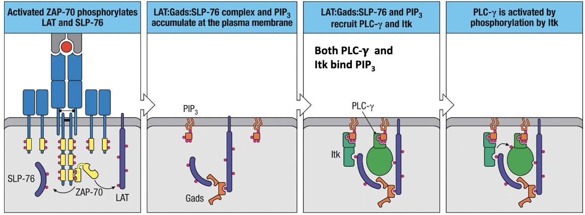
What are the two functions of activated (phosphorylated) ZAP-70?
1. phosphorylates LAT and SLP-76 (two adaptor proteins)
2. recruits PI3-K to cell membrane
2. recruits PI3-K to cell membrane
42
New cards
Gads
link LAT and SLP-76 together
43
New cards
Explain the activation pathway of PLC-γ with ZAP-70 and LAT/SLP-76
1. ZAP-70 phosphorylates LAT and SLP-76 which then come together by Gads
2. LAT:Gads:SLP-76 complex and PIP3 accumulate at plasma membrane
3. PIP3 recruits Itk and LAT:Gads:SLP-76 recruits PLC-γ and they bind via phosphorylated sections
4. PLC-γ is activated by phosphorylation by Itk
2. LAT:Gads:SLP-76 complex and PIP3 accumulate at plasma membrane
3. PIP3 recruits Itk and LAT:Gads:SLP-76 recruits PLC-γ and they bind via phosphorylated sections
4. PLC-γ is activated by phosphorylation by Itk
44
New cards
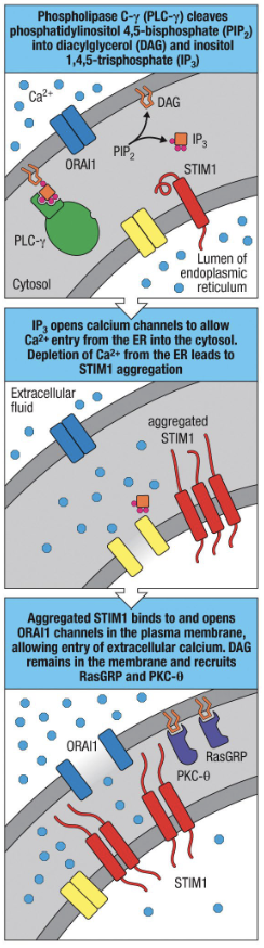
PLC-γ
adaptor protein that binds PIP3 and LAT:Gads:SLP-76 and is activated by Itk
cleaves PIP2 to make DAG and IP3
activates 3 signaling pathways that activate 3 different transcription factors
cleaves PIP2 to make DAG and IP3
activates 3 signaling pathways that activate 3 different transcription factors
45
New cards
What does cleaved PIP2 make and where do these pieces go?
DAG --> remains in membrane and recruits signaling molecules
IP3--> diffuses in cytosol
IP3--> diffuses in cytosol
46
New cards
What are the three signaling pathways that PLC-γ activates?
1. stimulation of Ca2+ entry = NFAT activation
2. Activation of Ras = activates MAPK = induces AP-1 expression
3. Activation of protein kinase C-θ (PKC-θ)= activates CARMA1 and induces NFκB activation
2. Activation of Ras = activates MAPK = induces AP-1 expression
3. Activation of protein kinase C-θ (PKC-θ)= activates CARMA1 and induces NFκB activation
47
New cards
Explain PLC-γ activation of Calcium
1. PLC-γ is activated and cleaves PIP2 to make DAG and IP3
2. IP3 diffuses into cytosol and binds to IP3 receptors on ER
3. These receptors are Ca channels so the binding stimulates the to open and release Ca into the cytosol
4. Low Ca in the ER induces STIM1 which binds to ORAI1 on plasma membrane
5. ORAI1 is Ca channel and opens to let Ca in from extracellular. The influx of Ca activates signaling pathway and replenishes ER ER Ca
48
New cards
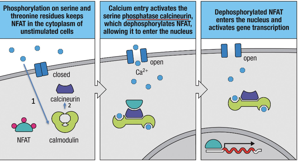
Explain activation of NFAT with activated PLC-γ
1. NFAT is phosphorylated and inactive
2. Ca enters cell and binds to calmodulin which binds to NFAT by phosphatase calcineurin. The phosphatase dephosphorylates NFAT
3. NFAT can now enter nucleus and active gene transcription
2. Ca enters cell and binds to calmodulin which binds to NFAT by phosphatase calcineurin. The phosphatase dephosphorylates NFAT
3. NFAT can now enter nucleus and active gene transcription
49
New cards
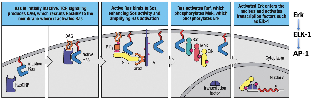
Explain Ras activation and expression of AP-1 by PLC-γ
1. DAG on membrane binds to RasGRP (GEF) and activates Ras
2. Ras binds to Sos and enhances its activity
3. Ras activates Raf, which phosphorylates Mek, which phosphorylates Erk
4. Phosphorylated Erk enters nucleus and activates transcription factor Erk-1 to produce AP-1
2. Ras binds to Sos and enhances its activity
3. Ras activates Raf, which phosphorylates Mek, which phosphorylates Erk
4. Phosphorylated Erk enters nucleus and activates transcription factor Erk-1 to produce AP-1
50
New cards
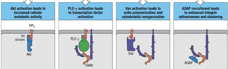
What for activation pathways does ZAP-70 and LAT and SLP-76 initiate?
1. activation of Akt
2. Activation of PLC-γ
3. Vav activation
4. ADAP activation
2. Activation of PLC-γ
3. Vav activation
4. ADAP activation
51
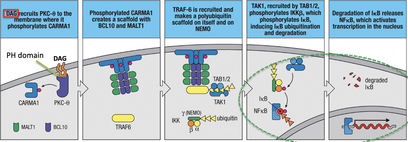
New cards
Explain how PLC-γ activation results in activation of PKC-θ (C-θ) and induces NFkB production
1. PKC-θ binds to PH domain of DAG which is in membrane
2. PKC-θ phosphorylates CARMA1 which creates a scaffold for TRAF-6 which is polyubiquitinated
3. TAK1 phosphorylates IKKβ, then IκB which induces IκB ubiquitination and degradation which releases NFκB which activates transcription in the nucleus.
2. PKC-θ phosphorylates CARMA1 which creates a scaffold for TRAF-6 which is polyubiquitinated
3. TAK1 phosphorylates IKKβ, then IκB which induces IκB ubiquitination and degradation which releases NFκB which activates transcription in the nucleus.
52
New cards
What does the activation of these 4 proteins do?
PLC-γ
Akt
Vav
ADAP
PLC-γ
Akt
Vav
ADAP
PLC-γ = transcription factor activation
Akt = increased cellular metabolic activity and promotes cell survival
Vav = actin polymerization and cytoskeletal reorganization
ADAP = enhanced integrin adhesiveness and clustering
Akt = increased cellular metabolic activity and promotes cell survival
Vav = actin polymerization and cytoskeletal reorganization
ADAP = enhanced integrin adhesiveness and clustering
53
New cards
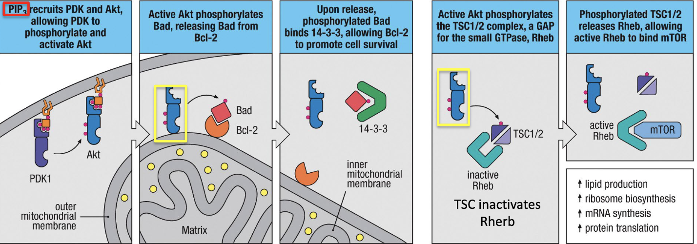
Explain Akt activation from ZAP-70 and LAT/SLP-76
1. Akt and PDK1 bind to PIP3 in the cell membrane
2.PDK1 phosphorylates Akt
3. Phosphorylated Akt phosphorylates Bad which releases Bad from Bcl-2.
4. Bad binds 14-3-3, which promotes cell survival
5. Akt phosphorylates a GAP for Rheb which allows Rheb to bind to mTOR for increased cellular metabolic activity
2.PDK1 phosphorylates Akt
3. Phosphorylated Akt phosphorylates Bad which releases Bad from Bcl-2.
4. Bad binds 14-3-3, which promotes cell survival
5. Akt phosphorylates a GAP for Rheb which allows Rheb to bind to mTOR for increased cellular metabolic activity
54
New cards
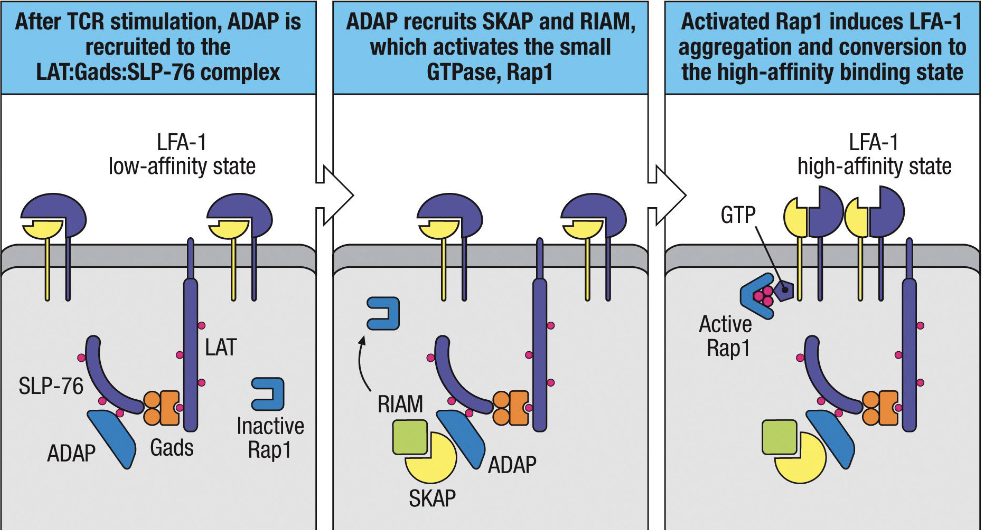
Explain ADAP activation from ZAP-70,SLP-76 and LAT
1. ADAP recruited to LAT:Gads:SLP-76 complex and binds to phosphorylated parts
2. ADAP recruits other proteins that activate Rap1 (GTPase)
3. RAP1 induces LFA-1 aggregation and conversion to the high-affinity binding state which increases adhesiveness and promotes stability of immune synapse
2. ADAP recruits other proteins that activate Rap1 (GTPase)
3. RAP1 induces LFA-1 aggregation and conversion to the high-affinity binding state which increases adhesiveness and promotes stability of immune synapse
55
New cards
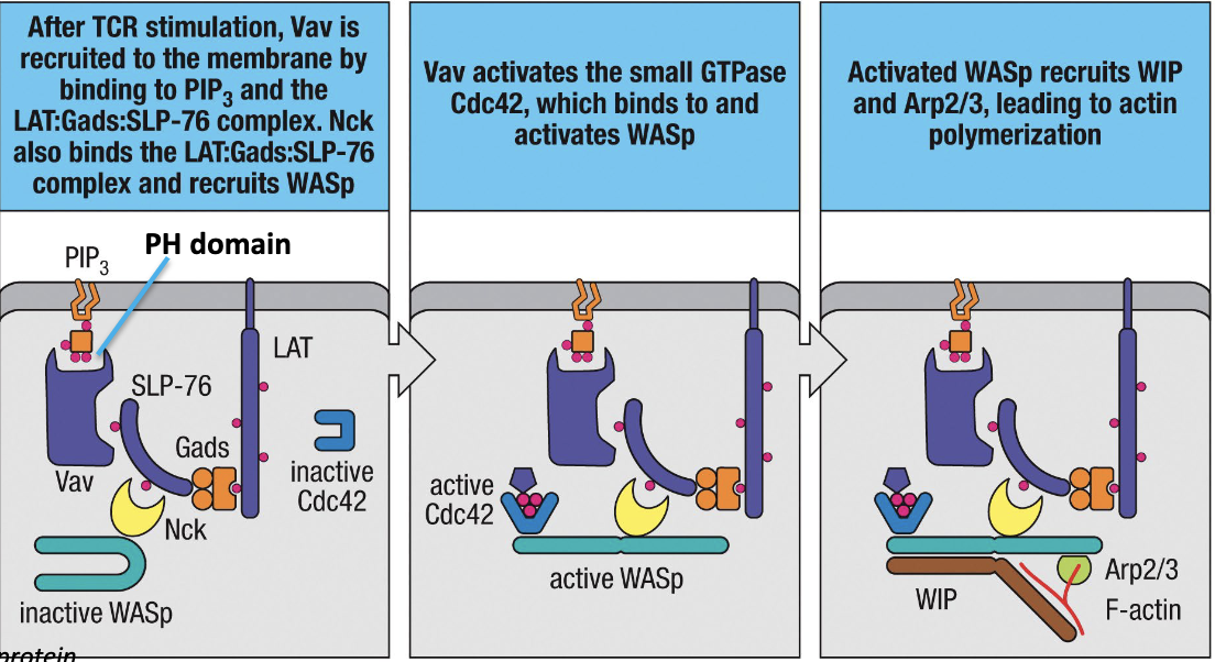
Explain Vav activation from ZAP-70, SLP-76 and LAT
1. Vav recruited to cell membrane and binds to PIP3 and the LAT:Gads:SLP-76 complex
2. Vav activates Cdc42 (small GTPase) which binds to and activates WASp
3. Activated WASp recruits others proteins that lead to actin polymerization
2. Vav activates Cdc42 (small GTPase) which binds to and activates WASp
3. Activated WASp recruits others proteins that lead to actin polymerization
56
New cards
What does defects in WASp cause?
Wiscott-Aldrich syndrome - defect in the formation of immune synapse and defects in antibody response
57
New cards
What happens to ADAP deficient cells?
impaired proliferation and cytokine production upon TCR stimulation
58
New cards
B7.1
Co-stimulatory ligand that binds to CD28 on T cells that help with activation
59
New cards
B7.2
Co-stimulatory ligand that binds to CD28 on DCs
60
New cards
What does B7 binding with CD28 cause?
PI3-kinase activation and recruitment of Lck

61
New cards
IL-2
-essential for T cell activation
-TCR activation leads to low IL-2 and CD28 activation leads to increased IL-2
-NFAT, AP-1, and NFkB all bind to promoter of IL-2 to induce expression
-TCR activation leads to low IL-2 and CD28 activation leads to increased IL-2
-NFAT, AP-1, and NFkB all bind to promoter of IL-2 to induce expression
62
New cards
What proteins phosphorylate ITAMs in BCR activation?
Blk, Fyn and Lyn
63
New cards
What protein transduces the signal in BCR activation?
Syk
64
New cards
What forms the B-cell co-receptor complex?
CD19, CD21 and CD81
65
New cards
Bkt
Binds to PIP3 on membrane and activates PLC-γ
66
New cards
What does a deficiency in Bkt cause?
X-linked agammaglobulinemia, characterized by lack of antibodies
67
New cards
Explain BCR activation by CD40 and CD40L
CD40 L on T cell and CD40 on B cell bind which ultimately activated NFkB
68
New cards
CTLA-4
inhibitory receptor on T cells for B7
has slightly higher affinity for B7 than CD28, so it reduces CD28 co-stimulation
has slightly higher affinity for B7 than CD28, so it reduces CD28 co-stimulation
69
New cards
PD-1
-inhibitory receptor on T and B cells
-has ITIM on tail (inhibitory motif)
-has ITIM on tail (inhibitory motif)
70
New cards
What happens when ITIM is phosphorylated
recruits SHP and SHIP, two phosphatases
71
New cards
SHP
tyrosine phosphatase that turns off signaling
72
New cards
SHIP
inositol phosphatase that removes phosphate from PIP3 to produce PIP2 to reduce Akt recruitment and activation
73
New cards
Checkpoint Blockade
inhibiting inhibitory receptors to enhance T cell responses
74
New cards
Where are B and T lymphocytes made?
Central (primary) lymphoid tissues (PLO)
B cells: bone marrow
T cells: thymus
B cells: bone marrow
T cells: thymus
75
New cards
Where do mature B and T cells go after PLO?
Secondary/Peripheral Lymphoid Organs (SLO) such as the lymph nodes, spleen, etc.
76
New cards
Where are antigens? SLO or PLO?
SLO
77
New cards
What is the ancestor cell of B and T cells?
Multipotent hematopoietic stem cell
78
New cards
Negative v. Positive selection of lymphocytes
Negative selection = BCRs and TCRs that interact strongly with self-antigens are eliminated to prevent autoimmune reactions
Positive selection = BCRs and TCRs that interact weakly with self-antigens survive
Positive selection = BCRs and TCRs that interact weakly with self-antigens survive
79
New cards
Where does BCR and TCR selection occur?
selection for BCR in bone marrow and selection for TCR in thymus
80
New cards
What cells do activated B cells give rise to?
plasma cells or memory B cells
81
New cards
Explain the B Cell Life Cycle
1. B cell precursor (bone marrow), Ig genes rearrange to make BCR
2. negative selection and immature B cell formation
3. migration to SLO and mature B cell forms by binding to antigen
4. activated B cell differentiates into plasma cell or memory cell
2. negative selection and immature B cell formation
3. migration to SLO and mature B cell forms by binding to antigen
4. activated B cell differentiates into plasma cell or memory cell
82
New cards
Stromal Cells
specialized network of non-lymphoid connective tissue that are essential in lymphocyte development
83
New cards
Steps of B-Cell Development
1. Lymphoid-Myeloid primed multipotent progenitor binds to FLT3 to ligand on bone marrow stromal cell to make CXCL12 and Il-7 receptor
2. CLP (on stromal cell) binds to VCAM-1 and CAM
3. Kit on B cell binds to SCF on stromal cell which activates kinase and proliferation of B cell progenitors
4. Late pro-B cell
5. Pre-B cell
6. Immature B cell release from stromal cell with IgM as receptor
2. CLP (on stromal cell) binds to VCAM-1 and CAM
3. Kit on B cell binds to SCF on stromal cell which activates kinase and proliferation of B cell progenitors
4. Late pro-B cell
5. Pre-B cell
6. Immature B cell release from stromal cell with IgM as receptor

84
New cards
What does a deficiency in IL-7 and the receptor cause?
block in B cell development and less T cells
85
New cards
During B cell development, which chains of the receptor rearrange first and second?
Heavy chain first and then light chain
86
New cards
Heavy chain VDJ rearrangement
D-J
V-DJ
VDJ
V-DJ
VDJ
87
New cards
Light chain VJ rearrangement
V-J
VJ
VJ
88
New cards
What does the B cell receptor do to test for correct rearrangement?
Makes two 'surrogate' proteins that have a structural resemblance to the light chain and can pair with μ chain
89
New cards
What happened to the child that inherited defective alleles of the λ5 gene?
-no pre-B-cell receptor development
-B-cell immunodeficiency so many infections
-B-cell immunodeficiency so many infections
90
New cards
What do BLNK and Btk do in B-cell signaling?
suppress RAG-1 and RAG-2 expression
91
New cards
Deficiency of BLNK
block of B-cell development at pro-B cell stage
92
New cards
Mutation in Btk
Bruton's X-linked agammaglobulinemia (XLA)- no mature B cells
93
New cards
Steps of making sure Ig genes were rearranged correctly in B cells
1. pre-B cell tests heavy chain gene
2. VpreB and λ5 made and bind to heavy chain to make pre-B cell receptor
3. complex associates with other receptors and dimerizes by surrogate invariant chains
4. dimerization phosphorylates ITAMs
5. BTK and BLNK suppress RAG 1 and 2
2. VpreB and λ5 made and bind to heavy chain to make pre-B cell receptor
3. complex associates with other receptors and dimerizes by surrogate invariant chains
4. dimerization phosphorylates ITAMs
5. BTK and BLNK suppress RAG 1 and 2
94
New cards
Allelic exclusion
only one allele of a gene is expressed while the other allele is silenced
95
New cards
Results of Heavy chain and light chain rearrangement. What can be done if non-productive rearrangement occurs in either?
Heavy chain produces both productive and non-productive rearrangements (50% of pre-B cells survive)
Light chain produces non-productive and can rescue it by further rearrangement (multiple attempts)
Light chain produces non-productive and can rescue it by further rearrangement (multiple attempts)
96
New cards
Isotypic exclusion
only one type of light chain is used
97
New cards
What are the two checkpoints to determine fate of B cell
1. capacity of heavy chain to bind the surrogate light chain and make pre-B cell receptor
2. capacity of binding the light chain to pre-B cell heavy chain and then asses B-cell receptor
2. capacity of binding the light chain to pre-B cell heavy chain and then asses B-cell receptor
98
New cards
What are glycoproteins, proteoglycans, and glycolipids
self-antigens
99
New cards
What is tolerance
reactivity to self-antigens
100
New cards
What does the fate of the immature B cell depend on when it comes to tolerance?
signal delivered from sIgM
-no strong reactivity to self-antigen --> mature
-strong reactivity to self-antigen --> clonal deletion or receptor editing
-no strong reactivity to self-antigen --> mature
-strong reactivity to self-antigen --> clonal deletion or receptor editing