Lecture 4 - Methods to study brain function
1/22
There's no tags or description
Looks like no tags are added yet.
Name | Mastery | Learn | Test | Matching | Spaced | Call with Kai |
|---|
No study sessions yet.
23 Terms
What are the main methods used to study brain function?
Electrophysiological recordings (e.g., EEG, ERPs)
Functional imaging (e.g., fMRI, PET, MEG)
Lesion studies (examining loss of function after brain damage)
Electrical stimulation of the brain (stimulating specific brain regions to observe behavioral effects)
The Wada Test (temporarily inactivating one hemisphere to determine lateralization)
Split-brain studies (examining patients with severed corpus callosum to study hemispheric specialization)
What does an Electroencephalogram (EEG) measure?
It measures electrical activity in the cortex via electrodes placed on the scalp (typically the 10-20 system with 19 electrodes).
Explanation: EEG captures voltage fluctuations generated by synchronized neuron firing. It has very high temporal resolution and detects rhythmic and transient activity.
What are the main uses of EEG?
To distinguish different brain states (e.g., sleep vs. wake), detect rhythms, study cognitive responses to stimuli, and monitor disorders such as epilepsy.
Explanation: EEG can show patterns linked to alertness, relaxation, sleep, and pathology.
How is the EEG typically described? - TA BORT
The EEG is typically described in terms of:
Rhythmic activity refers to ongoing, repeating wave patterns such as delta, theta, alpha, beta, and gamma rhythms.
Transient activity refers to short-lived, event-related responses such as event-related potentials (ERPs) that occur in response to specific stimuli.
What are the main EEG frequency bands and their Hz ranges?
Delta: up to 3 Hz
Theta: 4–7 Hz
Alpha: 8–12 Hz
Beta: 12–30 Hz
Gamma: 30–100 Hz
Explanation: Different frequencies are associated with different brain states (e.g., deep sleep vs. focused thinking).
When are delta waves most commonly seen?
During deep (slow-wave) sleep in adults and normal wakefulness in infants.
Explanation: High-amplitude, slow waves linked to restoration and recovery in sleep.
What characterizes theta waves?
4–7 Hz waves associated with drowsiness, meditation, and childhood brain activity.
Explanation: They often appear in transitional states between wake and sleep.
What do alpha waves indicate?
8 to 12 Hz. The basic rhythm in the posterior regions of the brain, with higher amplitudes on the dominant side. Found at central sites during rest. Strongly correlated with visual input. Emerge with eye closure and with relaxation. Also prominent indicator of a coma.
What do beta waves indicate?
Active thinking, concentration, problem solving.
Explanation: High-frequency but lower amplitude. Changes can reflect drug effects.
What are gamma waves thought to represent?
Coordination and binding of neural populations for complex cognitive processing.
Explanation: Often linked to attention, memory, and sensory integration.
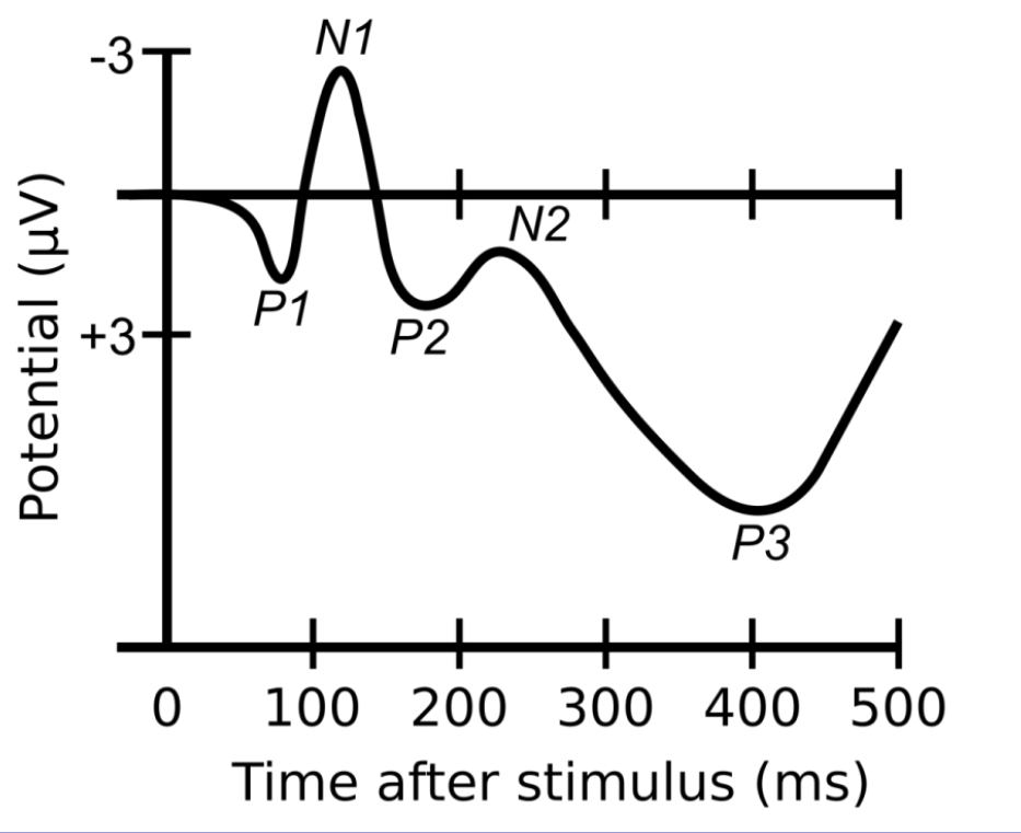
What are Event-Related Potentials (ERPs)?
Stimulus-linked changes in EEG activity.

What is the principle of fMRI (functional Magnetic Resonance Imaging)?
Neural activity requires oxygen; oxygenated hemoglobin is detected because it is diamagnetic.
Explanation: fMRI measures blood flow changes to infer which brain areas are active.
How does Positron Emission Tomography (PET) work?
A radioactive glucose tracer is injected; active brain areas use more glucose, producing photons that the scanner detects.
Explanation: PET has slow temporal resolution (~30 seconds).
What does Magnetoencephalography (MEG) measure?
Measure magnetic changes in the brain. When action potential is transported through the neuron, magnetic fields are produced around the neuron. Measure magnetic fields in the cortex; it cannot detect deeper inside the brain. Room must be insulated from outside magnetic fields – metal cage
Real time measurements.
What are lesion studies used for?
To determine which brain regions are necessary for specific functions.
Explanation: Damage or deliberate lesions reveal function-loss relationships.
Create lesion of the brain, either by cutting it out, inserting heat electrodes into specific brain regions, etc.
What is a stroke and what are its two main types?
A stroke is death of neurons due to poor blood supply.
Ischemic: lack of blood flow due to clotted arteries
Hemorrhagic: lack of blood flow due to bursting of an artery (and thus due to bleeding)
Why is electrical stimulation of the brain used during surgery and how?
To map functional regions such as Broca’s area.
Inducing microvolts into the brain to stimulate. If tumor or similar around, for example, brochas area, during surgery, while patient is awake, have conversation and use electrodes in brain to make sure exactly where brochas area is in their brain.
What is Transcranial Magnetic Stimulation (TMS)?
A technique that uses magnetic fields to induce electrical activity in specific brain areas.
Explanation: Electromagnetic induction; makes electricity flow by inducing magnetic field. TMS generate precise magnetic field to specific part of brain to electrically induce stimuli. E.g. inducing in occipital lobe – see blink of a light. Can help certain types of epilepsy if continuous pulses.
Why insert electrodes to inject pharmacological agents directly into the brain?
Electrodes can be inserted to specific part of brain to provide microinjections of pharmacological agents, e.g. testosterone or estrogen, to alter behavior.
What is the Wada Test (Intracarotid amobarbital test) used to determine?
Right hemisphere only get blood from right carotid, vice versa. Possible to inject substance to only right or left carotid to impair the right or left hemisphere. Impairing left hemisphere – giving patient a spoon, ask what it is, can’t answer, BUT can point at an image of spoon with left arm.
What is a callosotomy( Split brain) and why is it performed?
Cutting the corpus callosum to separate the hemispheres, used as a last-resort treatment for severe epilepsy.
Explanation: Results in split-brain processing where each hemisphere functions more independently.
What are the key functional differences between the left and right cerebral hemispheres?
Left hemisphere:
Sequential processing
Analytical thinking
Logical reasoning
Verbal abilities (words, grammar)
Right hemisphere:
Simultaneous/holistic processing
Intuitive thinking
Visual-spatial and imaginative abilities
Context and emotional tone in language (intonation)
What did Roger Sperry conclude about the hemispheres after split-brain research?
Each hemisphere is capable of conscious experience, including perceiving, thinking, remembering, reasoning, and feeling, even independently of the other.
Explanation:
In split-brain individuals, the left and right hemispheres can process information separately and may even hold different thoughts or experiences at the same time, since the corpus callosum connection is cut.