NEURO 3000 EXAM 3
1/112
There's no tags or description
Looks like no tags are added yet.
Name | Mastery | Learn | Test | Matching | Spaced | Call with Kai |
|---|
No analytics yet
Send a link to your students to track their progress
113 Terms
cerebrum
two hemispheres that receive input contralaterally
cerebellum
large structure of hindbrain that controls fine motor skills
- ipsilateral (same)
brainstem
regulates body temp, breathing, and consciousness
- essential for life (relay center)
spinal cord
conducts sensory and motor nerve impulses to and from the brain
- spinal nerves part of PNS
pyramidal decussation
motor fibers from the medulla cross the midline; continue into spinal cord as corticospinal tract
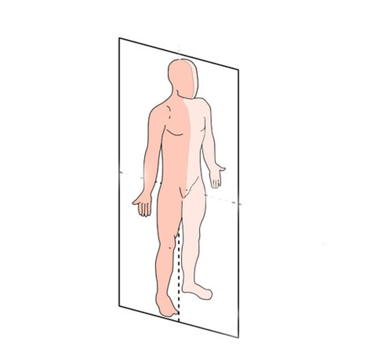
sagittal section
divides the body into left and right parts

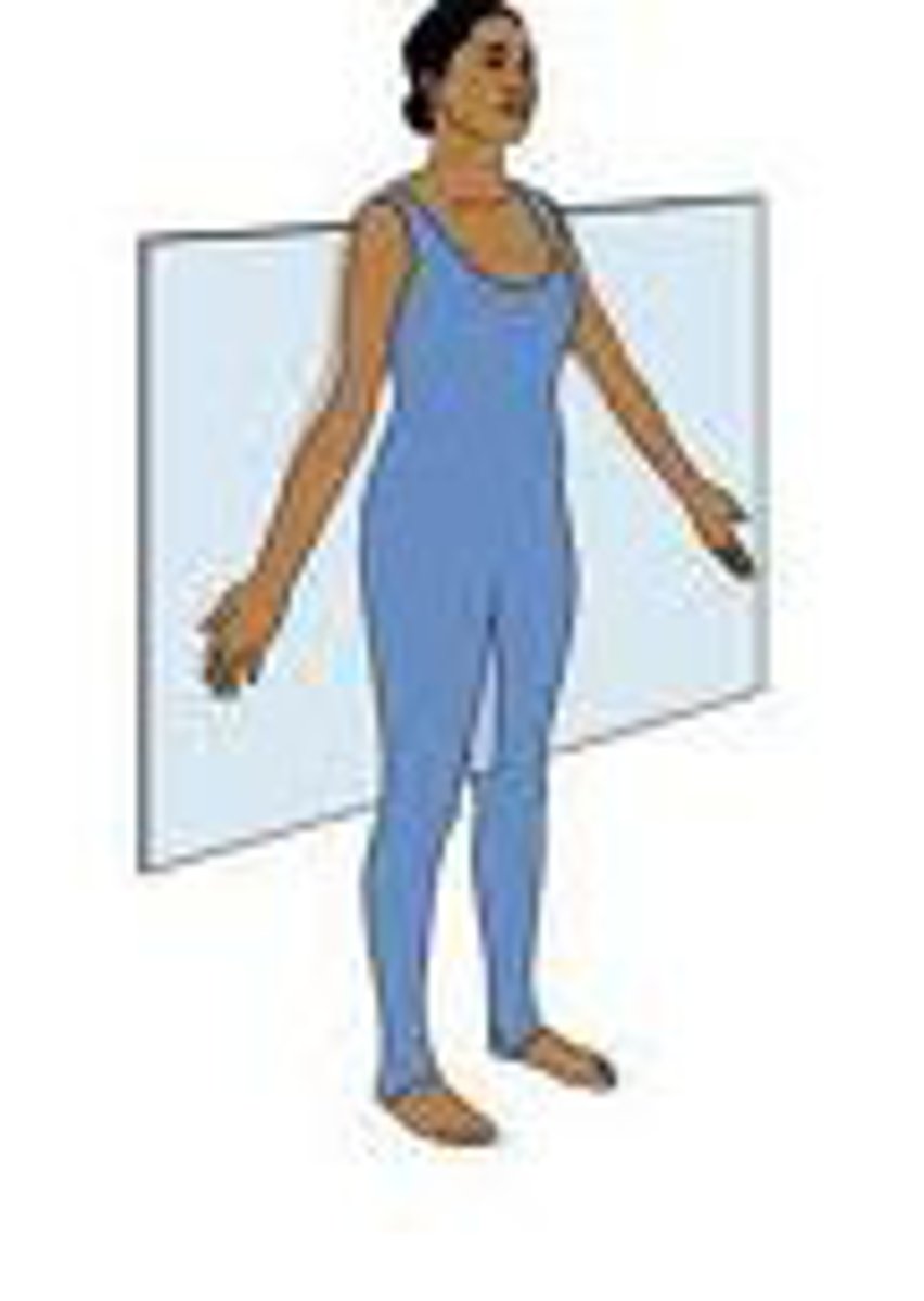
coronal/transverse section
divides body into front and back

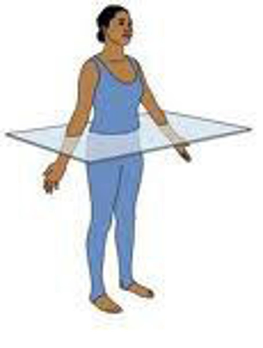
horizontal section
divides body into upper or lower sections

gyri
ridges of the brain
sulci
valleys between ridges
fissure
deep groove; deeper than sulci
- major divisions (left & right hemis, cerebrum from cerebellum)
grey matter
unmyelinated neuron cell bodies (cortex; superficial)
white matter
myelinated axons
nucleus
mass of neurons; usually deep in brain (deep cerebellar nucleus, etc.)
dorsal roots
sensory input to cord
ventral roots
motor neuron axons that exit the spinal cord
ganglion
collection of nerve cell bodies in the PNS
somatic nervous system
division of PNS that controls the body's skeletal muscles; voluntary movements
- cell body of motor neurons in CNS, axons in PNS
autonomic nervous system
controls involuntary actions
- mostly smooth muscles, heart muscles, and glands
sympathetic preganglionic neurons
originate in thoracic and anterior lumbar regions of spinal cord
parasympathetic preganglionic neurons
originate from various anterior cranial nerves and posterior sacral regions
internal capsule
white matter tract that carries outgoing fibers from cortex, and incoming from thalamus to cortex
postganglionic neurons PNS
project to glands
cleavage to blastocyst stage
2-10 days post fertilization
neural induction
gastrulation; days 11-15
- embryo cells move and form 3 germ layers
- mesoderm is formed & induces neurectoderm to neural fate by noggin
somites
generate bone and muscle related to spinal vertebrae
- generated from mesoderm
neural tube formation
neurulation; 16-25 days
- neural tube becomes brain and spinal cord
- neural crest become sensory and autonomic neurons, etc.
regionalization/patterning
28+ days; formation of brain vesicles (become forebrain, midbrain, hindbrain)
- AP and DV patterning
anterior posterior patterning
forebrain, midbrain, hindbrain, spinal cord
- controlled by retinoic acid
dorsal-ventral patterning
determines ventral and dorsal cell types
- controlled by sonic hedgehog, shh
notochord
becomes part of bone and muscle in spinal cord
- releases shh
sonic hedgehog
ventralizing signal
neurogenesis
proliferation, migration, differentiation
- at first, symmetric cell division of radial glia, then asymmetric after
day 36
forebrain expands & adds telencephalic vesicles
- hindbrain develops into metencephalon and myelencephalon
days 49-90
forebrain develops into diencephalon and telencephalon
forebrain
telencephalon and diencephalon
- perceptions, awareness, cognition, voluntary action
midbrain
mesencephalon
hindbrain
metencephalon and myelencephalon
- dorsal becomes cerebellum, ventral becomes pons
- medulla
- 4th ventricle
telencephalon
cerebral cortex, basal ganglia, hippocampus, lateral & 3rd ventricles
Diencephalon
thalamus and hypothalamus, part of 3rd ventricle
Mesencephalon
tectum, substantia nigra, cerebral aqueduct
Metencephalon
pons and cerebellum
Myelencephalon
medulla
cortical neurons
send axons back to brainstem through internal capsule, project on corticospinal tract, or project to basal ganglia (includes substantia nigra)
tectum
the dorsal part of the midbrain; includes the superior (visual) and inferior (auditory) colliculi
Tegmentum
The ventral part of the midbrain; VTA, substantia nigra
cerebral aqueduct
connects the third and fourth ventricles
pons
connects cerebral cortex to the cerebellum
medullary pyramids
carry corticospinal projections towards spinal cord
symmetric division
generate more radial glia that divide
asymmetric division
generates one postmitotic (farthest from ventricle) and one precursor
- postmitotic cell migrates and becomes neuron or glia
inside out development of cortex
cells that form each layer migrate past the ones that preceded them
- deeper layers diff into pyramidal neurons
- intermed. zone becomes white matter
- VZ disappears
differentiation
postmitotic precursor reaches destination & differentiates into neuron w/ dendrites and axon
semaphorin 3A
guidance molecule expressed in marginal zone
- dendrites attracted & axon is repulsed by (responsible for polarity of pyramidal cell)
synaptogenesis
formation of synapses
target dependent cell death
more neurons are generated than are actually needed
synaptic pruning
the elimination of neurons as the result of nonuse or lack of stimulation
- microglial role
frontal lobe
planning, organization, impulse control, decision making
- primary motor cortex (voluntary); caudal
- Broca's area
temporal lobe
language, hearing, memory
- auditory cortex
- wernicke's area
- ventral stream: what pathway
parietal lobe
receives sensory input for touch and body position
- primary somatosensory cortex; rostral
- dorsal stream: where pathway
occipital lobe
visual processing
corpus callosum
band of neural fibers (white matter) between brain hemispheres and carries info
thalamus
receives info from sensory systems and relays to cortical areas
hypothalamus
controls emotion and motivated behaviors; homeostasis
- has control of pituitary gland
meninges
three protective membranes that surround the brain and spinal cord
subarachnoid space
a space in the meninges filled with CSF supplied from ventricles
- CSF in space can be absorbed by blood vessels
ventricles
hollow inferior of NS filled with CSF
CSF
produced by choroid plexus in ventricles, exits ventricular system through apertures, and enters subarachnoid space
ventricular system
CSF comes from arterial blood, and removed by venous system
MRI
general brain structure (static)
DTI
scans water movement (static)
- white matter tracks
PET scan
identifying functional brain regions
- 2DG, measures metabolic function; correlates with electrical activity
fMRI
ability to detect changes in blood flow
- detect changes in brain regions among patients
tastants
chemicals that stimulate gustatory receptor cells
papillae
taste buds
- not true neurons
- detect chemicals in food at microvilli that project into taste pores
- constantly replaced (basal cells are precursors)
physiology of taste cells
chemicals cause depolarizing receptor potential; activates Ca2+ to release NT
- usually only respond to only 1 of 5 tastes
sour and salty taste cells
release serotonin
sweet, bitter, and umami cells
release ATP
sour taste mechanisms
Na+ sensitive channels are always open
- generated by acidic foods
sweet, bitter, and umami mechanisms
all GPCRs
- G protein activates PLC, which cleaves PIP2 into DAG and IP3 (2nd messengers)
- IP3 binds to its receptor which releases Ca2+
- Na+ channel depolarizes which releases ATP (ATP channel non-vesicular)
bitter taste receptors
heterodimers of T1R (3) and T2R (25)
sweet taste receptor
heterodimer T1R2 + T1R3
umami taste receptor
T1R1 and T1R3
gustatory sensory neuron cell bodies
cranial nerves 7, 9, 10
- receive input from taste cells, transmitted to cell body, then projects to gustatory nucleus
- ipsilateral system
olfactory epithelium
thin layer of tissue within nasal cavity
- contains the receptors for smell
olfactory receptors
neurons with cilia at one end that receive odorants
- axon projects to olfactory bulb
- expresses one receptor gene, but can bind to multiple odorant MCs
vomernasal organ
detects pheromones (in animals)
- controversial in humans
olfactory signal transduction
1. odorant dissolves in mucus
2. binds to receptor proteins on cilia (GPCR)
3. activates Golf in olf. cells
4. activates adenylyl cyclase, inc cAMP, open channel
5. Ca2+ and Na+ enter & depolarize
6. Cl channels leave cell and enhances depolarize
Axel and Buck
won the Nobel Prize in 2004 for discovering olfactory receptors
olfactory glomeruli
clusters of neurons near the surface of the olfactory bulbs
- bilaterally symmetric
lateral inhibition in olfactory bulb
granule cells in bulb help sharpen mitral cell responses in selected glomeruli
- granule cell: top down inputs
olfactory pathways
olfactory bulb, olfactory tubercle, thalamus, frontal cortex; mediates perception of odors
- olf bulb, primary olf cortex, amygdala (emotional & social behaviors)
sound
vibrations that travel through the air or another medium
frequency
the number of complete wavelengths per second
- pitch
amplitude
the height of a wave's crest
- loudness
tympanic membrane
eardrum
- sound waves move it
ossicles
three tiny bones in the middle ear
- move the membrane at the oval window
- amplify force
oval window
membrane at the enterance to the cochlea
- ossicles transmit vibrations
cochlea
tube in the inner ear through which sound waves trigger nerve impulses
- contains 3 fluid filled chambers
- fluid movement causes response in hair cells
organ of corti
contains hair cell and primary auditory nerve fibers
- hair cells arranged along basilar membrane
- have stereocilia projecting into endolymph