biology - topic 2 - a,b,c,d,e(human),g(plants),i,j
1/100
There's no tags or description
Looks like no tags are added yet.
Name | Mastery | Learn | Test | Matching | Spaced | Call with Kai |
|---|
No analytics yet
Send a link to your students to track their progress
101 Terms
what is the level of organisation
cells, tissues, organs, organ system, organism
describe the cell structure of a plant cell
- nucleus
- cell membrane
- cytoplasm
- ribosomes
- mitochondria
- chloroplasts
- permanent vacuole
- cell wall
describe the cell structure of an animal cell
cytoplasm, shape is not rigid, nucleus, ribosomes, mitochondria, cell membranes.
what are the differences between plant and animal cells
plant cell has chloroplasts, cell wall and vacuole
what does the nucleus do
It contains genetic material and controls the activities of the cell
what does the cytoplasm do
Its a gel-like substance where most of the chemical reactions happpen
what does the cell membrane do
It holds the cell together and controls what goes in and out
what does the cell wall do
Protects and supports the cell
what does mitochondria do
This is where most of the reactions for respiration take place. Respiration releases energy that the cell needs to work.
what do chloroplasts do
This is where photosynthesis occurs, which makes food for the plant. They contain chlorophyll - a green pigment - which absorbs light needed for photosynthesis
what do ribosomes do
protein synthesis
what does the vacuole do
It contains cell sap and helps maintain the shape of the cell + stores food water and waste
what is the importance of cell differentiation in the development of specialised cells
allows cells to specialize and perform specific functions in an organism. This allows for the formation of tissues and organs, and is necessary for the proper functioning of the body.
advantages of using stem cells in medicine
Can be used to heal things like certain types of cancer. It could also be used to regrow organs in the future.
disadvantages of using stem cells in medicine
-Ethical issue
-Long term effects are unknown
-Adult stem cells have a fixed cell they will turn into
-Obtaining stem cells is a difficult process
-Very costly
-Limitations to research possibilities
what are the molecules in carbohydrates
made up of sugar molecules
what are the molecules in lipids
made up of glycerol and fatty acids
what are the molecules in proteins
amino acids
what are the chemical elements in proteins
carbon, hydrogen, oxygen, nitrogen
what are the chemical elements in lipids
carbon, hydrogen, oxygen
what are the chemical elements in carbohydrates
carbon, hydrogen, oxygen
how to investigate the presence of glucose
Benedict's reagent can be used to test for glucose. The test involves heating a solution of the sugar to be tested with Benedict's reagent and observing the colour change of blue to orange.
how to investigate the presence of starch
We can test for starch with the help of an iodine test. Soak any food item, namely potato, in drops of iodine for some time. If it turns blue-black afterwards, it contains starch. The liquid used for testing starch is an iodine solution.
how to investigate the presence of proteins
The biuret test is used to detect peptide bonds in proteins. Add Biuret solution A to a solution of the food being tested and mix carefully. Then trickle a little Biuret solution B down the side of the tube. Look for a purple colouration where the solutions meet.
how to investigate the presence of fat
Shake or crush the food to make it dissolve. Filter or dilute the food and ethanol mix so that you get a clear liquid (a solution of fat in ethanol). Add this to a test tube of water. A white (milk-like) emulsion indicates the presence of fats or oils.
what is the role of enzymes
they are biological catalysts that speed up the rate of a chemical reaction
how does temperature change affect enzyme function
the enzyme's active site will denature therefore cannot bind to anything
what binds with an enzyme
substrate
experiment about how temp affects enzyme activity
Set up water baths at various temperatures (e.g. 0°C, 20°C, 40°C, 60°C and 80°C).
Add starch solution to 5 test tubes.
Add amylase solution to another 5 test tubes.
Place one starch and one amylase test tube into each water bath for 5 minutes - to allow the enzyme and substrate to reach the desired temperature.
Place 1 drop of iodine into each dimple on a spotting tile.
Add the amylase to the starch in the 0°C water bath.
Start the timer.
Every minute remove a sample of the starch-amylase solution and add it to a drop of iodine on the spotting tile.
Repeat step 8 until the iodine no longer changes colour - meaning that there is no starch present, in other words the amylase has broken all starch down.
Repeat steps 6-9 for each of the temperatures.
Record results.
Draw a graph to show the time taken for starch to be digested at different temperatures.
how can enzymes be affected by pH
if the pH is too high, the enzymes will denature and the active sites will lose shape
experiment on how pH affects enzymes
Set up a Bunsen burner, heatproof mat, tripod and gauze.
Place a beaker of water on the gauze and adjust the flame to keep the water at about 35°C.
Now put two drops of iodine solution into each spot of a spotting tile.
Add 2 cm3 of amylase enzyme solution to a test tube.
Place 2 cm3 of starch solution into the same tube.
Finally add 1 cm3 of pH solution to the tube. This will keep the pH constant.
Mix the solution in the test tube and place it into the beaker of water on the Bunsen burner.
Use a pipette to remove a few drops of solution every 20 seconds from the test tube and put them into a different well of the spotting tile.
Repeat until the iodine solution stops turning black.
Record the time this takes.
Repeat with different pH solutions.
What is diffusion?
Movement of molecules from high to low concentration
what is osmosis
Diffusion of water through a semi- permeable membrane
What is active transport?
movement of molecules into or out of a cell against a concentration gradient
which factors affect the rate of movement of substances into and out of cells and why
Surface Area to Volume Ratio:
Larger surface area allows more substances to diffuse at once, increasing the rate of movement.
Smaller cells typically have a higher surface area-to-volume ratio, enabling efficient diffusion.
Concentration Gradient:
The greater the difference in concentration between the inside and outside of the cell, the faster the movement of substances (e.g., in diffusion or osmosis).
Temperature:
Higher temperatures increase the kinetic energy of molecules, leading to faster diffusion rates.
Distance:
Shorter diffusion pathways allow substances to move more quickly.
Investigate diffusion in non-living systems
Diffusion in Non-Living Systems:
Experiment: Use agar jelly containing a pH indicator (like phenolphthalein) and immerse it in dilute acid.
Observation: Acid diffuses into the agar jelly, changing its color as it neutralizes the indicator.
Conclusion: Diffusion is the movement of particles from a region of higher concentration to lower concentration.
investigate osmosis in living systems
Osmosis in Living Systems:
Experiment: Place a piece of potato in distilled water and another in a concentrated sugar solution.
Observation: Potato in water becomes turgid (gains mass) due to water entering by osmosis, while the potato in sugar solution becomes flaccid (loses mass).
Conclusion: Osmosis is the movement of water across a semi-permeable membrane from a dilute solution to a concentrated solution.
investigate osmosis in non-living systems
Osmosis in Non-Living Systems:
Experiment: Fill a dialysis tube (semi-permeable membrane) with sugar solution, immerse it in distilled water, and observe the volume increase.
Observation: Water moves into the tube by osmosis.
Conclusion: Water moves through a semi-permeable membrane to balance solute concentrations.
Balanced diet should include?
Carbohydrates, proteins,lipids, vitamins, minerals, water and dietary fibre
Source & function of carbohydrate
Function = source of energy, from bread, cereals, pasta…
Sources and function of protein
Source - meat, fish, pulses, function - growth and repair
Sources and function Of lipids
Sources - butter,oil,nuts function - insulation and energy storage
Sources and function Of dietary fibre
Function - provides b luck for the intestine to push food through it, source - vegetables, whole grains
Sources and function Of vitamins and minerals
Function - needed in small quantities to maintain health, source - fruit and veg, meats, dairy products
Sources and function Of water
Function - needed for chemical reactions in the body, source - water, fruit, juice
vitamins and minerals + examples & uses
Examples:
calcium - needed for strong teeth and bones and involved in blood clotting - deficiency = osteoporosis - in milk
Vitamin D - helps body absorb calcium - in oily fish
Vitamin A - needed to make the pigment for retina for vision - in meat
Vitamin C - collagen protein which makes up skin, hair, gums and bones - deficiency = scurvy - found in greens and citruses
Iron - needed to make haemoglobin - in red meat
Variation in energy requirements - age
amount of energy young people need increases towards adulthood as this energy is needed for growth, children need a higher proportion of protein in their diet as it is required for growth - energy needs of adults decrease as they age
Variation in energy requirements - activity levels
the more active, the more energy required for movement as muscles are contracting more and respiring faster
Variation in energy requirements - pregnancy
during pregnancy, energy requirements increase as energy is ended to support the growth of the developing foetus as well as the larger mass that the mother needs to carry around, extra calcium and iron are needed in the diet to help build the blood, bones and teeth of the foetus
For breastfeeding mother, energy requirements increase and extra calcium is needed to make high-quality milk
Why do men need more energy than women
They have a larger proportion of muscle compared to fat
What is digestion
A process in which large, insoluble molecules in food such as starch are broken down into smaller, soluble molecules that can be absorbed into the bloodstream and delivered to cells in the body which can be used for growth, repair and function
Structure of alimentary canal
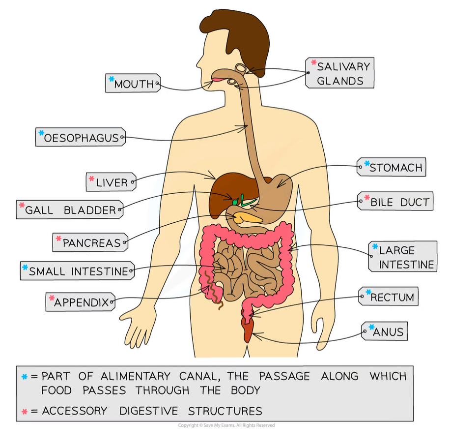
Function of mouth/salivary gland
Mechanical digestion: teeth chew food to break it into smaller pieces and increase its surface area to volume ratio
Chemical digestion: amylase enzymes in saliva start digesting starch into maltose
The food is shaped into a bolus (ball) and lubricated by saliva so it can be swallowed easily
Function of oesophagus
The tube that connects the mouth to the stomach
Wave-like contractions take place to push the food bolus down without relying on gravity
Function of stomach
Food is mechanically digested by churning actions while protease enzymes start to chemically digest proteins.
Hydrochloric acid is present to kill bacteria in food and provide the optimum pH for protease enzymes to work.
Function of small intestine
The first section is called the duodenum; this is where digestion of the food exiting the stomach is completed by enzymes that are present in the duodenum lining and secreted by the pancreas
The pH of the small intestine is slightly alkaline; around pH 8-9.
The second section is called the ileum and is where the absorption of water and digested food molecules takes place; the ileum is long and lined with villi to increase the surface area over which absorption can take place
Function of large intestine
Water is absorbed from the remaining material in the colon to produce faeces
Faeces are stored in the rectum and exit the body via the anus
Function of pancreas
Produces all three types of digestive enzymes: amylase, protease and lipase
Secretes enzymes in an alkaline fluid into the duodenum for digestion; this raises the pH of fluid coming out of the stomach
Function of liver
Amino acids that are not used to make proteins are broken down here (deamination), producing urea
Produces bile to emulsify fats (break large droplets into smaller droplets), an example of mechanical digestion
Function of gall bladder
Stores bile to release into the duodenum
Explain peristalsis
Peristalsis is a mechanism that helps moves food along the alimentary canal
Firstly, muscles in the walls of the oesophagus createwaves of contractions which force the bolus along
Once the bolus has reached the stomach, it is churned into a less solid form, called chyme, which continues on to the small intestine
Peristalsis is controlled by circular and longitudinalmuscles
Circular muscles contract to reduce the diameter of the lumen of the oesophagus or small intestine
Longitudinal muscles contract to reduce the lengthof that section the oesophagus or the small intestine
Mucus is produced to continually lubricate the food mass and reduce friction
Dietary fibre provides the roughage required for the muscles to push against during peristalsis
Stages of food breakdown
Food taken into the body goes through six different stages during its passage through the alimentary canal(the gut):
Ingestion - the taking in of substances, e.g. food and drink, into the body through the mouth
Mechanical digestion - the breakdown of food into smaller pieces without chemical change to the food molecules
Chemical digestion - the breakdown of large, insoluble molecules into small, soluble molecules
Absorption - the movement of small food molecules and ions through the wall of the intestine into the blood
Assimilation - the movement of digested food molecules into the cells of the body where they are used, becoming part of the cells
Egestion - the passing out of food that has not been digested or absorbed (as faeces) through the anus
Overall purpose of digestive enzymes
The purpose of digestion is to break down large, insoluble molecules into smaller, soluble molecules that can be absorbed into the bloodstream
What do lipids, proteins and carbohydrates all go through as first stage of break down
Mechanical digestion in the mouth, chewed with teeth and tongue, moistened by saliva. |
Stages of carbohydrates breakdown steps 2-7
|
|
|
|
|
|
Stages of proteins breakdown
Hydrochloric acid in the stomach breaks food down further. |
Muscular churning of stomach. |
Protease released from the stomach to break protein down into amino acids. |
Bile is released into the duodenum to neutralise the acidic chyme to provide the optimum pH for enzymes in the small intestine. |
Protease from the pancreas enters the duodenum and continues to break down protein into amino acids. |
Protease is released from the small intestine to continue break down of protein into amino acids. |
Stages of lipids breakdown
Hydrochloric acid in the stomach breaks food down further. |
Muscular churning of stomach. |
Bile is released into the duodenum to neutralise the acidic chyme to provide the optimum pH for enzymes in the small intestine. |
Bile is released from the gall bladder and it emulsifies lipids into smaller droplets over which lipase can act. Lipase from the pancreas enters the duodenum and breaks down lipids into fatty acids and glycerol. |
Lipase is released from small intestine to continue break down of lipids in FAs and glycerol. |
2 things bile does
Neutralises and emulsifies
Adaptations of small intestine for absorption
very log and has a highly folded surface with millions of villi = increase the surface area = faster and more absorption
Villi: increase surface area - microvilli on surface of villi
A short diffusion distance = villi have one cell thick walls
Well supplied with a network of blood capillaries = steep concentration gradient e.g lacteals runs through the centre of the villus to transport fatty acids and glycerol + enzymes produced in the walls of the villi assist with chemical digestion + villi waft
Practical : testing the energy content in a food sample - method + results
Method : Use the measuring cylinder to measure out 25 cm3 of water and pour it into the boiling tube
Record the starting temperature of the water using the thermometer
Record the mass of the food sample
Set fire to the sample of food using the bunsen burner and hold the sample 2 cm from the boiling tube until it has completely burned
Record the final temperature of the water
Repeat the process with different food samples
E.g. popcorn, nuts, crisps
Results:
The larger the increase in water temperature, the more energy is stored in the sample
We can calculate the energy in each food sample using the following equation:
Practical - investigating respiration - method + results
Method
Measure out 10 cm3 of hydrogencarbonate indicator into 3 boiling tubes
Put in a layer of cotton wool
Place 10 germinating seeds in tube A
Place 10 boiled/dead seeds in tube B
Place 10 glass beads in tube C
Seal each tube with a rubber bung
After 3 hours, observe the colour of the indicator
Results
In this investigation, we would expect to note the following
Tube A should turn yellow as the seeds are respiringand producing carbon dioxide
Tube B should remain red as the dead seeds produce no carbon dioxide
Tube C should remain red as there is no living material in there
Corms evaluation :
Change - We will change the content of the boiling tube (germinating seeds, dead seeds or glass beads)
Organisms - The seeds used should all be of the same age, size and species
Repeat - We will repeat the investigation several times to ensure our results are reliable
Measurement 1 - We will observe the change in the hydrogen carbonate indicator
Measurement 2 - ...after 3 hours
Same - We will control the volume of hydrogen carbonate indicator, the number of seeds/beads, the temperature of the environment
Practical - demonstrating the production of heat in respiration - method + results + evaluation
Method
Set up the flasks as shown in the diagram
Flask A with the dead seeds
Flask B with the germinating seeds
Make sure the cotton wool is plugging the top of each flask
Hold the thermometer in place with the cotton wool
Invert the flask
Record the initial temperature
After 4 days, record the final temperature
Results
The thermometer in the flask with the germinating seeds(Flask B) should show an increase in temperature
Flask A should remain at room temperature
This is because the seeds in flask B are respiring and producing heat energy in the process
This shows that respiration is an exothermic reaction
The seeds in flask A are not respiring because they are dead, so the temperature remains the same
Evaluation :
Change - We will change the content of the flasks (germinating seeds or dead seeds)
Organisms - The seeds used should all be of the same age, size and species
Repeat - We will repeat the investigation several times to ensure our results are reliable
Measurement 1 - We will observe the change in the temperature on the thermometer
Measurement 2 - ...after 4 days
Same - We will control the number of seeds, the starting temperature of the flasks, the material and size of the flasks
What is the role of diffusion in plants
Drives the process of gas exchange
Equation for photosynthesis
Carbon dioxide + water → glucose + oxygen
In order for plants to photosynthesise and respire..
Gas exchange of oxygen and carbon dioxide are needed and this requires diffusion of gases
Adaptations of leaf for gas exchange - spongy mesophyll
Have lots of air spaces to allow gases to diffuse in and out of cells faster, as it increases the surface area to volume ratio
Adaptations of leaf for gas exchange - Guard cells
Kidney- shaped cells that open and close the stomata by absorbing or losing water. When lots of water is available, the cells fill and open stomata
Adaptations of leaf for gas exchange - Stomata
Where gas exchange and loss of water by evaporation takes place - opens during the day and closes at night
Adaptations of leaf for gas exchange - Thin leaves
Short distance of diffusion for carbon dioxide to diffuse into the leaf and oxygen to diffuse out
Adaptations of leaf for gas exchange - Flattened shape
Increases surface area for absorption of light and carbon dioxide
… occurs at night and day as plants require energy at all times
Respiration
Why can photosynthesis only occur when light
It requires light
Why is there a net balance of gases during the day
As respiration generally occurs at the same rate as photosynthesis - but at night since there is no photosynthesis, more oxygen is taken into the plants and more carbon dioxide is released
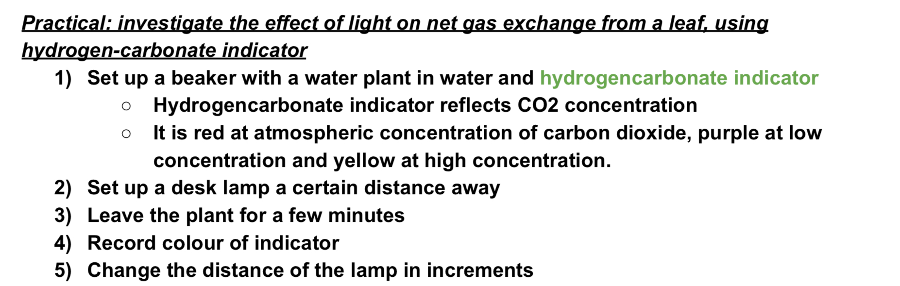
Practical - investigate the effect of light on net gas exchange from a leaf, using hydrogen carbonate indicator - method + results
Results = closer lamp is to plant = stronger light intensity = more photosynthesis = more CO2 taken into the plants by plant = low CO2 concentration = hydrogen carbonate indicator turns purple + if there is more CO2 present - indicator = yellow
Why do simple, unicellular organisms rely on diffusion for movement of substances
Bcs they have a relatively large surface area to volume ratio and so due to low metabolic demands, this diffusion across the surface of organisms is enough
Why do multicellular organisms rely on a transport system
They have a small surface area to volume ratio so instead need adaptations that allow molecules to be transported in and out of cells faster e.g root hair cells, alveoli, villi
Practical - investigate the role of environmental factors in determine the rate of transpiration from a leafy shoot
Set up potometer underwater to remove air bubbles in xylem so there is a continuous stream of water and the system is made airtight
Measure distance moved by air bubbles in xylem over time
Change a limiting factor
if air bubbles move faster = greater rate of water uptake and therefore higher rate of transportation
What is plasma important for
The transport of CO2, digested food, urea and hormones & heat energy
Adaptations of red blood cells
biconcave shapes = maximises SA for O2 to be absorbed
Flexible = fit into narrow blood vessels
No nucleus = creates more space for haemoglobin
Contains haemoglobin which is a red protein that combines with oxygen to allow oxygen transport
How does the immune systems respond to disease using white blood cells - phagocytosis
type of white blood cells which can undergo phagocytosis (phagocyte), where the pathogen is engulfed and killed and they have a non-specific function
How does the immune systems respond to disease using white blood cells - lymphocytes
produces antibodies (lymphocytes)
Each pathogen has an antigen on their surface and this antigen has a specific structure which is complementary to an antibody
Once antibodies begin to bind to pathogen, the pathogens start to clumps together, making it easier for white blood cells to find them
If you become infected again with the same pathogen, the specific complementary antibodies will be produced at a faster rate and the individual will not feel the symptoms of the illness, they are said to be immune
How does the immune systems respond to disease using white blood cells - antitoxins
producing antitoxins
WBCs can bind to the toxins released by the pathogen to neutralise them
What happens in vaccinations
Contain either a dead or inactivated form of the pathogen and when injected, they stimulate the white blood cells to produce the correct complementary antibody to combat it. Since the antigen is dead or inactivated, it doesn’t cause any symptoms to the individual
Some of these white blood cells remain int eh body as memory cells and if this person is exposed to real pathogen in future the memory cells procured antibodies much faster and lots more so it can be destroyed before any symptoms are felt
How are platelets involved in blood clotting
wound = platelets arrive to stop bleeding
Clotting cascade occurs within blood plasma
Platelets release chemicals that cause fibrinogen proteins to form a mesh of insoluble fibrin across wound = trapping red blood cells = clot
Clot develops into scab to protect wound from bacteria
Structure of arteries
Arteries carry blood AWAY from the heart = layers of muscle in the walls make them strong + contain elastic fibres which allow them to stretch + this helps the vessels withstand high pressure created by pumping of heart
Structure of veins
Carry blood TOWARDS the heart → lumen (tube which blood flows through) is wide to allow the low pressure blood to flow through + they have valves to ensure blood flows in right one way direction
Structure of capillaries
Allow blood to flow very close to cells to enable exchange of substances, capillary walls are one cell thick to create short diffusion pathway + permeable walls so substances can move across them
General structure of circulation system
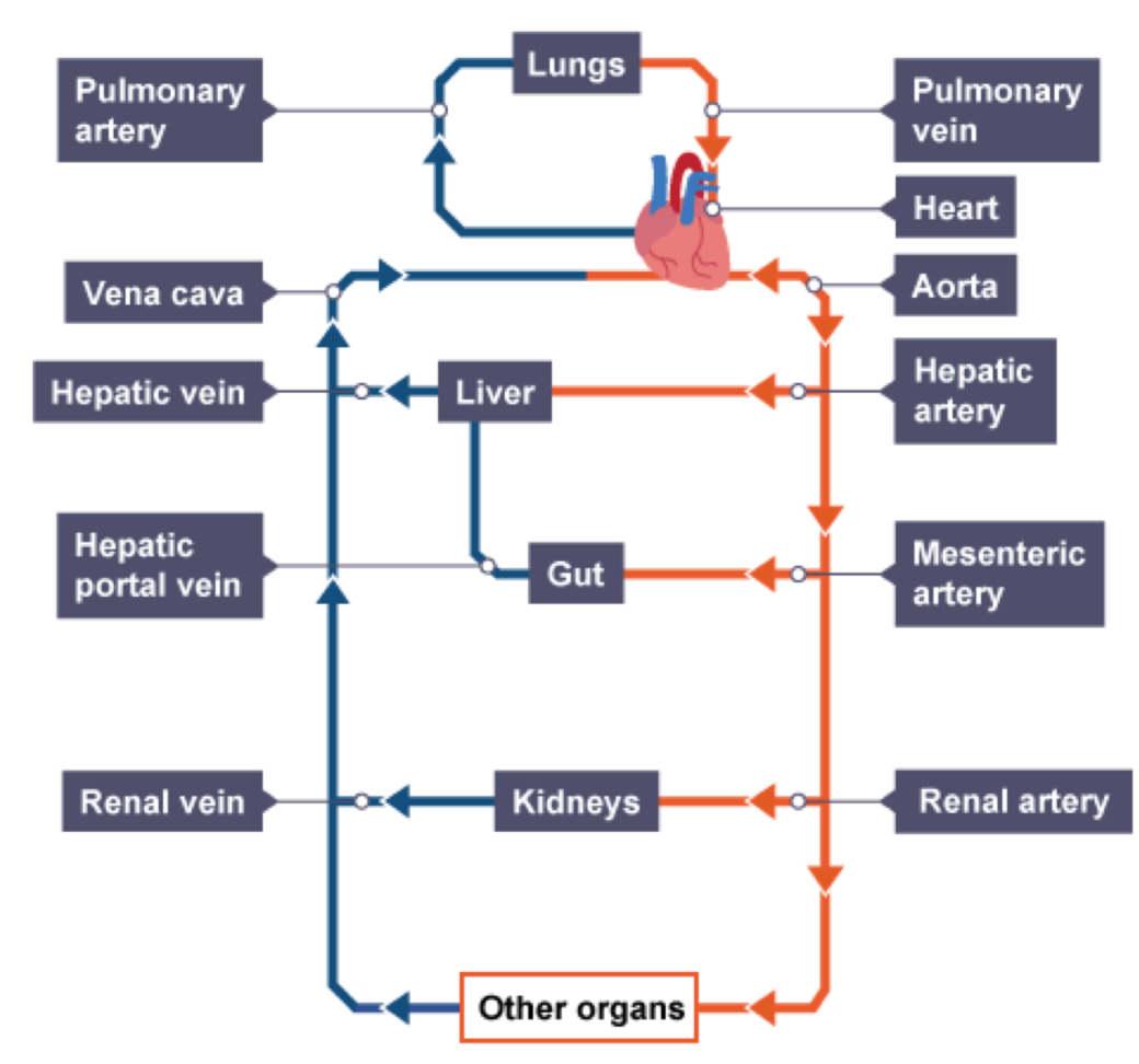
How does blood flow through heart
From vena cava, then RA
From pulmonary vein, then LA
Atria contract, forcing the blood into ventricles
Ventricles contract, pushing blood in RV to pulmonary artery (to go to lungs) and blood in LV into aorta (to be taken around body)
As this happens valves close