BISC 205 - Circulatory system
1/49
There's no tags or description
Looks like no tags are added yet.
Name | Mastery | Learn | Test | Matching | Spaced | Call with Kai |
|---|
No analytics yet
Send a link to your students to track their progress
50 Terms
What is a gastrovascular cavity and what are examples of organisms that have it?
A gastrovascular cavity is a central cavity that serves as a two-way digestive tract with a single opening for both food intake and waste removal.
Cnidarians (jellyfish, hydra) and flatworms
What are the primary functions of a circulatory system?
Transport of oxygen and nutrients to metabolizing tissues
Removal of carbon dioxide and other waste products from tissues
Transport of signalling molecules and immune cells throughout the body
Protection and healing (i.e., white blood cells, antibodies, platelets, etc.)
Regulation of body temperatures by controlled blood flow to the skin through vasodilation and vasoconstriction
What are the three basic components of open / closed systems?
a circulatory fluid
a set of tubes = blood vessels
a muscular pump = the heart
What are examples of circulatory fluids?
Interstitial fluid, blood, lymph, hemolymph
What is interstitial fluid?
extracellular fluid that directly bathes cells and tissues, delivering nutrients and collecting waste
What is blood?
fluid that circulates within a closed circulatory system
What is lymph?
fluid that circulates in the secondary system of vertebrates called the lymphatic system
What is hemolymph?
fluid (mixture of interstitial fluid and blood) that circulates within an open circulatory system
Describe an open circulatory system.
blood flows freely within body cavities and bathes the organs directly
all the blood in not enclosed in blood vessels
blood is pumped into a body cavity called hemocoel, where the blood mixes with the interstitial fluid (hemolymph)
as heart beats and the animal moves, hemolymph circulates to bathe organs within the hemocoel and then re-enters heart
more ideal for animals that have lower metabolic rates because the need for oxygen is less
blood pressure remains low, oxygen takes longer to reach body cells in all extremities
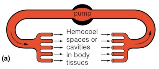
Describe the heart in an open circulatory system.
In an open circulatory system, the heart is usually sac-like or tubular.
it has ostia or lateral openings which are closed when the heart contracts and open when the heart relaxes (vacuum is created to suck blood into heart through ostia)
these hearts are known as suction pumps
the beating is set through nerve impulses (neurogenic hearts)
What are examples of animals that have an open circulatory system?
arthropods - lobsters, crabs, shrimp, insects, spiders, scorpions, centipedes and millipedes
bivalves
snails
Describe a closed circulatory system.
the blood is confined to blood vessels and is separate from the interstitial fluid (and does not come in direct contact with tissues)
blood travels in one direction in vessels (unliked in open circulatory system in which there is no unidirectional flow)
more ideal for animals with higher metabolic rates because need for oxygen is greater
blood pressure remains high, which means oxygen takes shorter time to reach body cells in all extremities

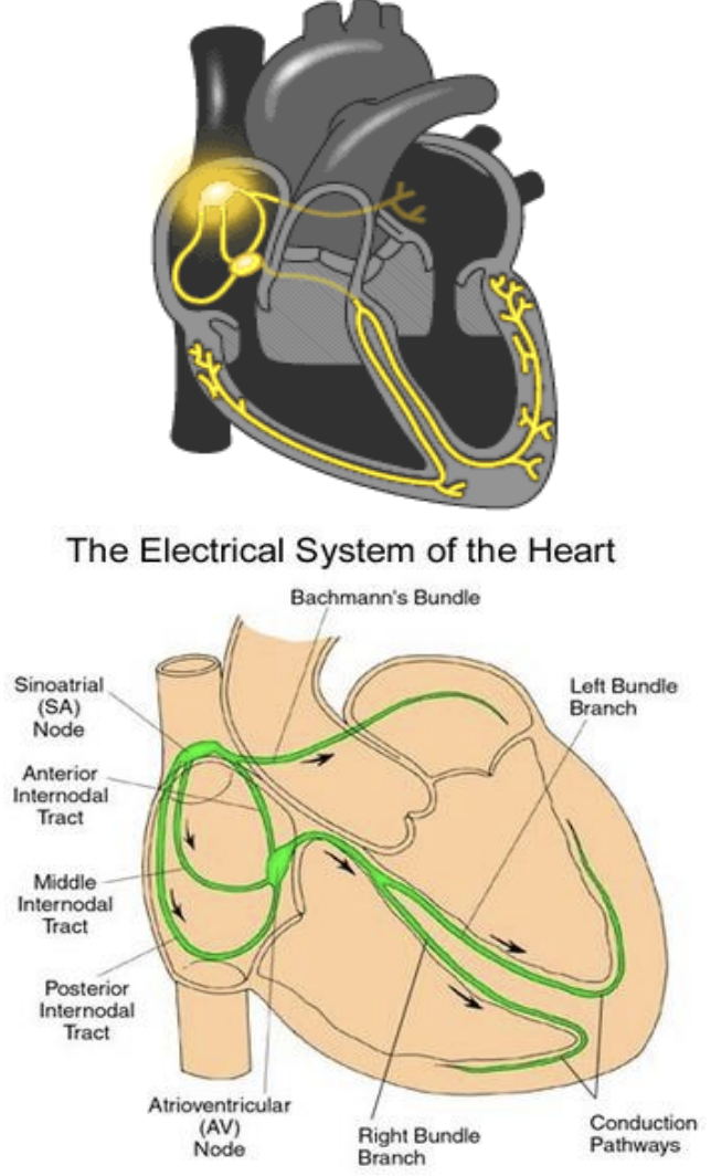
Describe the heart in a closed circulatory system.
chambered heart with muscular ventricles which pump blood in body with pressure
these hearts are known as pressure pumps
the rhythm is set in specialized neuromyocardial cells within heart
known as myogenic hearts since contractions are produced spontaneously without requiring external stimulation from nerve cells
What are examples of animals that have a closed circulatory system?
in all vertebrates (mammals, fish, birds/reptiles, amphibians)
some invertebrates (earthworms, octopuses and squids)
What are the advantages of an open circulatory system? (over closed system)
requires less energy for distribution of circulatory fluid
renders animals much less vulnerable to pressure (advantage for organisms that live at great depths because it prevents compression of their bodies)
less oxygen is needed since organisms with this system have a lower metabolic rate
What are the disadvantages of an open circulatory system?
less efficient in moving oxygen and nutrients to all body tissues and cells (not ideal for larger organisms)
has comparatively low blood pressure, which makes it difficult to reach extremities within a larger organism
limited capability to increase or decrease distribution and velocity of blood flow
better to sustain lower metabolic rates (results in slower movements and weaker adaptability to new environments)
What are the advantages of a closed circulatory system? (over open system)
more efficient in delivering oxygen and nutrients throughout organism to tissues and cells
provides more power in form of pressure so that blood reaches all extremities of the body
efficient distribution of antibodies, immune responses are stronger, helping body to fight off infection more effectively
more capability to increase or decrease distribution and velocity of blood flow
better to sustain higher metabolic rates (results in faster movement and better adaptability to new environments)
What are the disadvantages of a closed circulatory system?
requires more energy for distribution of blood (i.e., heart must work harder to pump blood faster, so it reaches destination quickly)
since blood is contained within vessels, a severe injurt can result in rapid and significant blood loss
a high-pressure system is susceptible to physical strain and blockages/blood clots within blood vessels, which can lead to serious health complications
What are the two types of closed circulatory systems?
singles circulation and double circulation
What are the differences between single circulation and double circulation?
Single circulation: blood passes through the heart once on each loop around the body, blood entering the heart is deoxygenated.
Double circulation: blood passes through the heart twice on each loop around the body, blood entering the heart can be oxygenated and deoxygenated.
List examples of organisms with single circulation:
determine the number of atria and ventricles present in the heart
identify the chambers where the blood enters and leaves
bony fish: 1 atrium, 1 ventricle, blood enters the heart at the sinus venosus and leaves through the bulbus arteriosus
elasmobranchs / cartilaginous fish: 1 atrium, 1 ventricle, blood enters the heart at the sinus venosus and leaves through the conus arteriosus
List examples of organisms that have double circulation.
amphibians, reptiles, mammals
For double circulation, define the following circuits: pulmocutaneous, pulmonary and systemic
pulmocutaneous circuit: oxygen poor blood flows through this circuit and picks up oxygen from the lungs and skin, found in amphibians.
pulmonary circuit: oxygen poor blood flows through this circuit and picks up oxygen through the lungs, found in mammals and reptiles.
systemic circuit: oxygenated blood flows through this circuit to deliver oxygen to the tissues, found in amphibians, reptiles and mammals.
Describe the adaptations of double circulation in amphibians.
2 atria and 1 ventricle, no septum
The atria receive blood from the 2 circuits (pulmocutaneous and systemic)
The sinus venosus bring deoxygenated blood from the body into the right atrium, while the pulmonary vein brings oxygenated blood from the lungs/skin into the left atrium
Mixing of oxygenated and deoxygenated blood occurs in the ventricle
This lowers the concentration of the oxygenated blood as it leaves and reduces the efficiency of oxygenation
The ventricle pumps blood via the conus arteriosus into both pulmocutaneous and system circuits (forked artery)
Spiral fold in the conus arteriosus that minimizes the mixing of blood and helps direct oxygenated blood into systemic circuit and deoxygenated blood into pulmocutaenous circuit
Describe the adaptations of double circulation in reptiles.
most reptiles (i.e., turtles and lizards) have 3 chambered hearts
2 atria and 1 ventricle
partial septum
less mixing of oxygenated and deoxygenated blood compared to amphibians
pulmonary circuit (lungs) and systemic circuit
crocodilian reptiles have 4 chambered hearts
2 atria and 2 ventricle
complete septum (no mixing within the heart)
foramen of Panizza, a small opening with a valve-like structure located at the base of right and left aortas which allows for mixing of the blood outside the heart
Describe the adaptations of double circulation in mammals and birds.
mammals and birds have a 4 chambered heart
2 atria and 2 ventricle
full septum (no mixing)
the left side of the heart pumps and receives only oxygen-rich blood, which the right side receives and pumps only oxygen-poor blood
pulmonary circuit (lungs) and systemic circuit
What are the differences between arteries and veins?
Arteries:
carry blood away from the heart (usually oxygenated)
under high pressure
thick-walled (compared to vein of similar size)
blood moves in one direction due to high blood pressure and smooth muscle contraction
situated deeper in body
smaller lumen compared to vein of similar size (to help maintain higher blood pressure)
Veins:
carry blood to the heart (usually deoxygenated)
under low pressure
thin-walled (compared to artery of similar size)
blood moves in one direction due to one-way valves, skeletal muscle contraction and smooth muscle contraction
situated near surface of skin
larger lumen compared to artery of similar size
Name the three layers of blood vessels.
tunica intima (innermost layer)
tunica media (middle layer)
tunica externa (outermost layer)
Describe the tunica intima and determine the differences between veins and arteries.
the inner lining is called the vascular endothelium, a sheet of epithelial cells which creates a frictionless pathway for blood
in arteries, there is a support structure called the internal elastic membrane, this is not present in veins
in arteries, the endothelium appears wavy/pleated due to more contraction of smooth muscle
in veins, the endothelium appears smooth
Describe the tunica media and determine the differences between veins and arteries.
composed of smooth muscle and elastin
controls vasoconstriction and vasodilation
normally the thickest layer in arteries to help withstand high blood pressure and maintain vessel shape
in veins, this layer is thinner than tunica externa. less smooth muscle reduces the structural rigidity of veins which leads to higher compliance
there is an external elastic membrane in arteries, but not in veins - lower elastin contributes to higher compliance in veins
Describe the tunica externa and determine the differences between veins and arteries.
primarily composed of collagen fibers to help support and reinforce shape of blood vessel
has vasa vasorum (tiny blood vessels) and nervi vasorum (small nerves)
this layer is normally thickest in veins:
prevents walls from collapsing under low blood pressure
provides protection from damage since veins are superficially located in body
Describe the structure and function of capillaries.
capillaries are the smallest of the body’s blood vessels that convey blood between arterioles and venules
the endothelium of capillaries are one cell layer thick (simple squamous layer), which makes it easier for exchange to occur between the blood and interstitial fluid which surrounds tissues
What is blood pressure?
Blood pressure is associated with pressure pushing on the walls of the capillary
not constant throughout the capillary, higher blood pressure on arterial side and lower blood pressure on venous side
What is osmotic pressure?
Osmotic pressure is associated with movement of water from a hypotonic environment (interstitial fluid) to a hypertonic environment (blood)
constant throughout the capillary
What is the law of bulk flow?
Q = ΔP / R
where:
Q = flow rate
ΔP = pressure gradient (perfusion pressure)
R = resistance
Bulk flow of fluid occurs when an external force is applied to fluid, setting it in motion, this helps in transport of substances across long distances far faster than would be possible by diffusion alone
Define flow rate.
Flow rate = volume of fluid that moves past a given point per unit time
What is the difference between perfusion pressure and transmural pressure?
Perfusion pressure = flow in and flow out
Transmural pressure = difference between internal and external pressure
What is the resistance formula and describe the 3 factors that affect resistance in circulatory system.
R = 8Lη / πr4
r = radius of vessel lumen
L = length of vessel
η = coefficient of blood viscosity (higher η means higher viscosity)
What is Poiseuille’s equation?
Q = ΔPπ r4 / 8Lη
What are the assumptions and violations of Poiseuille’s equations?
This equation assumes that tubes in circulatory system are unbranched and rigid, and the flow involves a simple fluid moving steadily through tubes.
In real circulatory systems, the vessels are branched and distensible, and the flow is often pulsatile. The fluid is also not simple as blood is made up of plasma and cells
What is the equation for velocity of blood?
Blood velocity = Q / A
determined by pressure and cross-sectional area
measured as distance per unit time
What is the law of LaPlace?
T = aPr
T = wall tension
a = a constant (1 for cylindrical blood vessel)
P = transmural pressure
r = radius of vessel lumen
the tension on walls of blood vessel is proportional to blood pressure and vessel radius (lumen)
walls of a blood vessel with a larger lumen radius will be exposed to higher tension than would blood vessels with a smaller lumen radius
Rewrite the law of LaPlace to consider thickness of blood vessel wall
σ = Pr / w
σ = wall stress
P = transmural pressure
r = radius of vessel lumen
w = blood vessel wall thickness
What is cardiac output and what is the formula?
cardiac output (CO) is a measurement of the amount of blood pumped out of each ventricle in one minute
CO = HR x SV
HR = heart rate, SV = stroke volume (the volume of blood pumped out of each ventricle per beat)
What is the formula for stroke volume?
SV = EDV - ESV
EDV = end diastolic volume, the maximum volume of blood in ventricle at rest
ESV = end systole volume, the volume of blood remaining in ventricle after contraction
What is ejection fraction and what is the formula?
EF = (SV / EDV) x 100
Ejection fraction is the percentage of blood ejected from the heart per beat
What is a normal ejection fraction?
typically between 55-70%
an EF below 50% can signal heart failure, indicates that the heart is not pumping strongly enough
What are the caused of a reduced ejection fraction?
weakness of the heart muscle, such as cardiomyopathy
heart attack that damaged the heart muscle
heart valve disease
viral infections like myocarditis
long-term, uncontrolled high blood pressure
Why doesn’t ejection fraction reach 100%?
EF will not reach 100% because the ventricles do not completely empty with each heartbeat.
What is the Frank-Starling effect?
represents relationship between stroke volume and end diastolic volume
a greater blood volume in the ventricles at rest leads to an increase in blood volume leaving the ventricles during systole (contraction)