Male Carnivore External Genitalia (Mod. 5)
1/29
There's no tags or description
Looks like no tags are added yet.
Name | Mastery | Learn | Test | Matching | Spaced |
|---|
No study sessions yet.
30 Terms
What are the 3 main products of the male reproductive tract?
1) Gametes / sperm
2) Androgenic hormones (testosterone)
3) Semen
The canine penis is termed as a “musculo-cavernous” type penis. What the fuck does this mean?
This means that the penis contains large amounts of cavernous sinus tissue
Erectile tissue
Distends with blood to form an erection
What are the 4 main parts of the canine penis?
1) OS penis - internal bone in the penis
2) Root
3) Body
4) Glans
Bulbus Glandis
Pars Longa Glandis
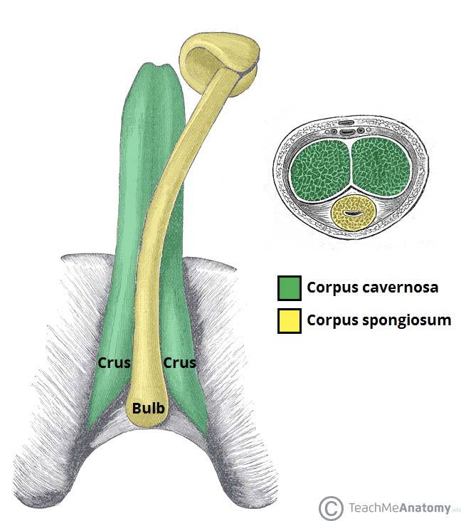
Describe the root and body structure of the penis… What are the three major components? What are these made up of, and how are they arranged?
The three major components are the bulb of the penis, and the two crura on either side.
The crura arise from the ischial arches of the pubic bone. As they get closer to the body of the penis, they converge and join to form a single body (see image)
Each crura consists of:
Corpus cavernosum (cavernous tissue core)
Encased in tunica albuginea (thick connective tissue)
*** Note: there is a ventral groove between the two crura to accommodate the urethra, which is encased in a vascular sleeve known as the corpus spongiosum
Since the previous card went more into the crura, describe the bulb of the penis… what is it made up of? How does this tissue change as it runs down the body of the penis?
Bulb is made up of an enlargement of corpus spongiosum tissue, which is covered by bulbospongiosus muscle
Originates at the pelvic outlet (see image)
Corpus spongiosum tissue carries on as thin, vascular sleeve surrounding the urethra as it moves down the body of the penis
At the distal end of the penis, corpus spongiosum expands AGAIN to form the glans
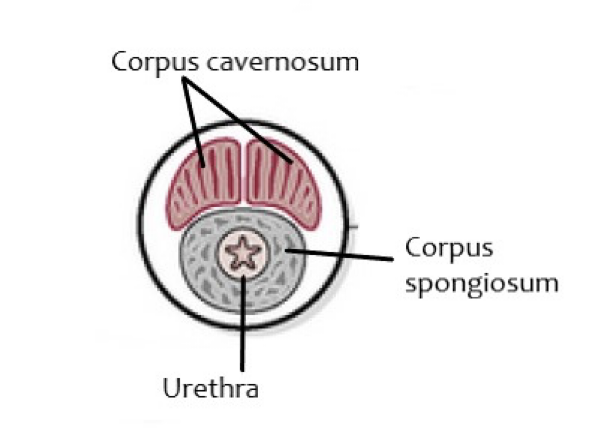
If you were to take a cross section of the penis, describe how it looks and what tissues you would see.
On the top of the cross section, you would see the two crura with their cavernous tissue core
These two will remain separated by a septum that runs between them
Encased in tunica albuginea
Beneath the two crura is the corpus spongiosum casing that surrounds the urethra
Quick recap… describe the purpose and function of the corpus cavernosum and corpus spongiosum of the penis.
Corpus cavernosum - Forms the core of the crura, and forms the bulk of spongy vascular tissue within the body and root of the penis
Corpus spongiosum - Mostly a thin, vascular sleeve around the urethra… the two enlargements of this tissue form the bulb and glans of the penis
Is MUCH spongier than the corpus cavernosum (hence the name), allowing for higher vascularity and higher potential for expansion
Is also more delicate
The distal end of the corpus cavernosum becomes what?
The OS penis
Initially starts out as a fibrous outgrowth of the corpus cavernosum
Describe the structure of the OS penis…. what is its most notable feature, and why is this important?
The most notable feature is its ventral groove
OS penis looks like a “U” shape in a cross section
This is where the urethra passes through the area of the glans
IMPORTANT TO REMEMBER, because this gives the urethra limited capacity to expand
Dogs passing bladder stones; stones often get stuck at the proximal end of the urethra due to restriction by the OS penis
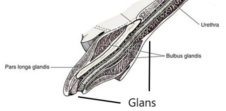
Describe the structure of the glans… what tissue is it made up of, and what are its 2 main components?
Found on the distal end of the penis; corpus cavernosum ends and glans formed completely by corpus spongiosum
Made up of 2 different glands…
1) Bulbus glandis -
Proximal portion of the glans that swells during erection and mating
Quite literally the “bulbous” looking portion of the tip of the penis
Responsible for the “tie” during mating in canines
Firmly attached to the OS penis
2) Pars longa glandis -
The distal portion of the glans; more elongated
Responsible for initial penetration and helps penis maintain rigidity during erection
Less attached to the OS penis
Has urethral orifice at tip
What is the “prepuce” of the penis? What are its 2 main layers?
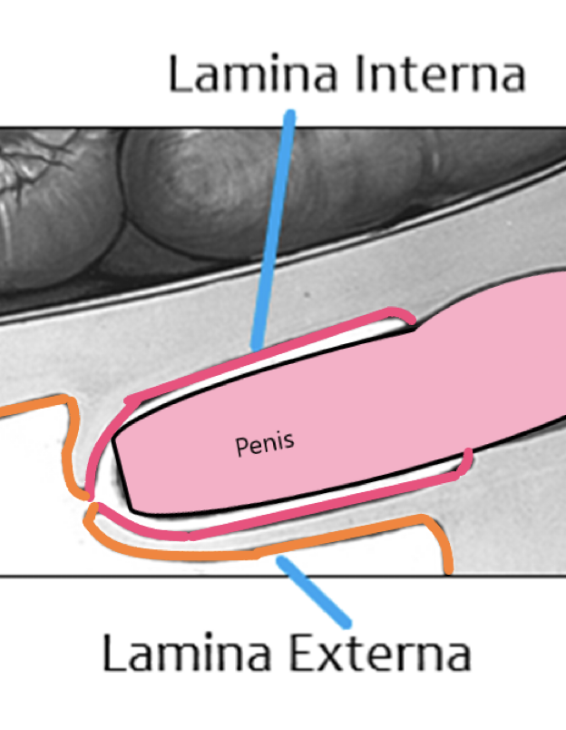
The prepuce is the invagination of the abdominal skin, which covers and protects the glans penis when not erect
Has 2 layers of tissue:
1) Lamina externa -
Outer layer; continuous with the outer skin, and is basically just skin
2) Lamina interna -
Inner layer; lines the preputial cavity and covers the free end of the penis
Lined with lymphoid tissue; not very surprising, structure = function
*** NOTE: PREPUCE ALSO HAS SMEGMA GLANDS
What are two clinical problems that the prepuce can cause?
1) Congenital narrowing of preputial orifice -
Prevents extrusion (exiting) of the penis from the prepuce… called “phimosis”
2) Paraphimosis -
The inability to retract the penis into the prepuce
Causes:
Orifice too narrow; penis goes out and swells, then can’t retract
OR fur wrapped around penis when extruded… causes stricture
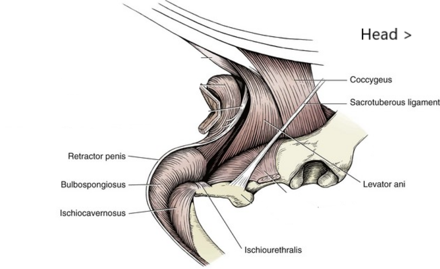
What are the 4 main muscles associated with the penis?
1) Bulbospongiosus (sinlge muscle)
2) Ischiocavernosi (paired and powerful; look at the name and think about what it could be associated with)
3) Ischiourethralis (single muscle; again, look at the name, gives away association)
4) Retractor penis (paired)
Is the bulbospongiosus smooth or skeletal muscle? Where is its origin, insertion, and what is its function?
SKELETAL muscle
Origin: Continuation of the urethralis muscle
Insertion: On corpus spongiosum
Function: Pumps blood into cavernous spaces
Is the ischiocavernosi smooth or skeletal muscle? Where is its origin, insertion, and what is its function?
SKELETAL muscle
Origin: Ischium (in the name)
Insertion: Encloses crura and inserts on corpus cavernosum (again, the name)
Function: Pumps blood into the cavernous space
***** So basically, the bulbospongiosum and ischiocavernosi are both associated with their specific erectile tissue, AND have the same function of pumping blood into cavernous spaces to allow for erection. Basically, think of these as the erectile MUSCLES, which function to ERECT the penis by pumping in more blood upon contraction
Is the ischiourethralis smooth or skeletal muscle? Where is its origin, insertion, and what is its function?
SKELETAL muscle
Origin: Ischium (name)
Insertion: Fibrous ring around penile veins
Function: SMALL BUT IMPORTANT; occludes dorsal VEIN during erection, preventing blood from draining from the penis
Is the retractor penis smooth or skeletal muscle? Where is its origin, insertion, and what is its function?
SMOOTH muscle
Origin: caudal VERTEBRAE
Insertion: PREPUCE
Function: DRAWS PENIS INTO PREPUCE… NOT INVOLVED IN ERECTION
Here’s a diagram to show where the muscles are in the penis…
What are the two major arterial supplies to the penis and prepuce? (think inside and outside)
Internal Pudendal Artery - Penis
External Pudendal Artery- Prepuce
Describe the blood supply to the penis, starting at the internal pudendal artery…
Goes from internal pudendal artery → Artery of Penis, which branches into three different arteries:
1) Dorsal Artery of Penis - Supplies the GLANS (both bulbus glandis and pars longa)
2) DEEP Artery of Penis - Supplies CORPUS CAVERNOSUM within the crura
3) Bulbourethral Artery - Supplies CORPUS SPONGIOSUM in proximal part of penis
** if you think about it, the names kinda relate to what they supply
Describe the penile venous drainage system… what roll does it have in erection? In what three areas of the penis is there venous drainage?
Veins in the penis have valves within them to PREVENT REFLUX during erection. The three main areas the penis is divided into are:
1) The pars longa and prepuce
2) Bulbus Glandis
3) Corpus cavernosum and bulb of penis
Describe the path of venous drainage from each of the 3 “areas” of the penis… what are the 3 main veins in the penis?
The 3 main veins are:
External Pudendal Vein
Dorsal Vein ***
Internal Pudendal Vein
Pars Longa and Prepuce drainage -
Can drain 2 different ways… either into the bulbus glandis, or into the external pudendal vein of the prepuce
Bulbus Glandis drainage -
Drains into the Dorsal Vein → Internal Pudendal Vein
Corpus cavernosum and Bulb of Penis drainage -
DORSAL vein runs along dorsal surface of penis… simply collects blood from corpus cavernosum and bulb of penis as it goes along
What is a notable feature of the Dorsal Vein?
The Dorsal Vein is the most important drainage route for the bulk of the penis.
Runs the length of the dorsal surface of penis
Has a FIBROUS RING ASSOCIATED WITH ISCHIOURETHRALIS MUSCLE (previously mentioned before)
Once this muscle contracts during erection, fibrous ring CONSTRICTS vein, preventing drainage
Describe the 2 stages of the canine erection and what happens during these stages…
1st Stage -
INCREASED blood flow into penis via arteries
OCCLUSION of VENOUS OUTFLOW via ischiourethralis ring and valves
Results in blood building up within cavernous spaces in penis (swelling) until arterial blood pressure is reached
2nd Stage -
Rhythmic CONTRACTION of ischiocanernosus and bulbospongiosus muscles
Forces blood into cavernous spaces above arterial pressure
Describe why the “tie” happens in mating dogs…
Corpus spongiosum expands more than corpus cavernosum, so as the male dismounts and faces away from the bitch, this reduces venous drainage even more and bulbus glandis remains swollen
“Ties” with vagina
Once bulbospongiosus relaxes, blood drains from bulbus glandis and swelling goes down, allowing the dogs to uncouple
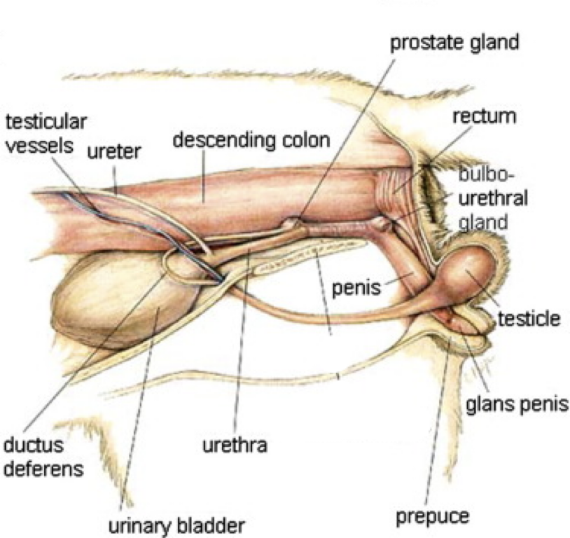
Describe the big differences in the FELINE penis compared to the canine penis…
CAUDOVENTRALLY DIRECTED (>) see image
Therefore, opening of cat’s prepuce faces caudally as well
Groove of OS Penis is DORSAL instead of ventral because of this reason as well
Penis is shorter
Glans is smaller, with keratinized spines (ouch)
Responsible for triggering induced ovulation in female
As erection occurs:
There is a curvature to this penis, so apex ends up directing CRANIOVENTRALLY for penetration
What is the overall importance of the scrotum? What is its most notable external feature?
Importance: thermoregulatory mechanism
Allows maintenance of testicular temperature, which should be slightly below body temp
Notable feature:
Median groove, dividing it into left and right halves
Correlates to an internal septum
What is the important muscle layer within the scrotum? Describe it and what it does.
Dartos:
Combination of smooth muscle and fibrous tissue
Closely adhered to the skin, along with testes and fascia
Is responsible for septum between the two testicles
In cold temperatures, the dartos actually contracts, drawing the testicles closer to the body to increase their temperature
So another thermoregulatory mechanism
Also supports the vaginal tunic, which is the outermost layer of the testes
Describe the vaginal tunic and its two layers… why is it clinically important to know about this? (think of neutering…)
The vaginal tunic is an outpouching of the parietal peritoneum through the inguinal canal.
Completely encloses testes, spermatic cord, and other structures that supply the testes
The two layers of the vaginal tunic:
1) Parietal outer layer - what we see
2) Visceral inner layer - CLOSELY ADHERED TO TESTES
******* There is a space between these two layers, filled with fluid… COMMUNICATES DIRECTLY WITH PERITONEAL CAVITY
This is why it is clinically significant… if neuter not completely sterile, can result in full-blown peritonitis
Quick review of inguinal canal… What is its function, and what are its two different rings?
Function:
Allows passage of testes to scrotum as animal develops
Testes develop inside the abdominal cavity and will descend into scrotum (remember, covered by vaginal tunic)
Two rings:
1) Deep inguinal ring - essentially the canal’s “opening”
Complex… associated with an additional muscle in males called the cremaster muscle
Detaches from internal abdominal oblique, and accompanies spermatic cord structures as testes drop
2) Superficial inguinal ring - the canal’s “exit”
Close to the attachment of the linea alba to pubic bone…. sits just in front of pelvis
Can be palpated in some patients