Surface Anatomy Of The Brainstem
1/24
There's no tags or description
Looks like no tags are added yet.
Name | Mastery | Learn | Test | Matching | Spaced | Call with Kai |
|---|
No analytics yet
Send a link to your students to track their progress
25 Terms
Name the three components of the brainstem in order from superior to inferior.
The three components of the brainstem from superior → inferior are:
Midbrain — the top part, connecting the brainstem to the diencephalon
Pons — the middle bulging segment
Medulla oblongata — the lowest part, continuous with the spinal cord
What is the tectum of the midbrain, and what does the term mean?
The tectum of the midbrain is the dorsal (“roof”) surface of the midbrain
Location: Forms the roof over the cerebral aqueduct at the midbrain level
Key feature: Contains the four colliculi (2 superior, 2 inferior)
What are the cerebral peduncles, and what structure lies between them?
Cerebral peduncles = the two big motor “stalks” on the front (ventral) midbrain carrying major descending motor tracts
Between them: the interpeduncular fossa
What is the interpeduncular fossa, and what emerges from it?
Interpeduncular fossa = the midline dip on the front (ventral) midbrain between the two cerebral peduncles
What emerges from it:
Oculomotor nerves (CN III) come straight out of this fossa
Summarise the key surface landmarks of the midbrain on both its dorsal and ventral surfaces.
Dorsal (back) surface of the midbrain:
Tectum = “roof”
Contains the four colliculi (2 superior + 2 inferior)
Ventral (front) surface of the midbrain:
Paired cerebral peduncles (big motor stalks)
Interpeduncular fossa between them
CN III (oculomotor nerve) exits from this fossa
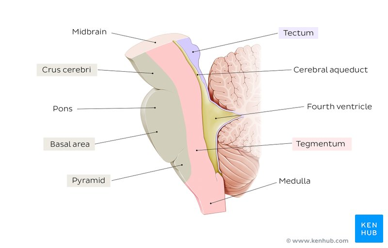
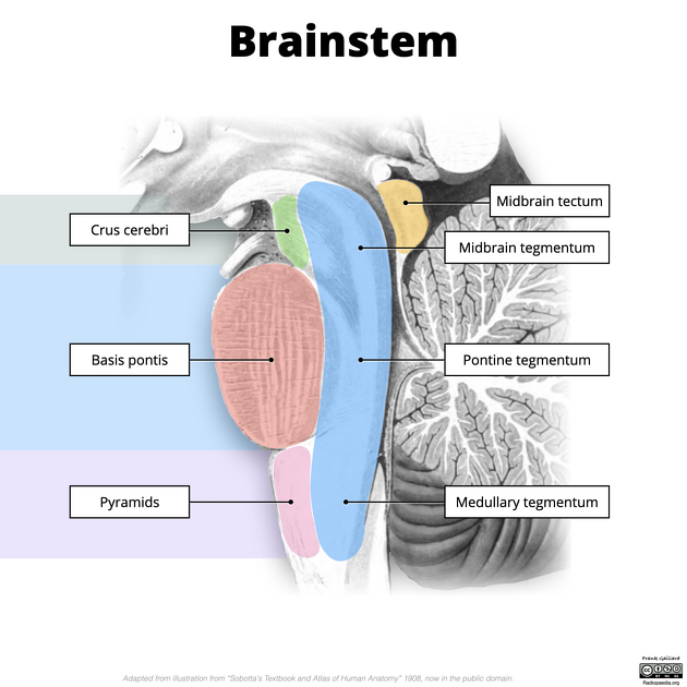
What are the two main parts of the pons?
The pons has two main parts:
Basal pons — the front (ventral) bulging part
Tegmentum — the back (dorsal) “covering” part
What is the tegmentum of the pons, and what structure does it contribute to?
The tegmentum of the pons is the back (dorsal) “covering” part of the pons
It forms part of the floor of the 4th ventricle together with the medullary tegmentum
It contains cranial nerve nuclei and ascending sensory tracts
What is the basal pons, and what is its distinguishing surface feature?
The basal pons is the front (ventral) bulging part of the pons
It creates the rounded, protruding surface you see on the ventral brainstem
It contains transverse pontocerebellar fibres and descending corticospinal fibres
What is the 4th ventricle, and which brainstem structures contribute to its floor?
The 4th ventricle is the CSF‑filled cavity behind the pons and medulla, connecting the cerebral aqueduct to the central canal
Its floor is formed by:
Pontine tegmentum → upper floor
Open/rostral medulla → lower floor
Describe the anatomical boundaries of the medulla oblongata.
The medulla oblongata sits between the pons and the spinal cord
Superior boundary: the inferior border of the pons
Inferior boundary: the foramen magnum, where it becomes the spinal cord
It is the lowest part of the brainstem
Distinguish between the "open" (rostral) and "closed" (caudal) portions of the medulla.
Open medulla = 4th ventricle present
Closed medulla = only the central canal
Open (rostral) medulla: the 4th ventricle is visible on the back → forms the lower floor of the ventricle.
Closed (caudal) medulla: the ventricle narrows into the central canal, just like the spinal cord.
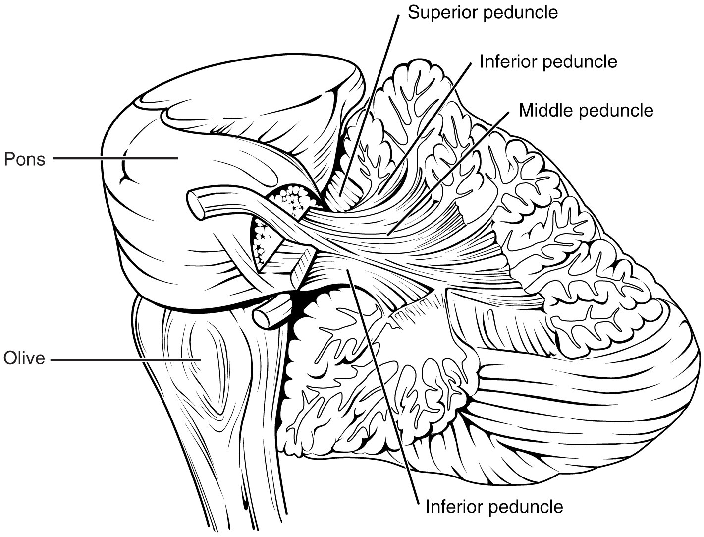
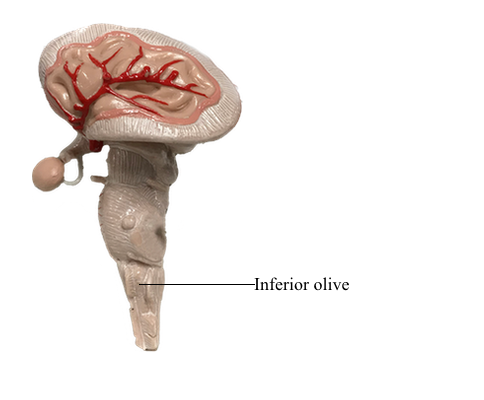
What is the inferior olive, where is it located, and why is it clinically important as a landmark?
The inferior olive is the oval bulge on the side of the medulla.
Why it matters: it’s the key landmark for locating four cranial nerves.
Above the olive: CN IX, X, XI
Below the olive: CN XII
What are the medullary pyramids, and what do they contain?
The medullary pyramids are the two long ridges on the front (ventral) surface of the medulla.
What they contain:
The corticospinal tract — the main motor pathway coming from the primary motor cortex.
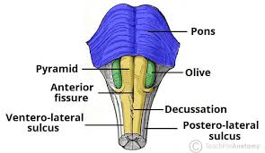
Summarise the key surface landmarks visible on the medulla, specifying whether each is on the anterior, lateral, or posterior surface.
Anterior (front) surface
Medullary pyramids — long motor ridges
Anterior median fissure — groove between the pyramids
Lateral surface
Inferior olive — the big oval bulge
Above the olive: CN IX, X, XI
Below the olive: CN XII
Posterior (back) surface
Upper part: forms the floor of the 4th ventricle (open medulla)
Lower part: becomes closed, like the spinal cord
Describe the CN I (olfactory nerve) — what structures are visible on the inferior brain, and does CN I actually attach to the brainstem?
CN I (olfactory nerve) does not attach to the brainstem.
What you actually see on the inferior brain:
Olfactory bulb — small oval structure sitting on the cribriform plate
Olfactory tract — the band running back from the bulb
Key point:
The true olfactory nerves are tiny filaments from the nose that synapse in the bulb. The bulb and tract belong to the inferior frontal lobe, not the brainstem.
Describe the CN II (optic nerve) landmarks visible in the inferior view, and where is the optic chiasm?
CN II (optic nerve) is seen from below as the two optic nerves coming from each orbit.
They meet at the optic chiasm — the X‑shaped crossing where nasal retinal fibres cross and temporal fibres stay on the same side.
The optic chiasm sits just above the pituitary gland.
Where does CN III (oculomotor nerve) attach to the brainstem?
CN III (oculomotor nerve) attaches to the midbrain.
It emerges from the interpeduncular fossa — the midline dip between the two cerebral peduncles on the ventral midbrain.
Then it travels forward toward the orbit through the superior orbital fissure.
Where does CN V (trigeminal nerve) attach to the brainstem?
CN V (trigeminal nerve) attaches to the lateral side of the pons.
It’s the largest cranial nerve
Has a big sensory root + small motor root
It’s the dominant nerve on the mid‑lateral pons
Where does CN VI (abducens nerve) attach to the brainstem?
CN VI (abducens) attaches at the pontomedullary junction.
It exits at the midline, just above the medullary pyramids
Sits right at the border between pons (top) and medulla (bottom)
Where does CN VII (facial nerve) attach relative to CN VI?
CN VII (facial nerve) attaches at the pontomedullary junction, just like CN VI — but more lateral.
CN VI: exits at the midline
CN VII: exits lateral to CN VI at the same level
Where does CN VIII (vestibulocochlear nerve) attach relative to CN VII?
CN VIII (vestibulocochlear) attaches right behind CN VII at the lateral pontomedullary junction.
CN VII: more anterior
CN VIII: just posterior/dorsolateral to CN VII
Which three cranial nerves attach to the lateral surface of the medulla, and what is their relationship to the inferior olive?
CN IX, X, XI all attach to the lateral medulla, and they all sit above the inferior olive.
They emerge as rootlets in the posterolateral sulcus
They lie just behind the olive’s bulge
Where does CN XII (hypoglossal nerve) attach in relation to the inferior olive?
CN XII (hypoglossal) attaches just below the inferior olive.
Sits in the anterolateral sulcus
Emerges as rootlets between the pyramid and the olive
Lies below and slightly medial to CN IX, X, XI
What do the medullary pyramids contain, and from where do their constituent fibres originate?
The medullary pyramids contain the corticospinal tract — the main voluntary motor pathway.
Their fibres come from primary motor cortex (precentral gyrus)
These fibres are the axons of upper motor neurons heading down to control contralateral body movement
A stroke affecting the anterior medullary surface on one side might damage the medullary pyramid. What type of motor deficit would this produce on the opposite side of the body?
A stroke hitting one medullary pyramid damages the corticospinal tract before it crosses.
Result: Contralateral UMN weakness with:
Spasticity
Hyperreflexia
Babinski sign
Clonus
Why: The fibres haven’t decussated yet, so injury on one side affects the opposite body.