HISTOLOGY LAB
1/106
There's no tags or description
Looks like no tags are added yet.
Name | Mastery | Learn | Test | Matching | Spaced | Call with Kai |
|---|
No analytics yet
Send a link to your students to track their progress
107 Terms
OVUM
slide 54b
cytoplasm
zona oellucida
cell membrane
nuclear membrane
nucleolus
nucleus
OVUM
Connective Tissue
Embryonal Tissue
(Umbilical cord)
slide #61
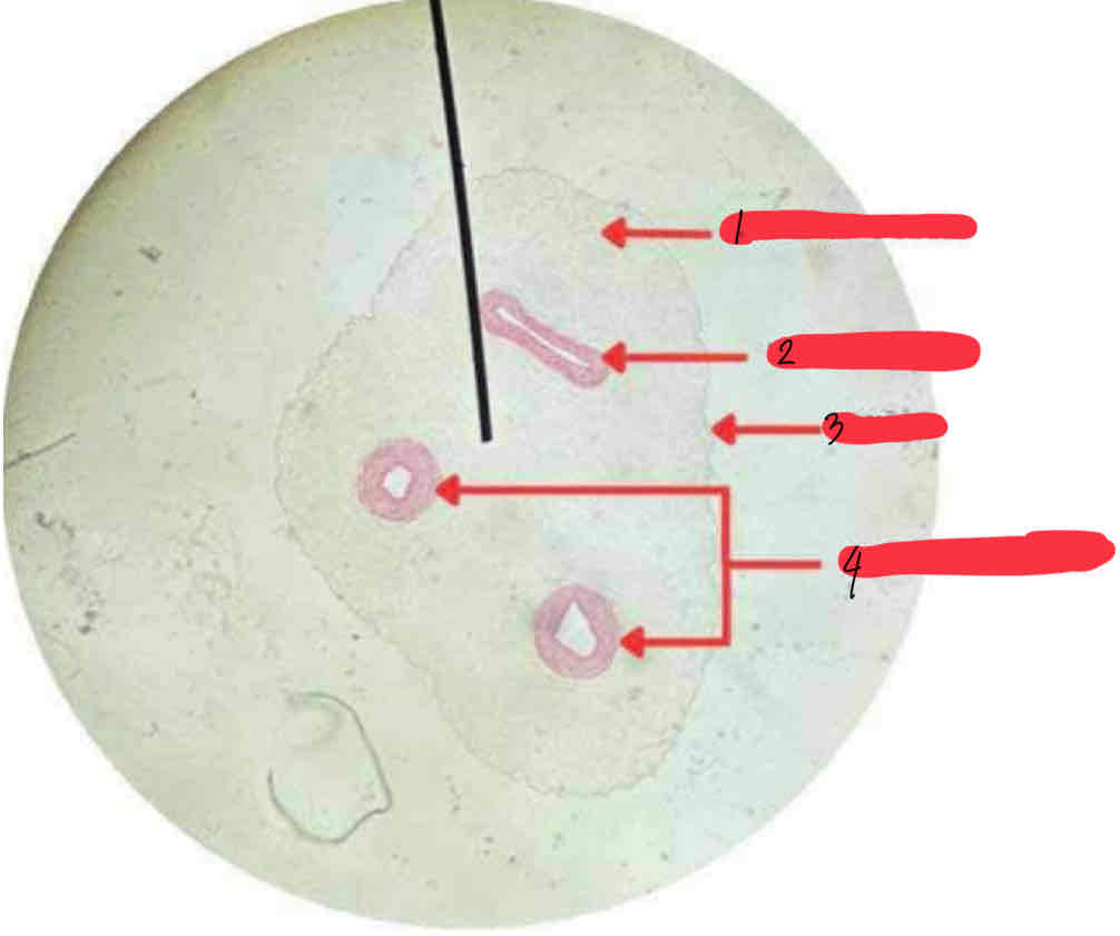
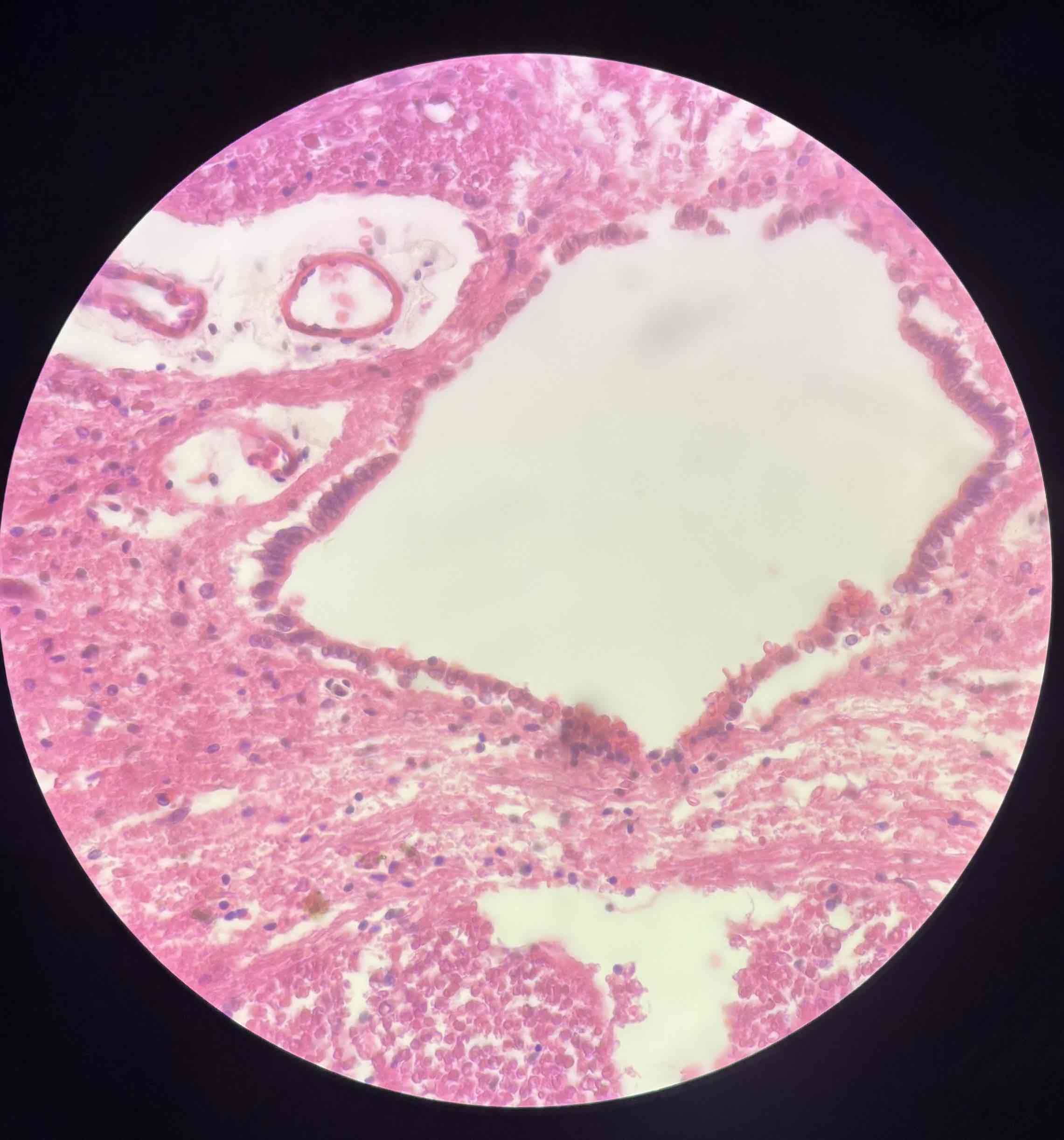
WHARTON'S JELLY
UMBILICAL VEIN
AMNION
UMBILICAL ARTERY
Umbilical Cord
Nerve cell
Spinal cord
slide # 7
Cell Membrane
Nuclear Membrane
Nucleolus
Cytoplasm
Nucleus
Nerve Cell

Transitional epithelium, with condensed border
Urinary Bladder
Slide 36
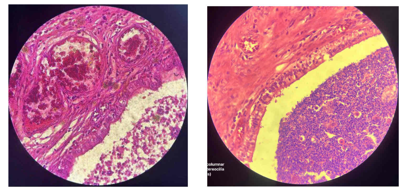
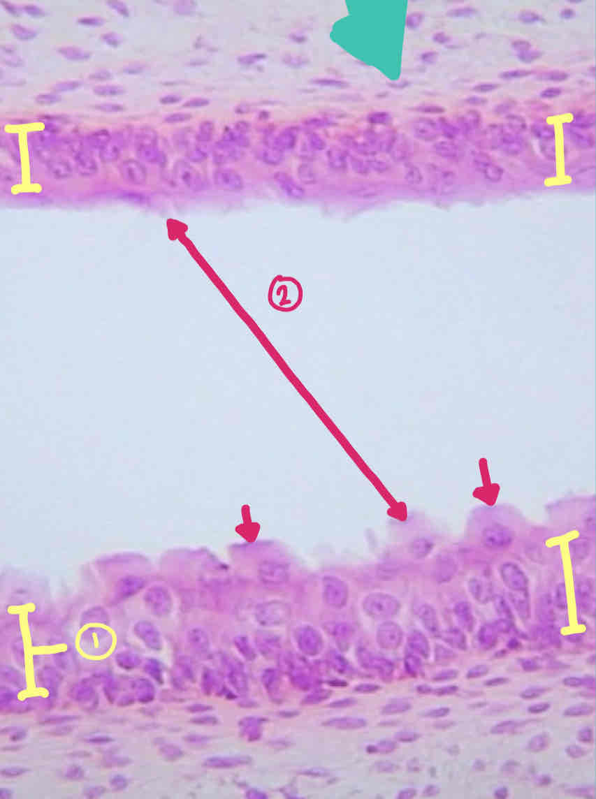
Pseudostratified columnar epithelium w/ cilia (larynx)
Slide 42
PSEUDOSTRATIFIED COLUMNAR EPITHELIUM
GOBLET CELLS
larynx
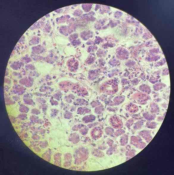
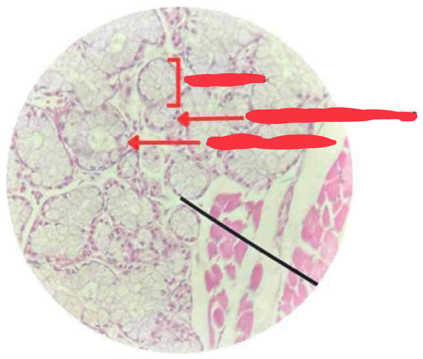
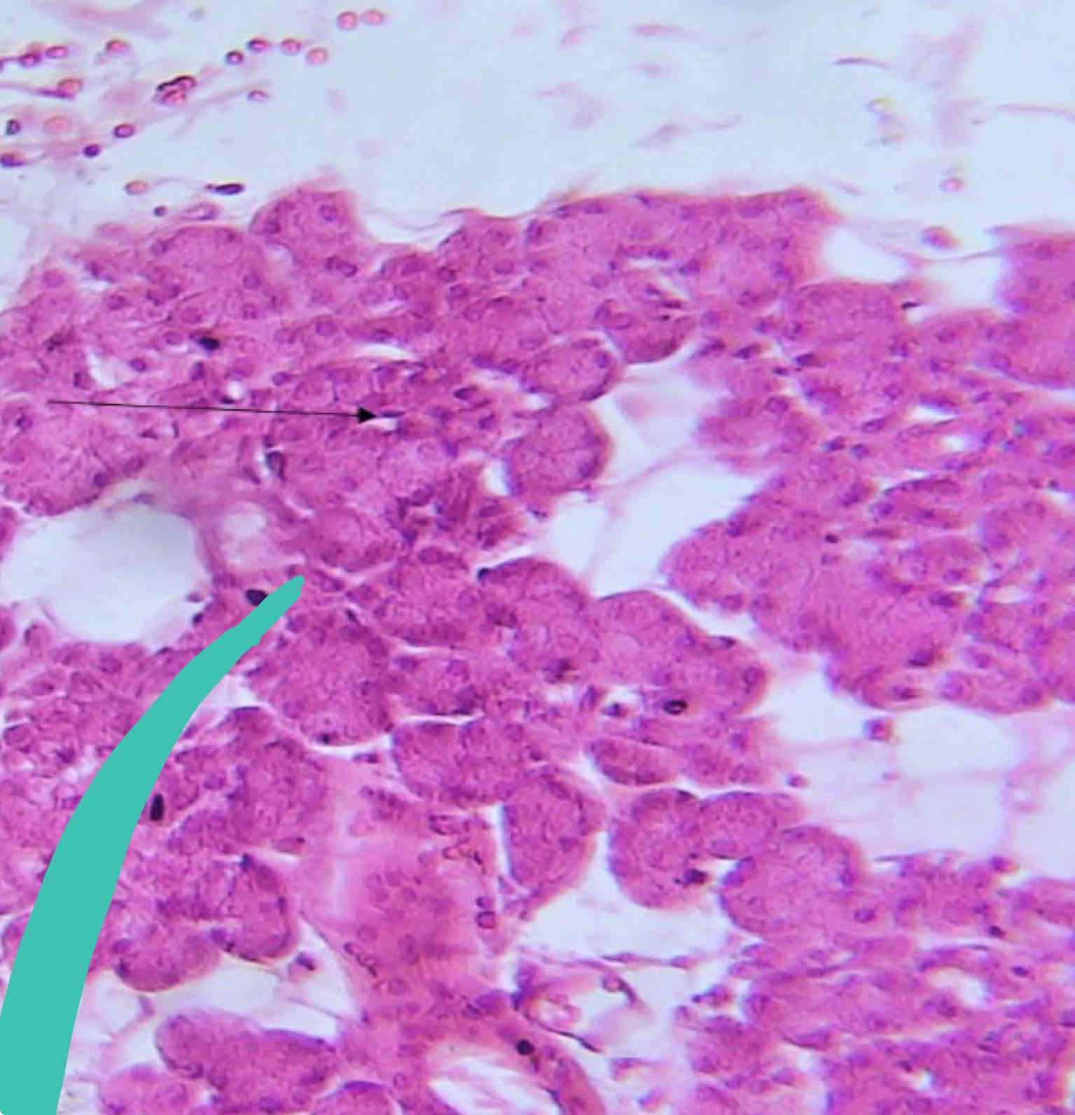
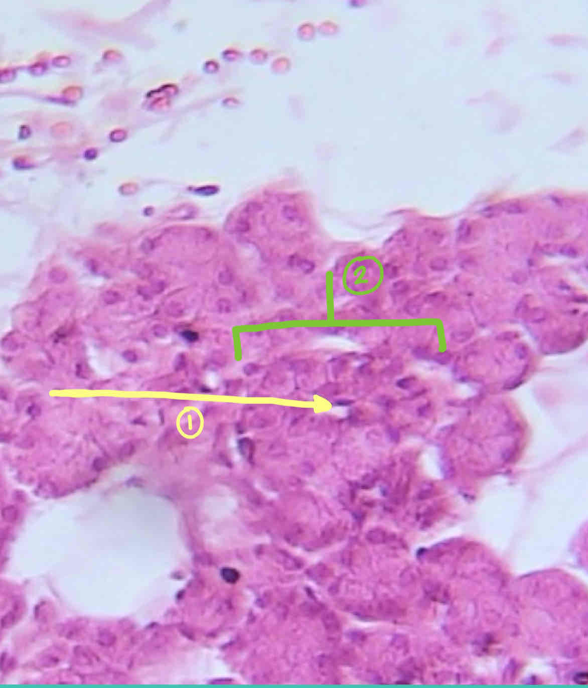
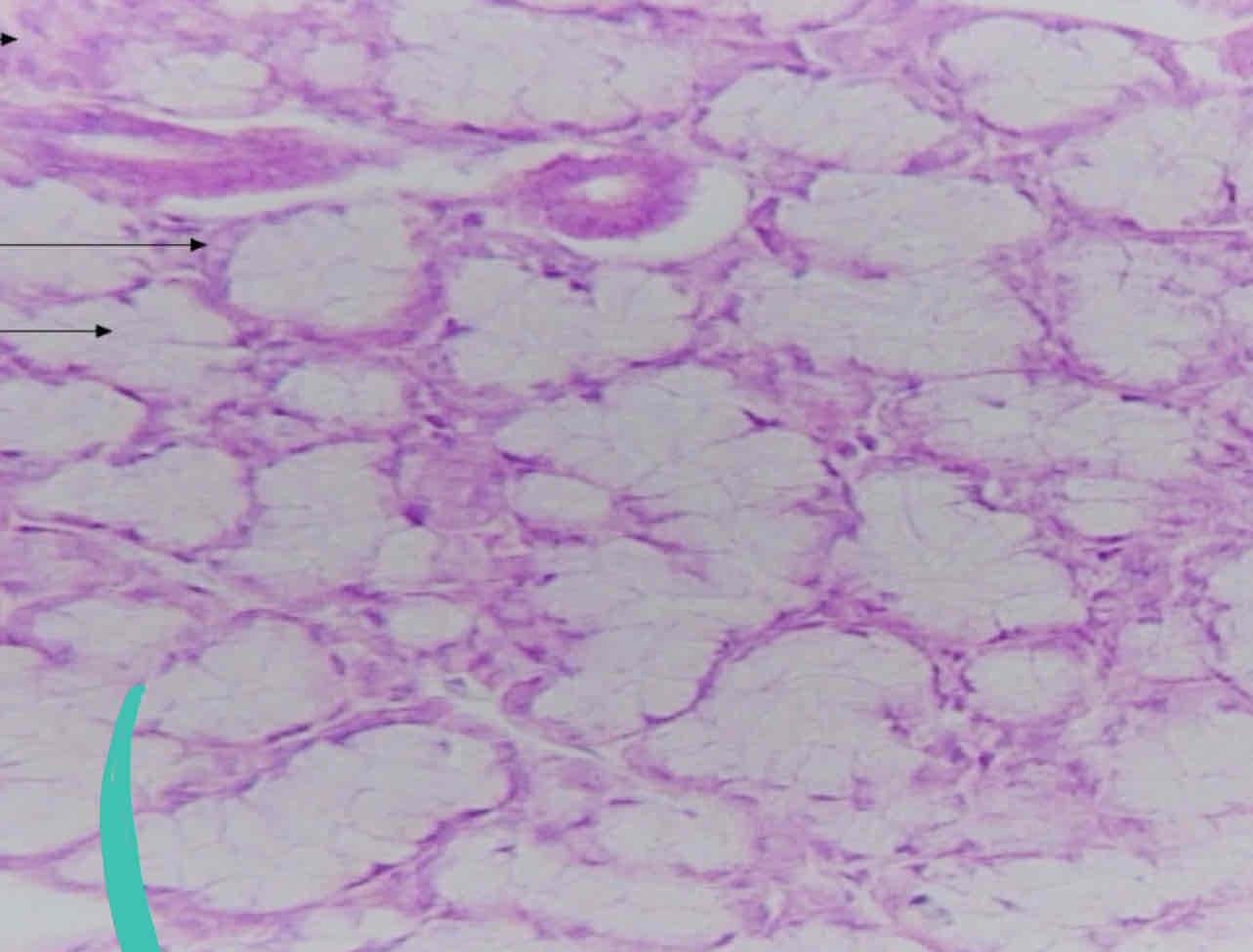
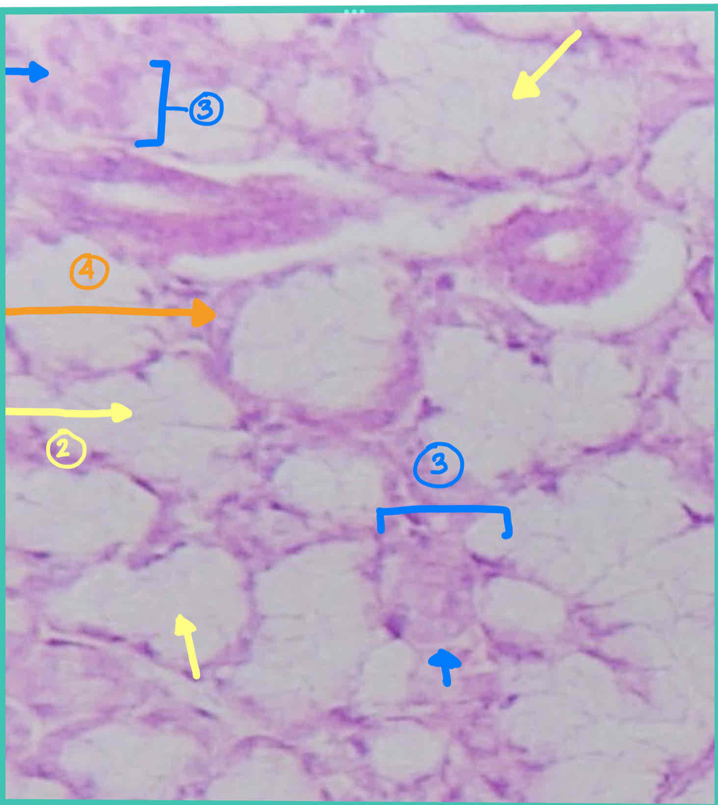
Serous glandular epithelium (parotid gland)
Slide 20
CENTRALLY LOCATED NUCLEUS
ACIDOPHILIC CYTOPLASM
SEROUS ACINUS
parotid gland
Larynx
Pseudo stratified columnar epithelium w/ cilia
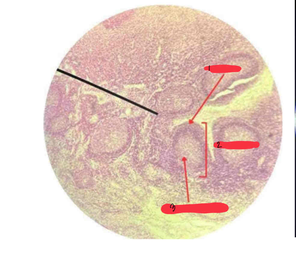
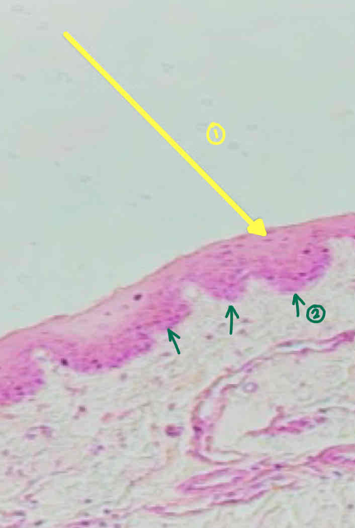
Stratified squamous epithelium, non-keratinized (esophagus)
(SLIDE #24)
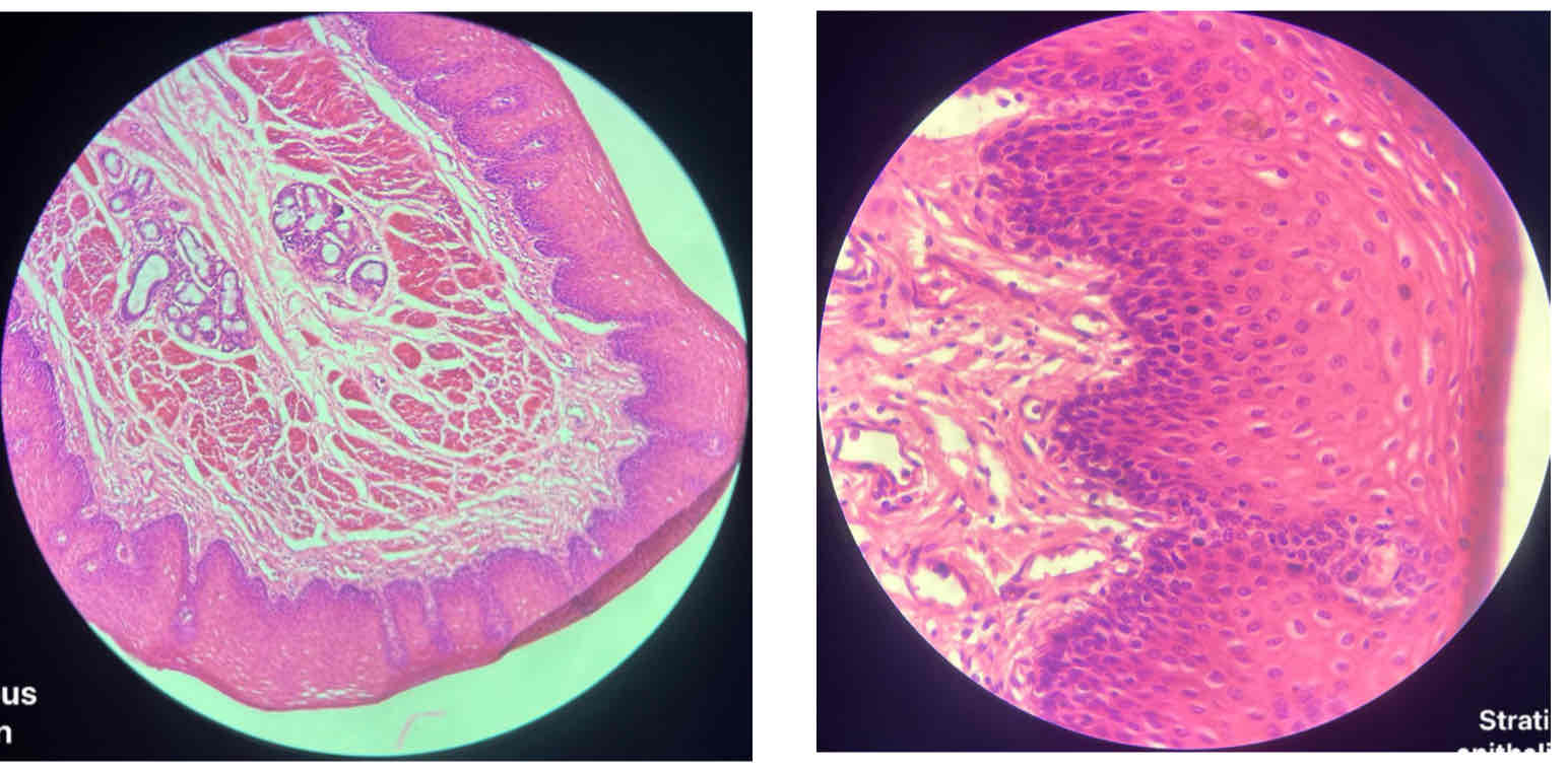
STRATIFIED SQUAMOUS EPITHELIUM
BASEMENT MEMBRANE
PAPILLA
LAMINA PROPRIA
Esophagus
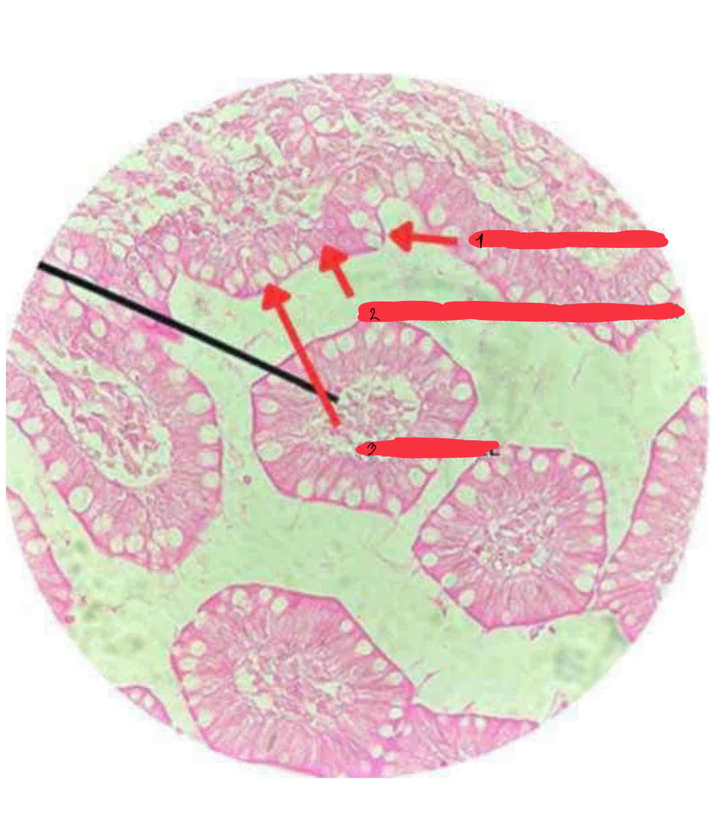
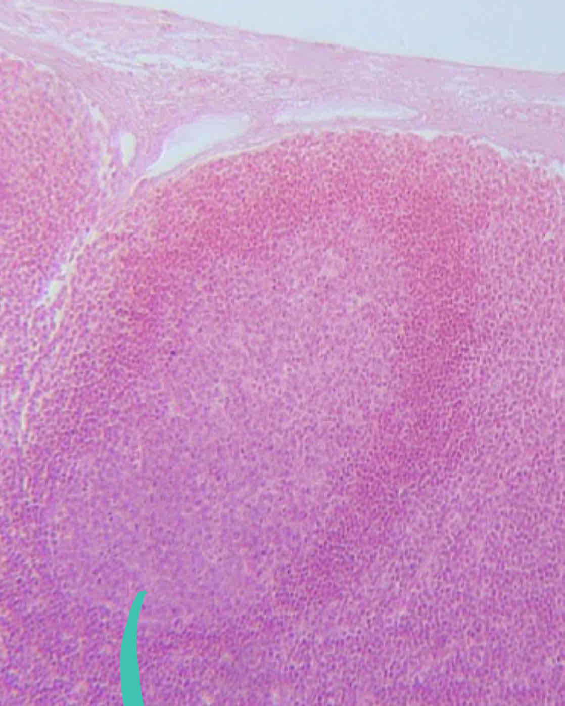
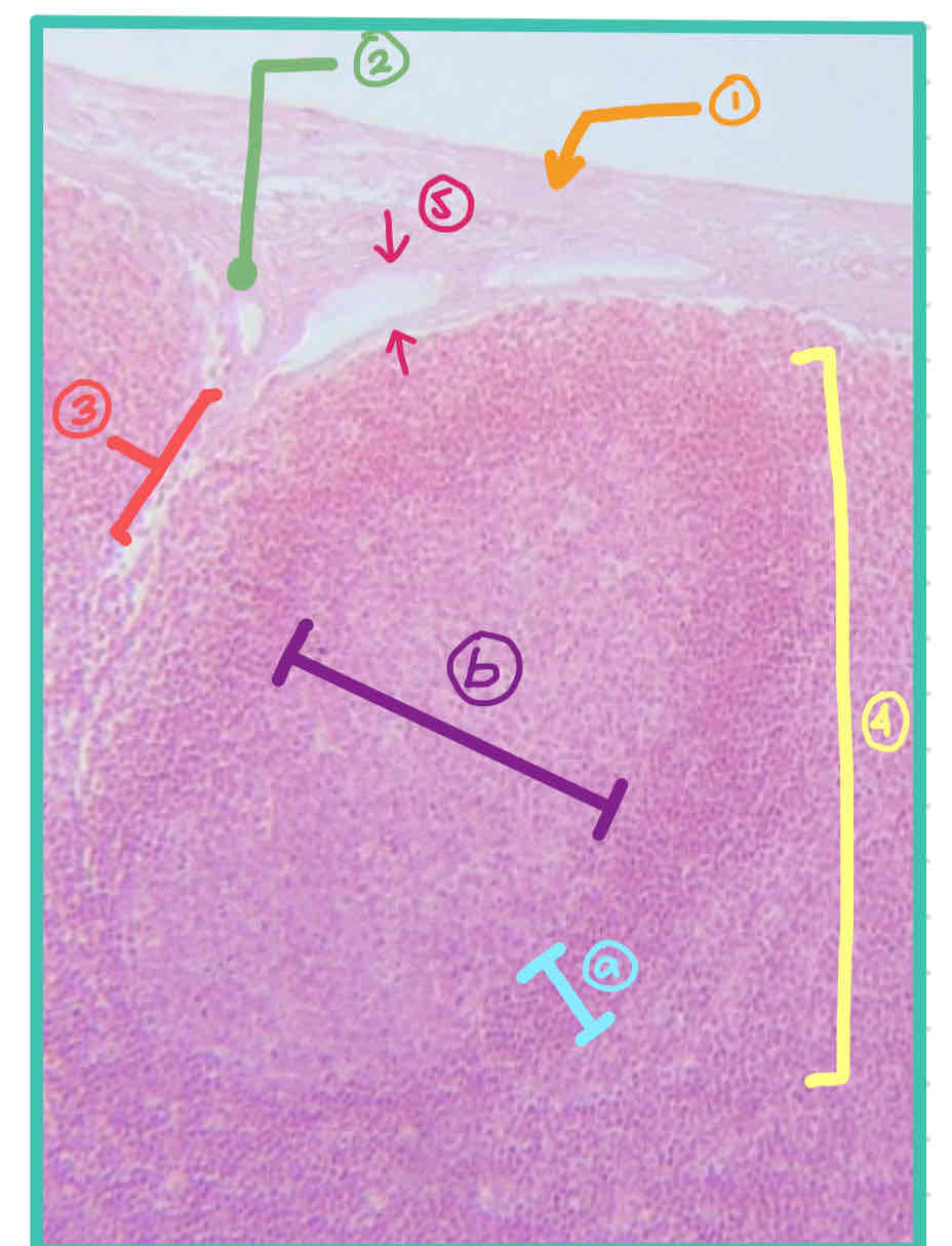
LYMPH NODE
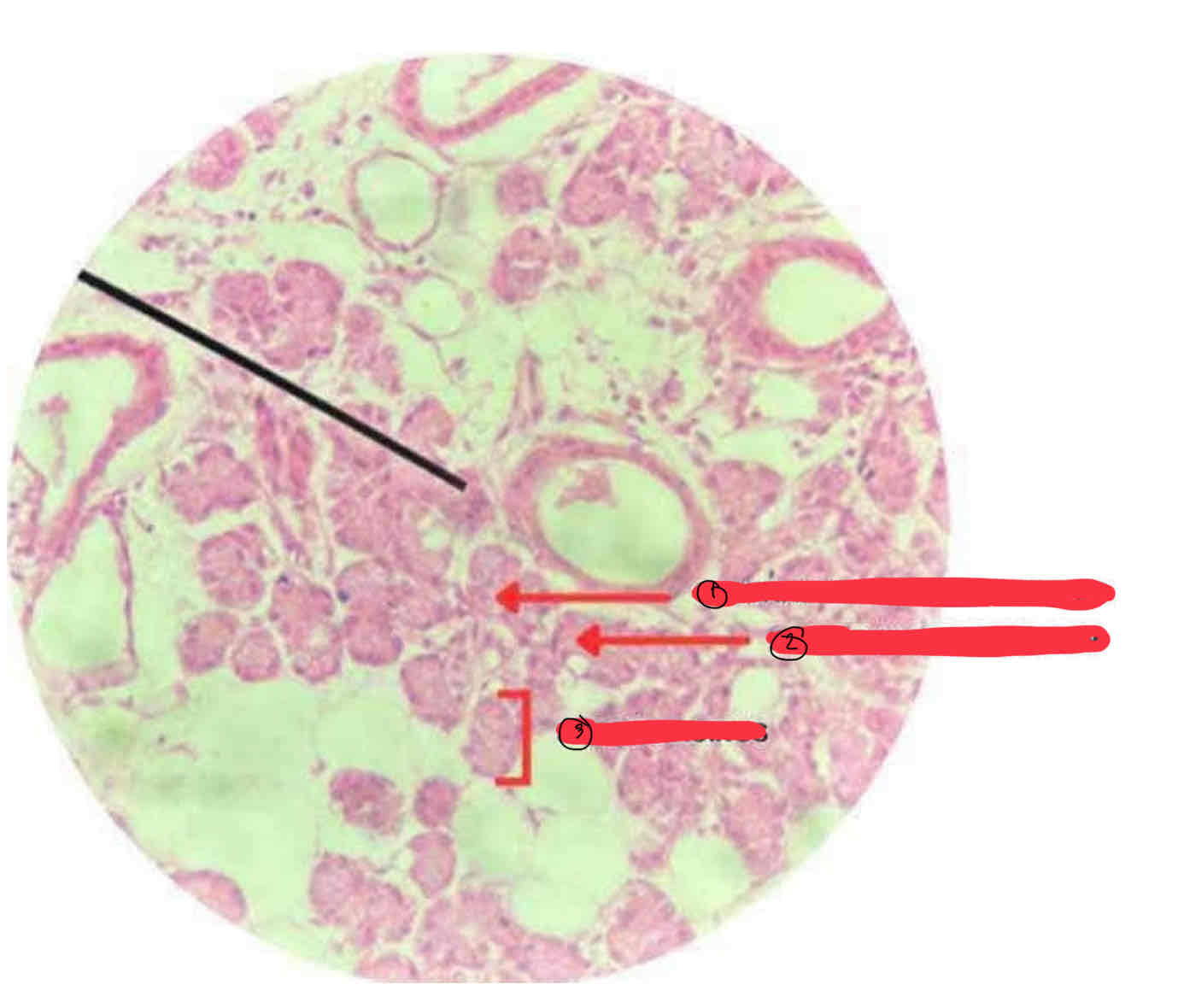
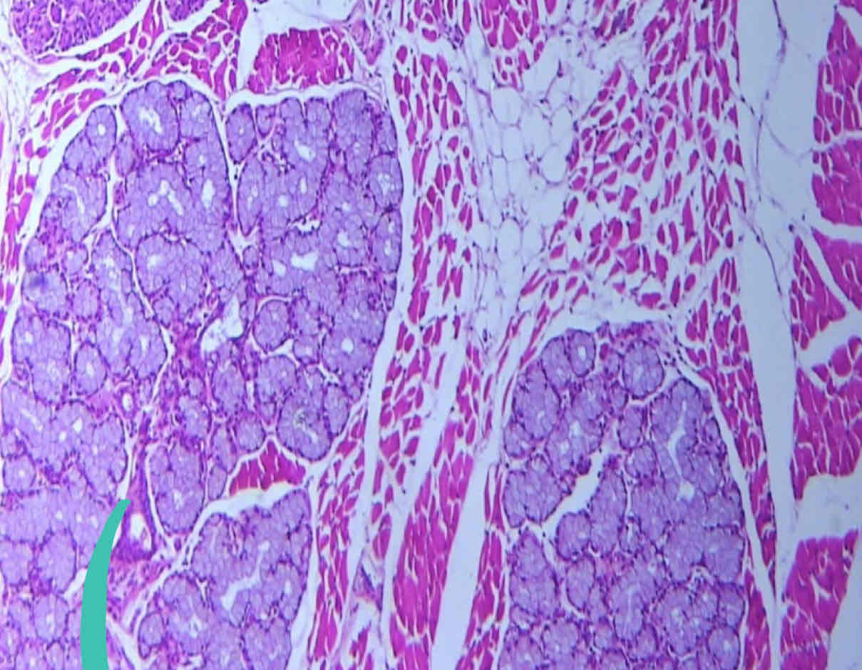
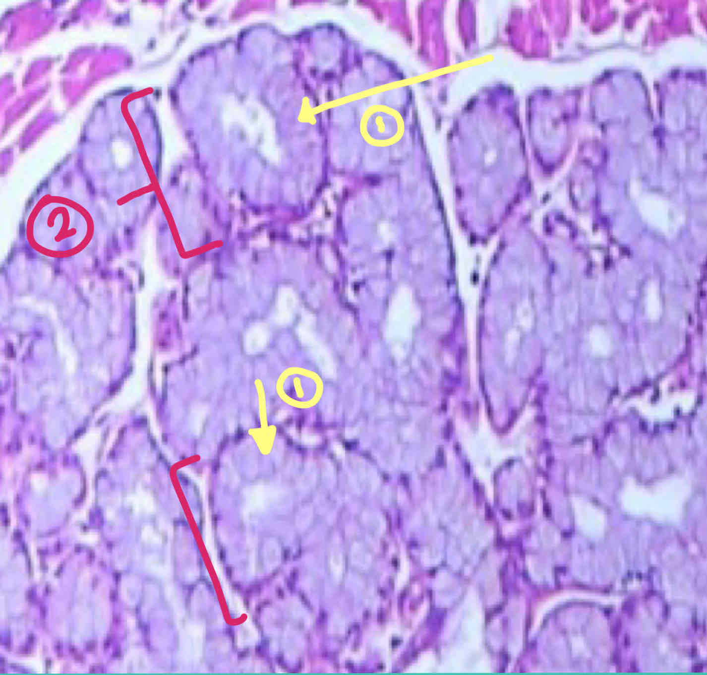
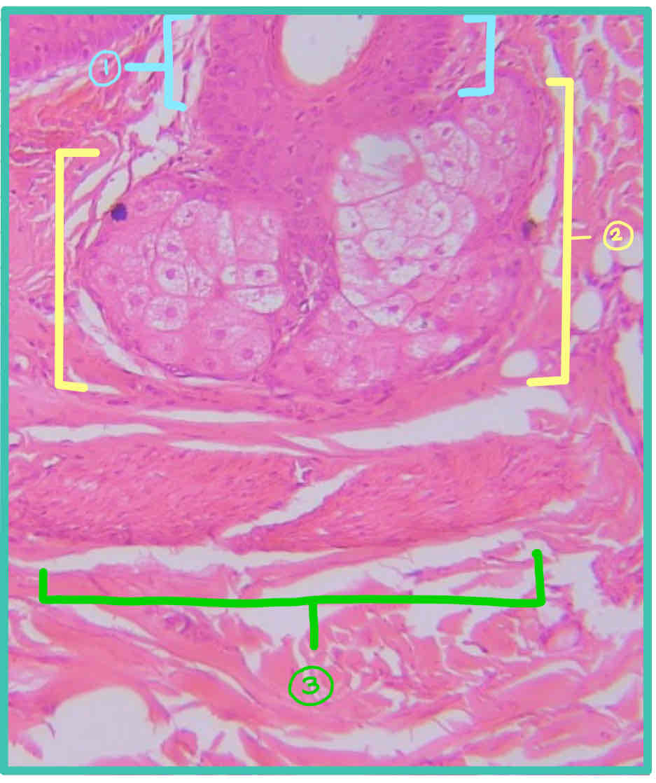
Sero-mucous glandular epithelium (sublingual gland)
(SLIDE #21B)
SEROUS ACINUS
SEROUS DEMILUNE OF GIANNUZZI
Sublingual Gland
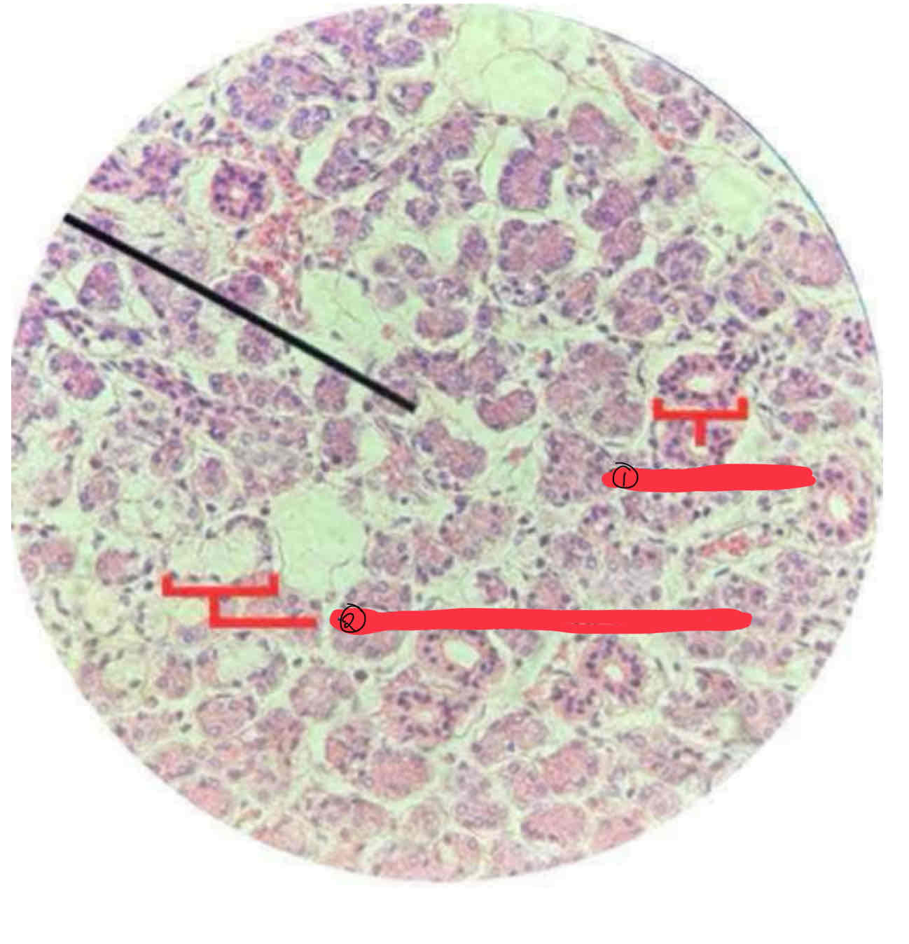
Serous glandular epithelium (parotid gland)
(SLIDE #20)
CENTRALLY LOCATED NUCLEUS
ACIDOPHILIC CYTOPLASM
SEROUS ACINUS
Parotid Gland
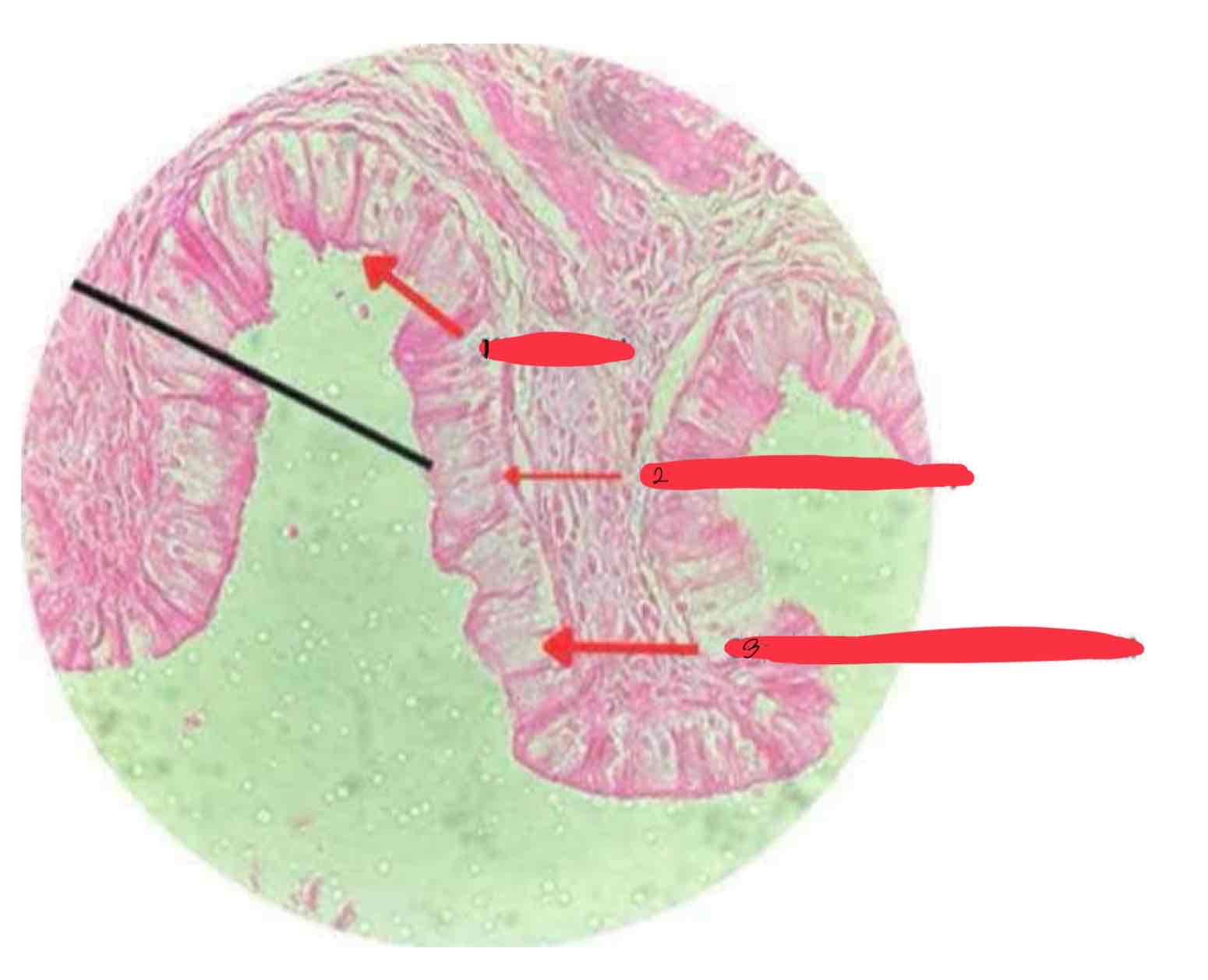
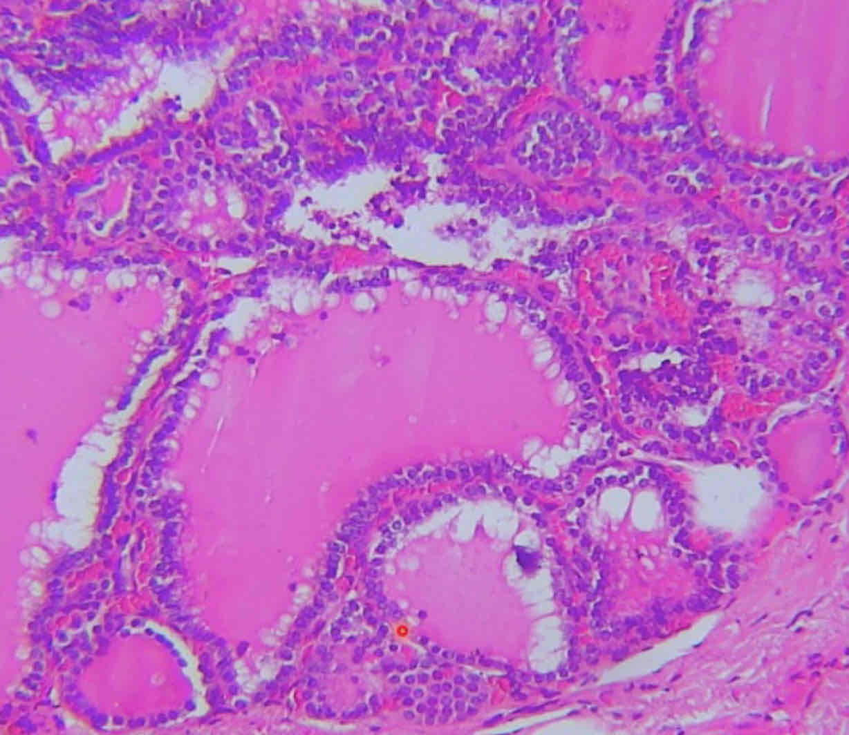
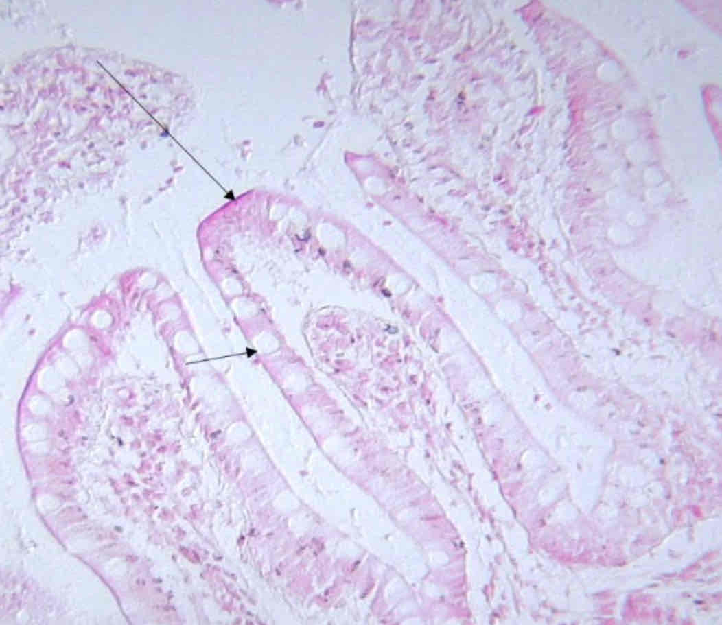
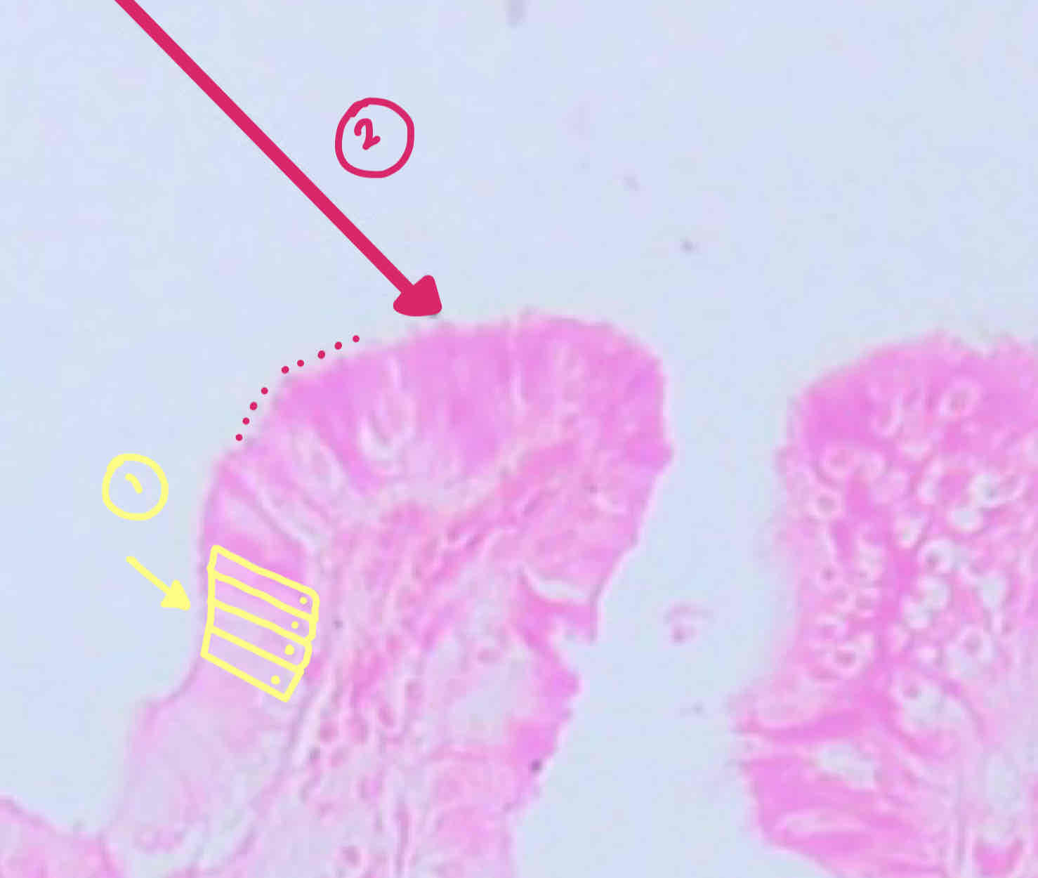
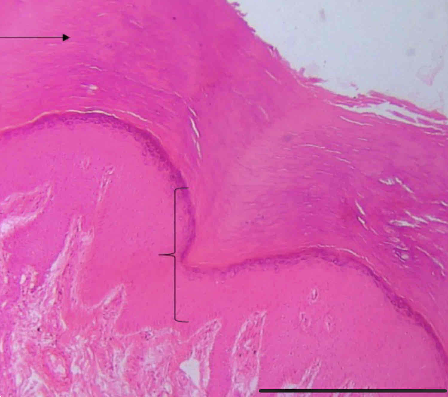
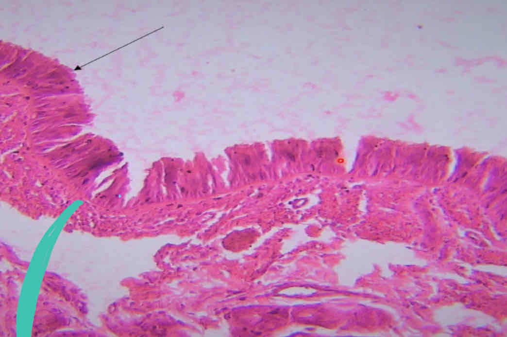
Simple columnar w/ microvilli (gall bladder)
slide 35
MICROVILLI
NUCLEUS (AT THE BASE)
SIMPLE COLUMNAR EPITHELIUM
Gall bladder
SIMPLE SQUAMOUS EPITHELIUM
SIDE VIEW
slide 38
SIMPLE SQUAMOUS EPITHELIUM
GLOMERULUS
SIMPLE CUBOIDAL EPITHELIUM
Slide 38
SIMPLE SQUAMOUS EPITHELIUM TOP VIEW
slide #5
Simple cuboidal
(SLIDE #51)
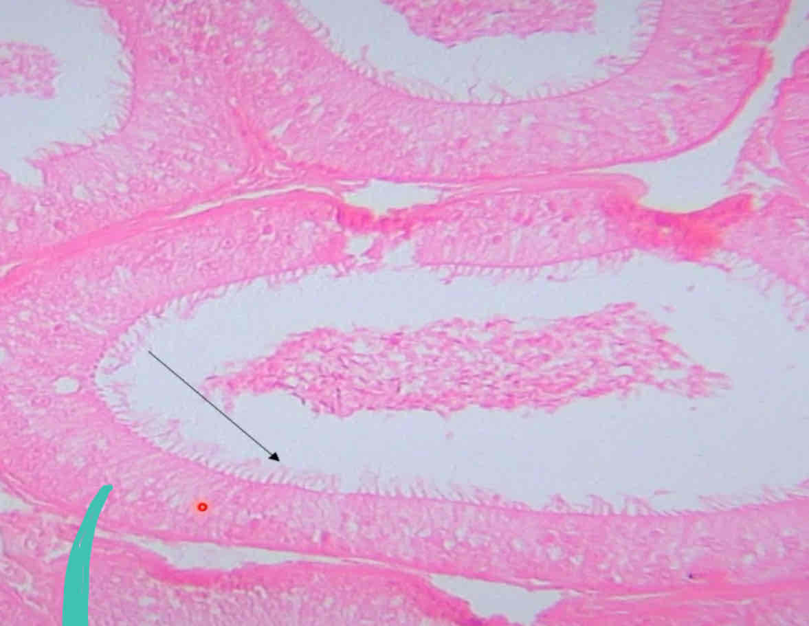
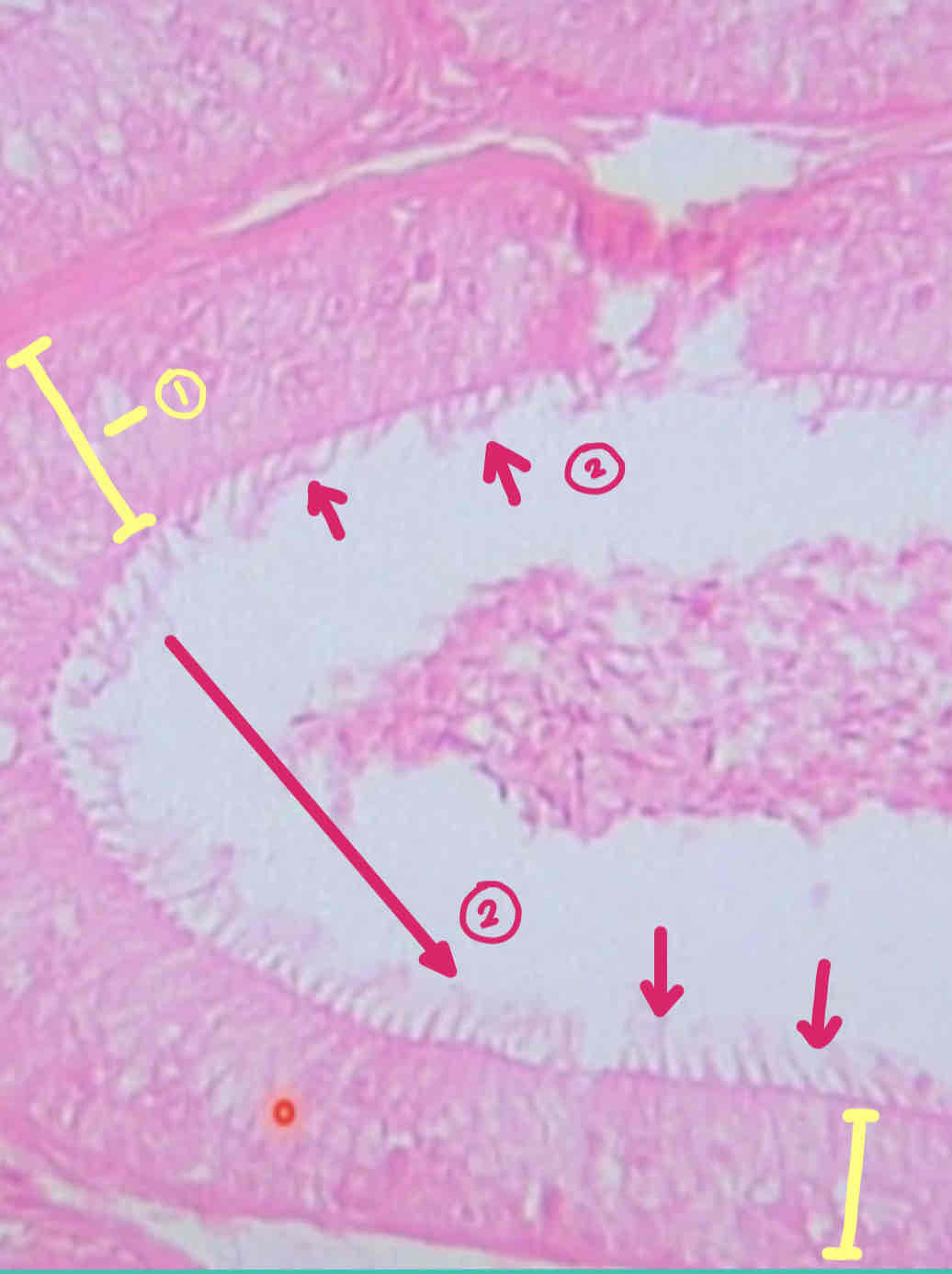
Simple columnar w/ striated border (ileum)
slide 35
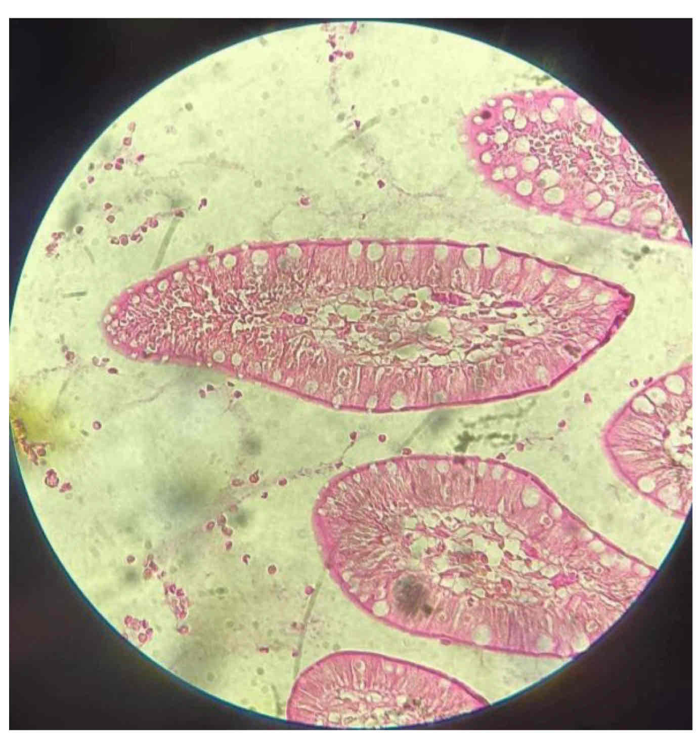
STRIATED BORDER
SIMPLE COLUMNAR EPITHELIUM
GOBLET CELL
Ileum
Mucous glandular epithelium (tongue)
Slide 22
MUCOUS ACINUS
PERIPHERALLY LOCATED NUCLEUS
BASOPHILIC CYTOPLASM
Tongue
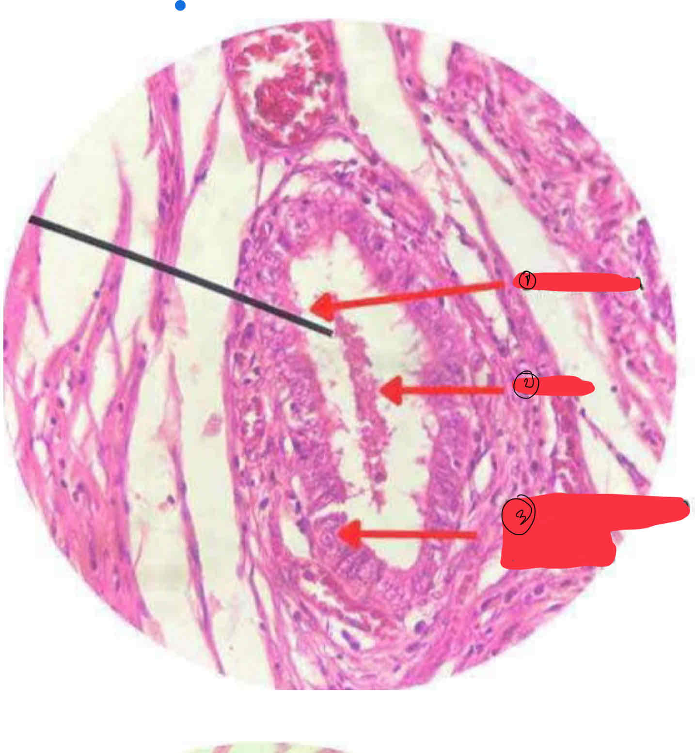
Pseudostratified columnar epithelium w/ stereocilia (epididymis)
(SLIDE #44B)
STEREOCILIA
SPERM
PSEUDOSTRATIFIED COLUMNAR EPITHELIUM
Epididymis
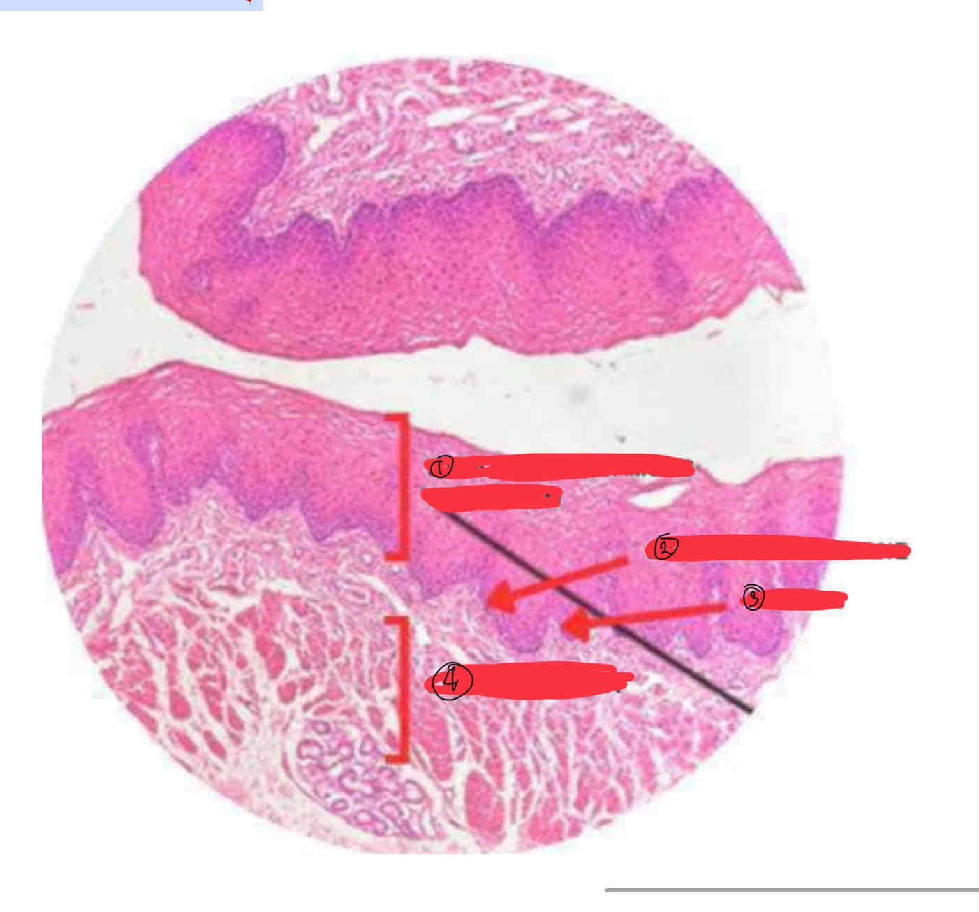
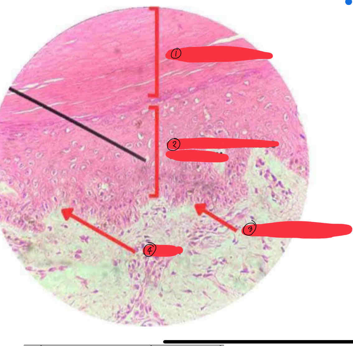
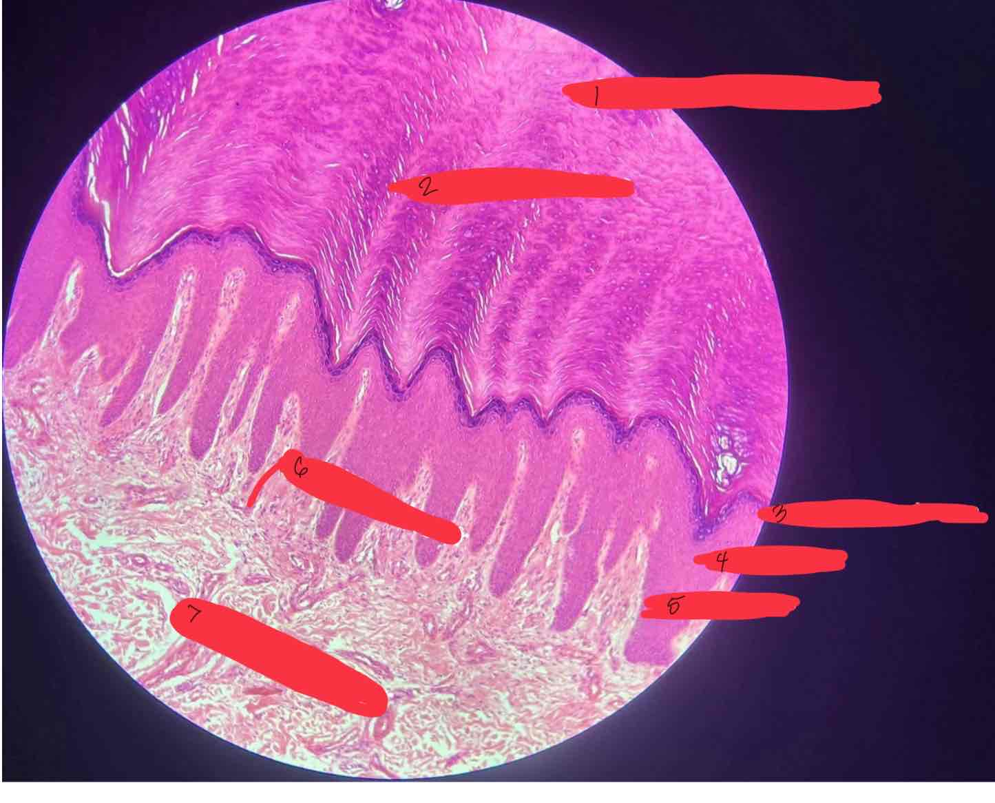
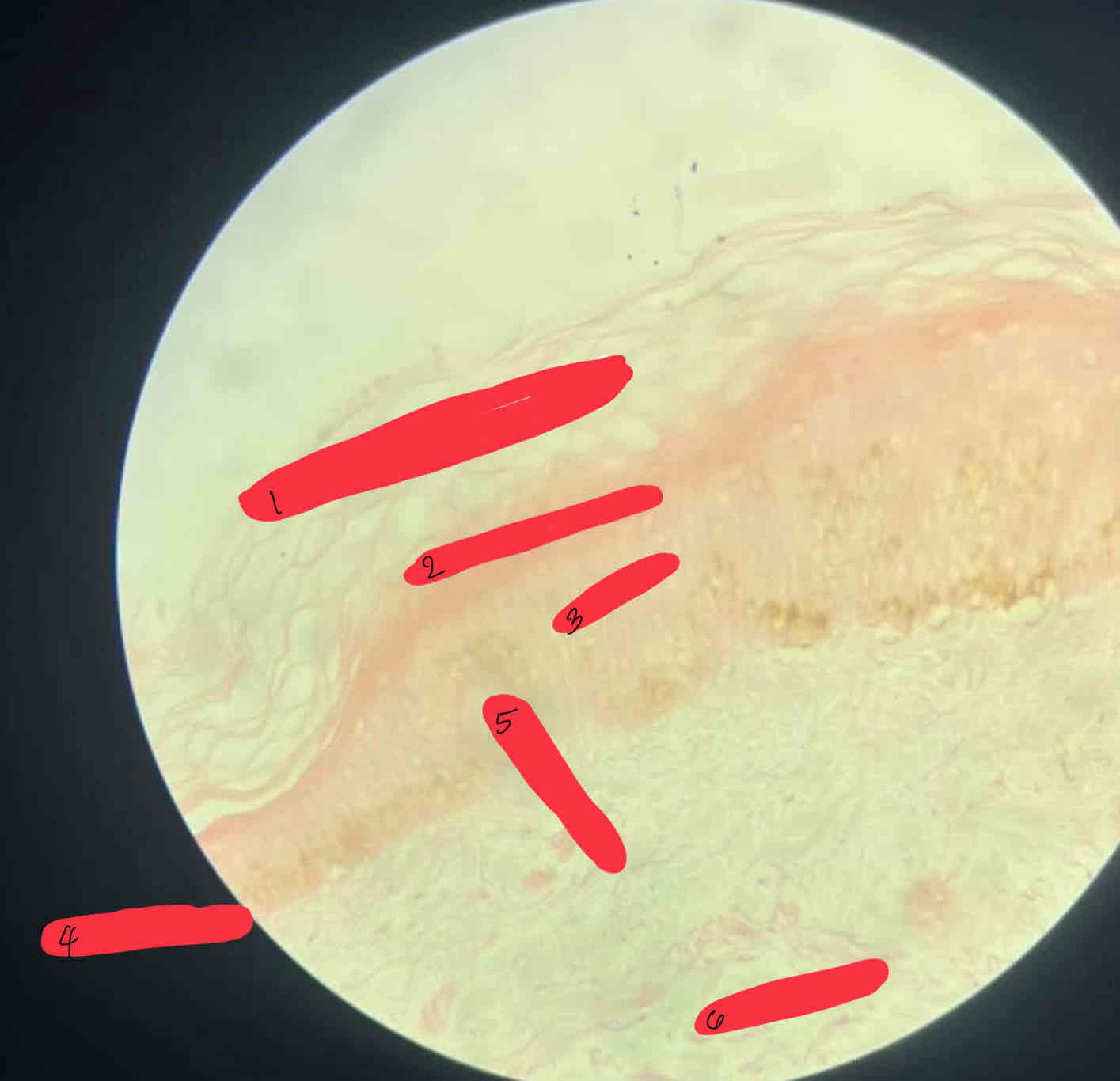
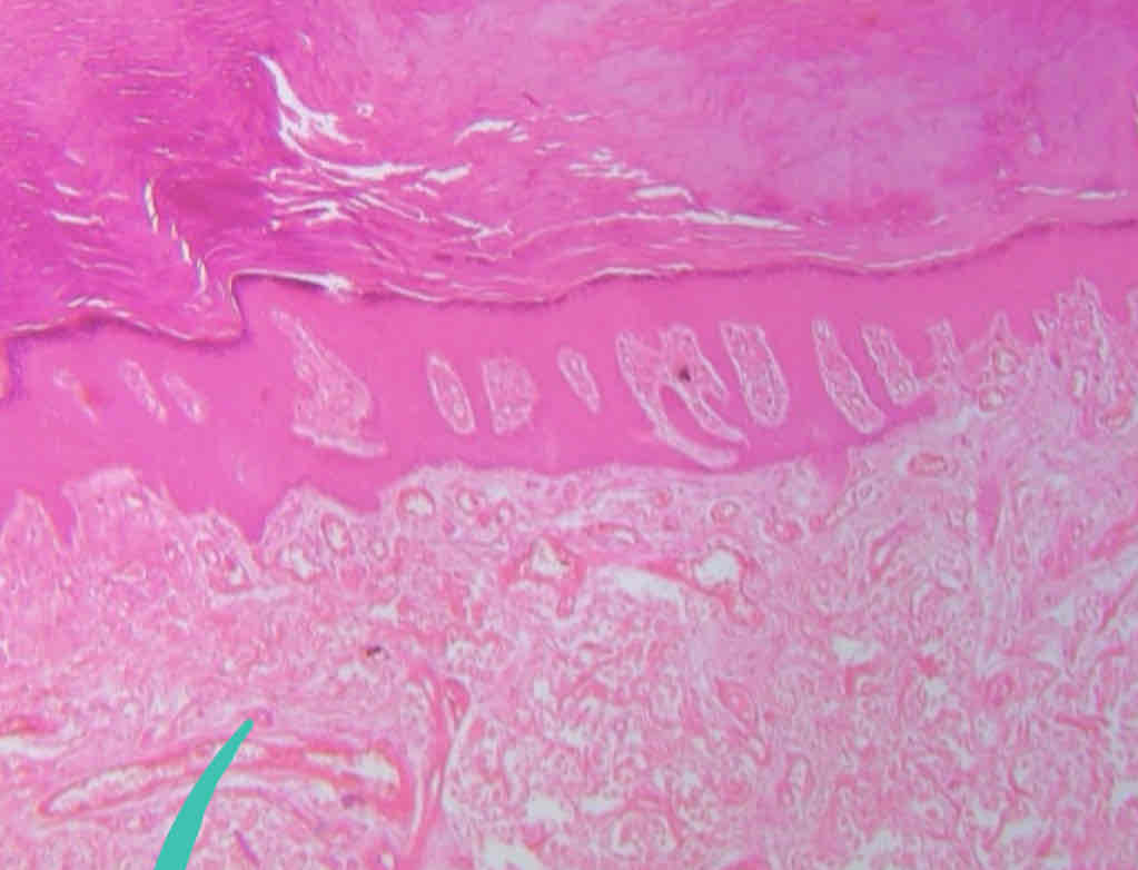
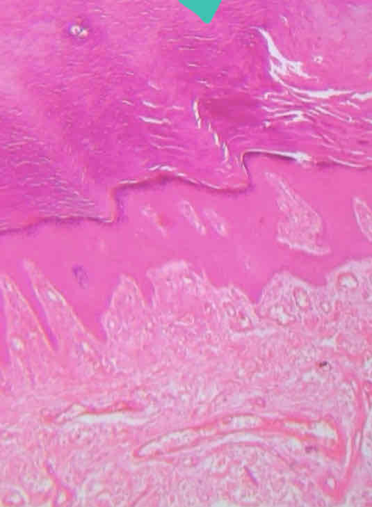
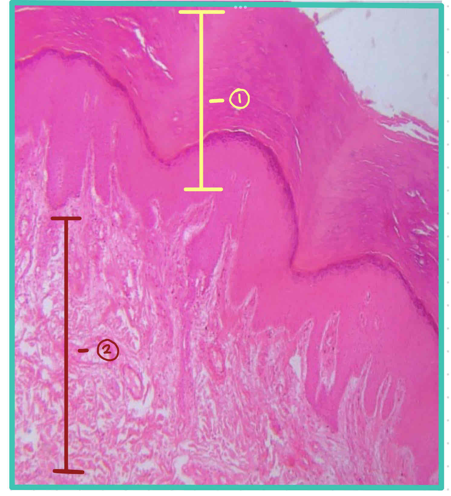
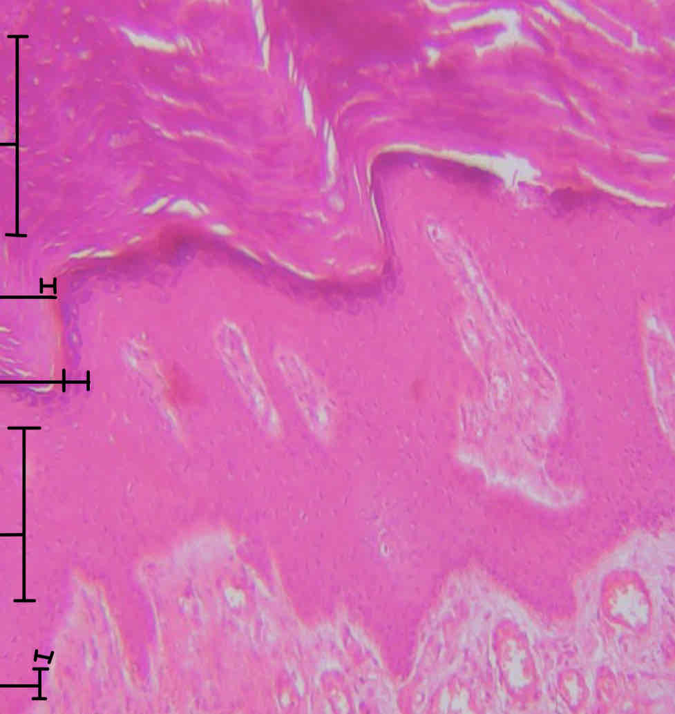
Stratified squamous epithelium, keratinized (thick skin)
(SLIDE #19)
KERATINIZED LAYER
STRATIFIED SQUAMOUS EPITHELIUM
BASEMENT MEMBRANE
PAPILLA
Slide 19
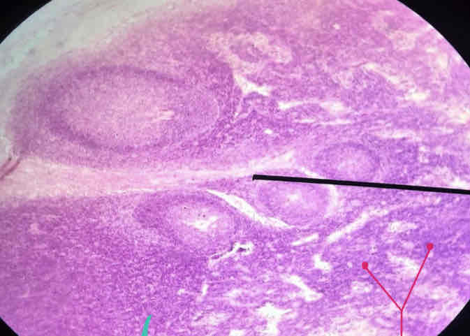
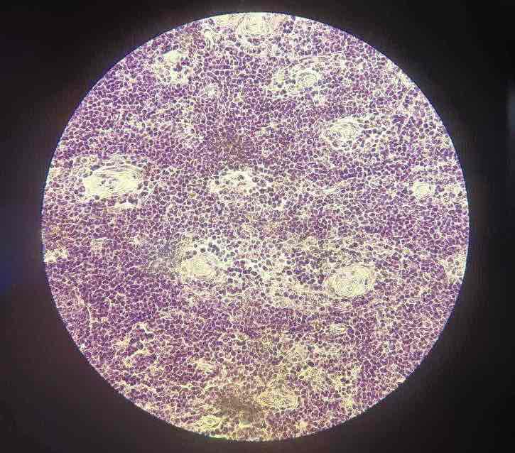
Lymphoid/ adenoid tissue
SLIDE #15
Corona
Lymph Node
Germinal Center
Lymphoid
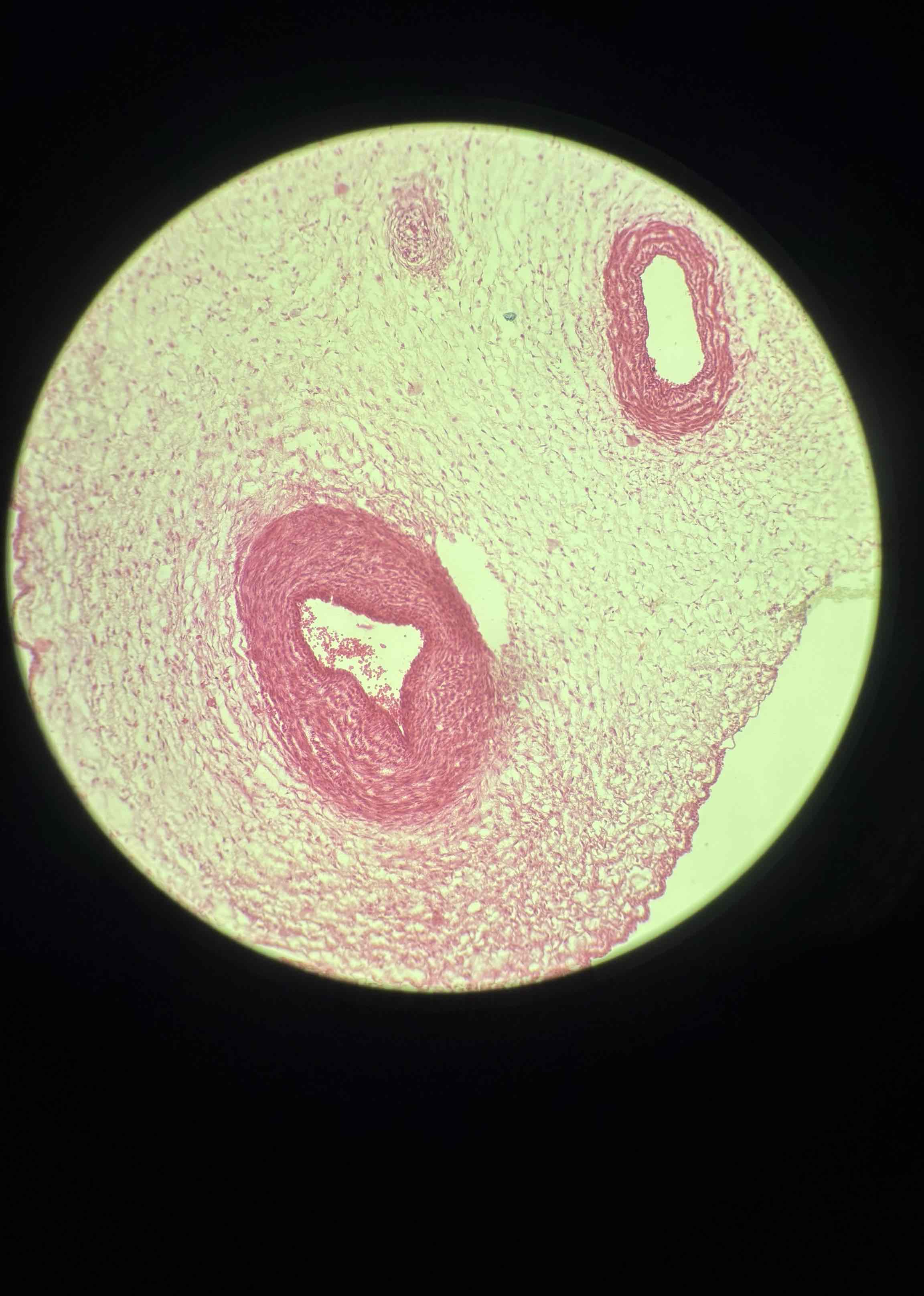
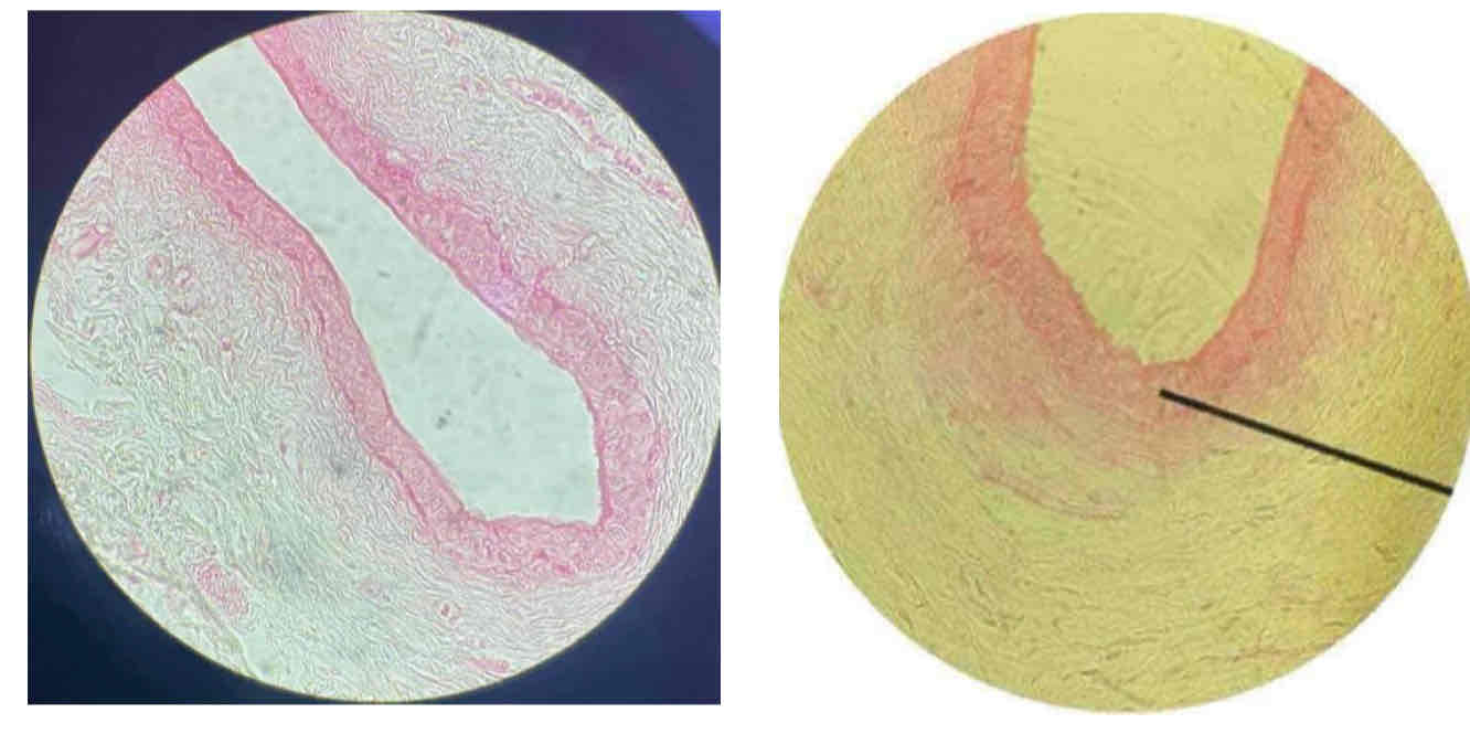
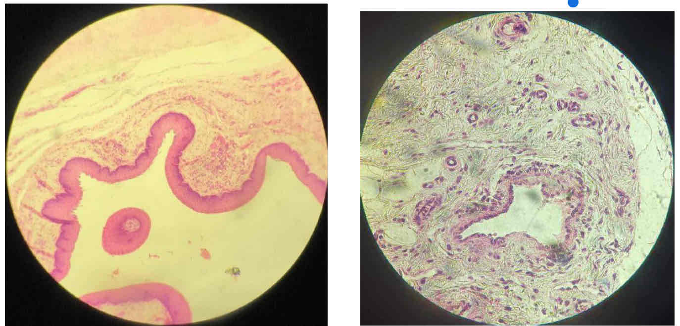
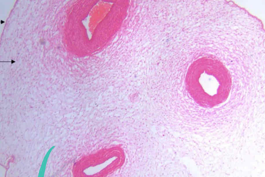
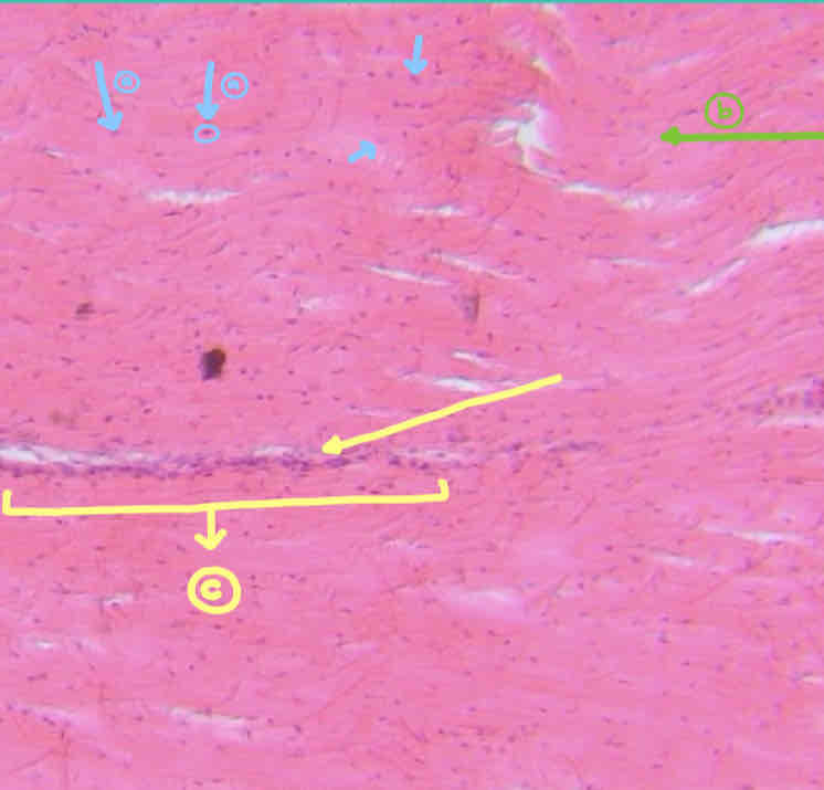
Dense elastic tissue (ligamentum nuchae)
slide #1
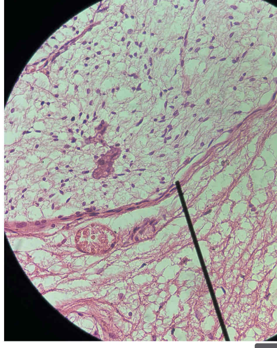
Developing embryo (mesenchyme)
SLIDE #6
NUCLEUS
CYTOPLASMIC PROCESSES
Mesenchyme
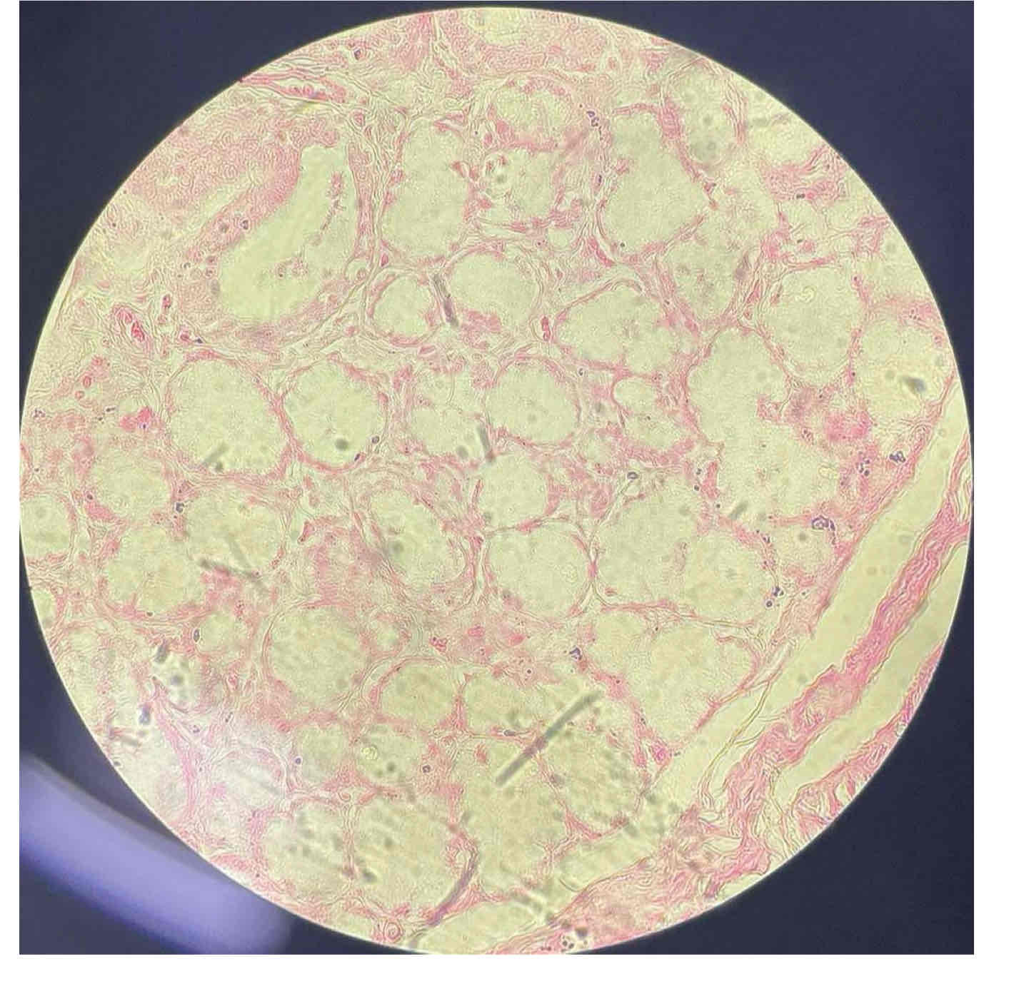
Adipose tissue (thin skin)
slide 16
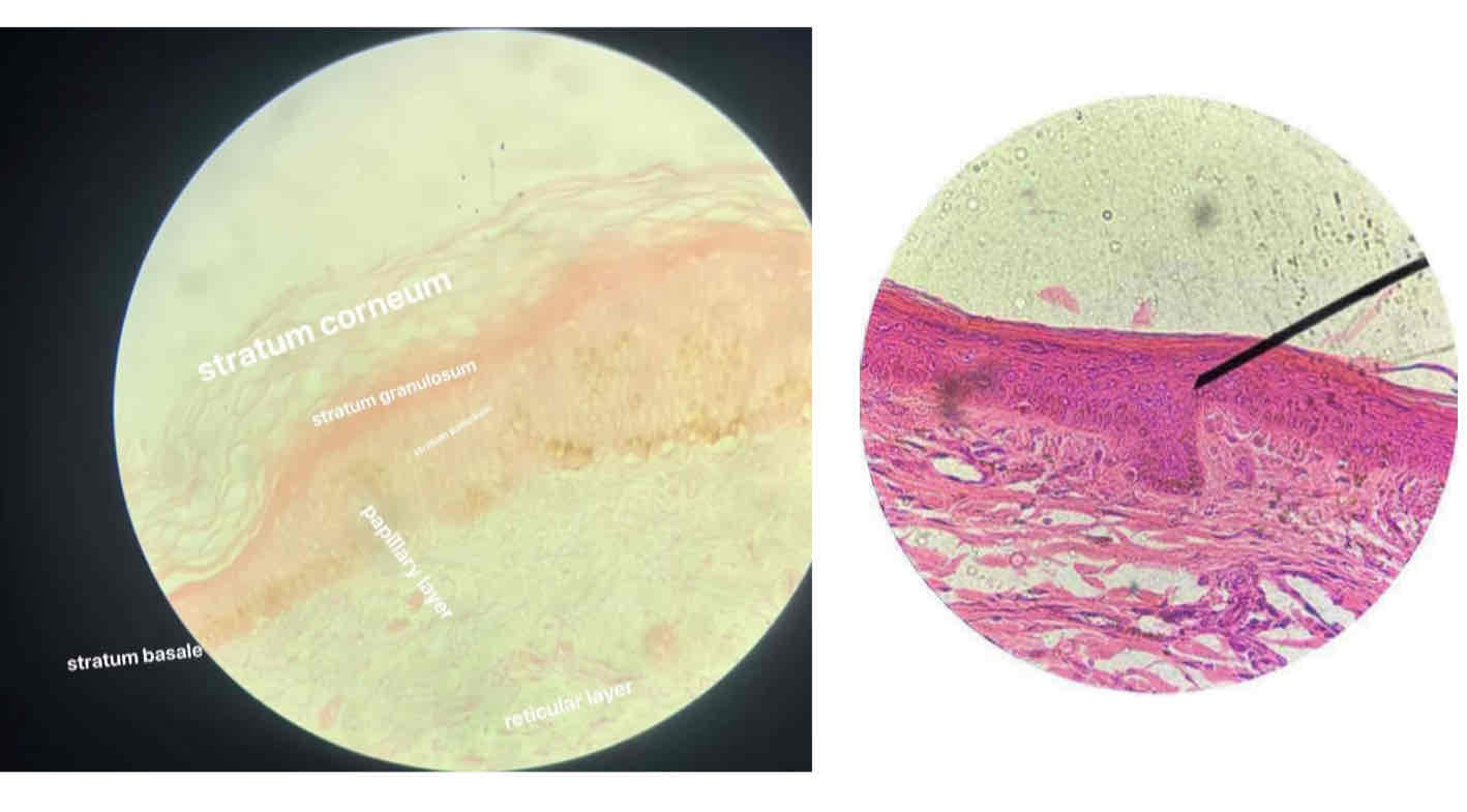
Thick Skin
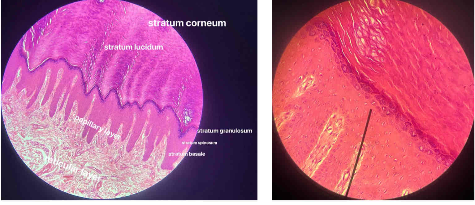
stratum corneum
stratum lucidum
stratum granulosum
stratum spinosum
stratum basale
papillary layer
Reticular Layer
Thin Skin
stratum corneum
stratum granulosum
stratum spinosum
stratum basale
papillary layer
reticular layer
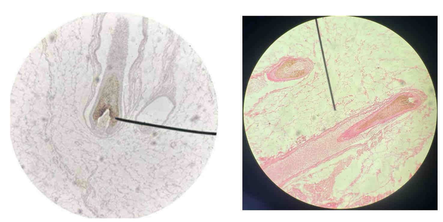
Hair follicle (top view)
Hair follicle (longitudinal section)
Developing Embryo (Specimen)
Mesenchyme (Tissue)
Connective Tissue
Mesenchymal Cells
Cytoplasmic Process
Reticular Fibers
Silver Stain
Preponderant Cells: 1&2
Preponderant Fiber: 3
Stain: 4
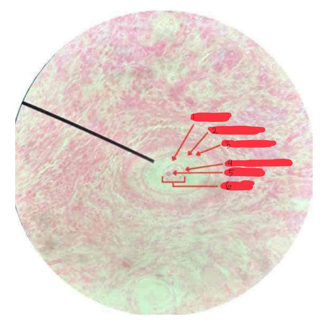
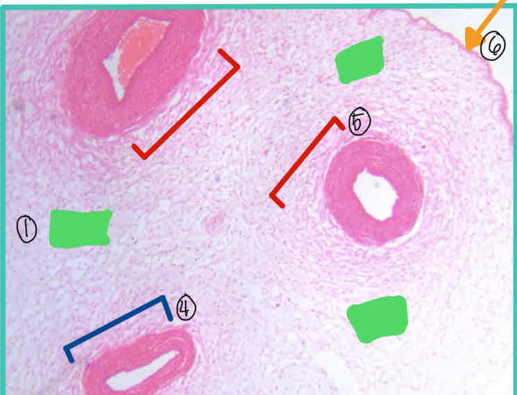
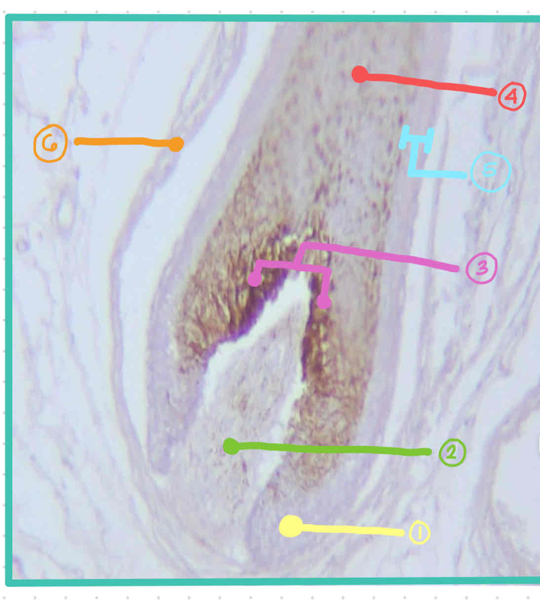
Umbilical Cord
Mucous Connective Tissue (Wharton’s Jelly)
Organ/Specimen:
Tissue:
1) Tissue: Mucous Connective Tissue / Wharton’s Jelly
2) Preponderant Cells: Fibroblast
3) Preponderant Fibers: Collagen Fibers
4) Umbilical Artery
5) Umbilical Vein
6) Amnion
1) Tissue:
2) Preponderant Cells:
3) Preponderant Fibers:
4)
5)
6)
Thick Skin
Areolar Tissue
Organ:
Tissue:
Fibroblast
Collagen Fibers
Areolar Tissue
Preponderant Cells:
Preponderant Fibers:
Tissue:
Thin Skin
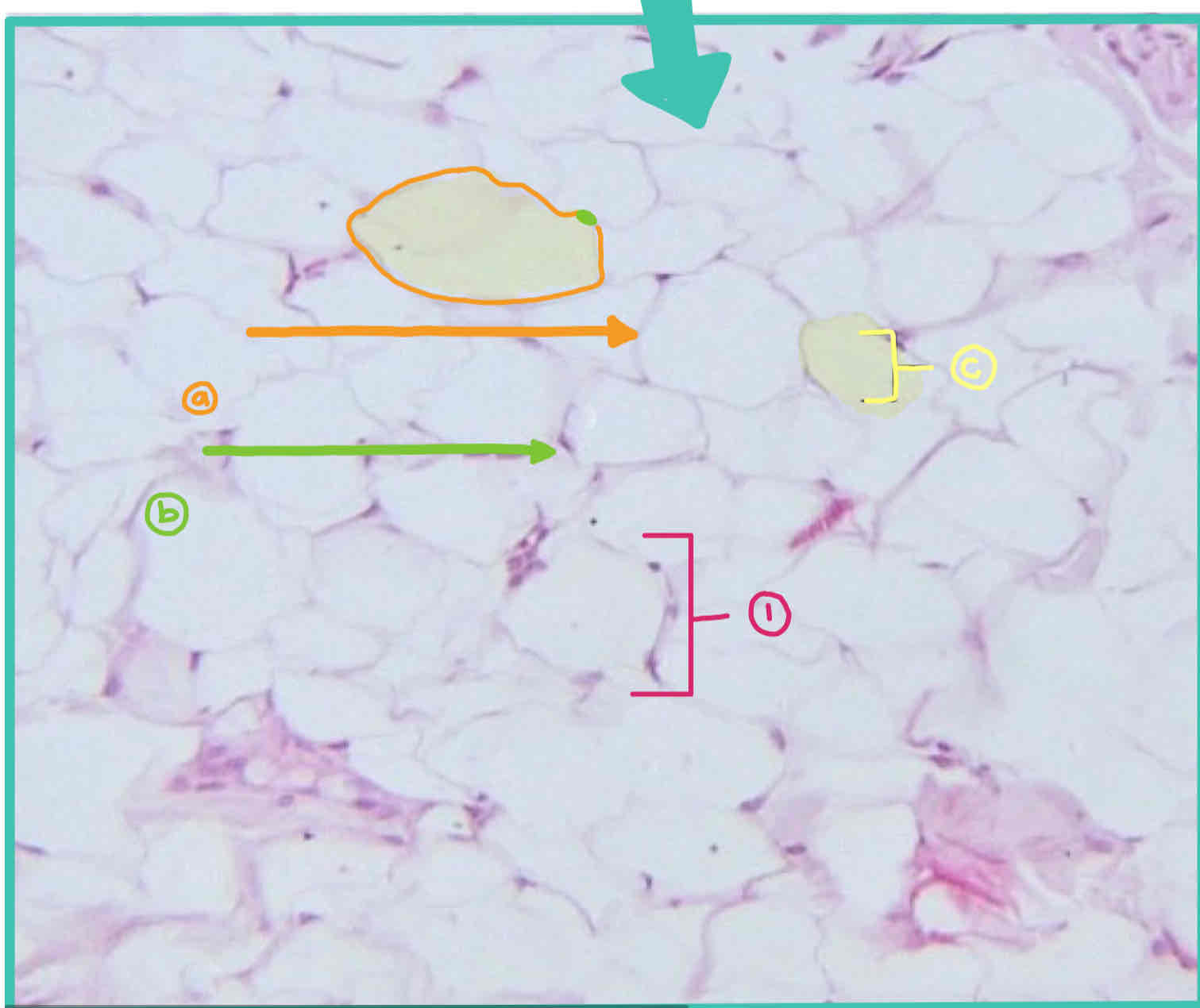
Adipose Tissue
Organ:
Tissue:
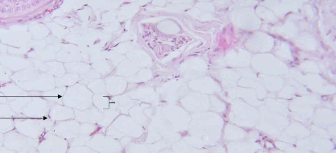
Preponderant Cell: Adipose Cell
a. cytoplasm
b. nucleus
c. fat droplet
Preponderant Fiber: Collagen and Reticular Fibers
Preponderant Cell:
a.
b.
c.
Preponderant Fiber:
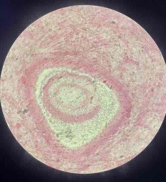
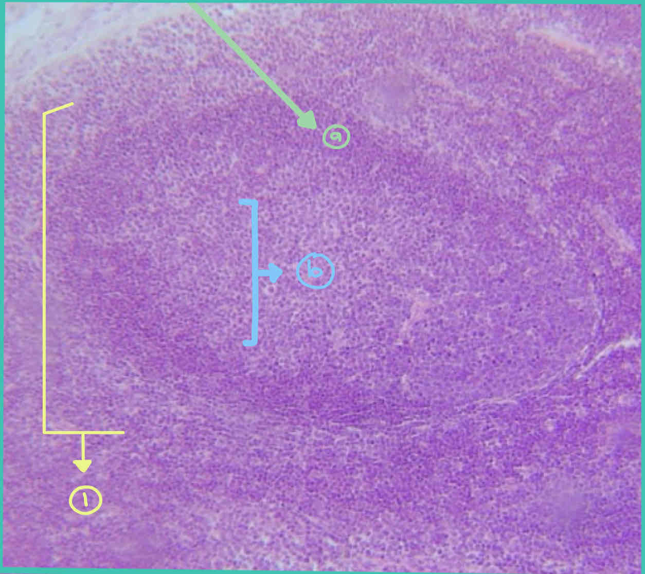
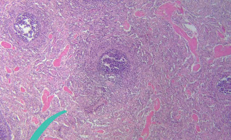
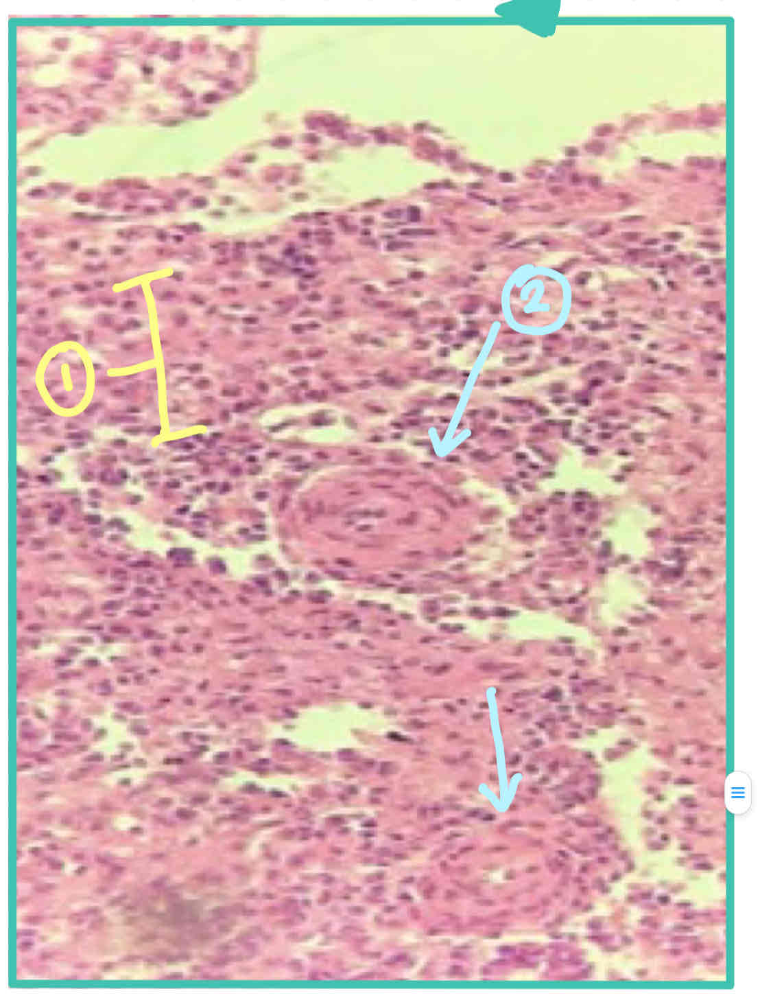
Lymph Node
Lymphoid/Adenoid Tissue
Organ:
Tissue:
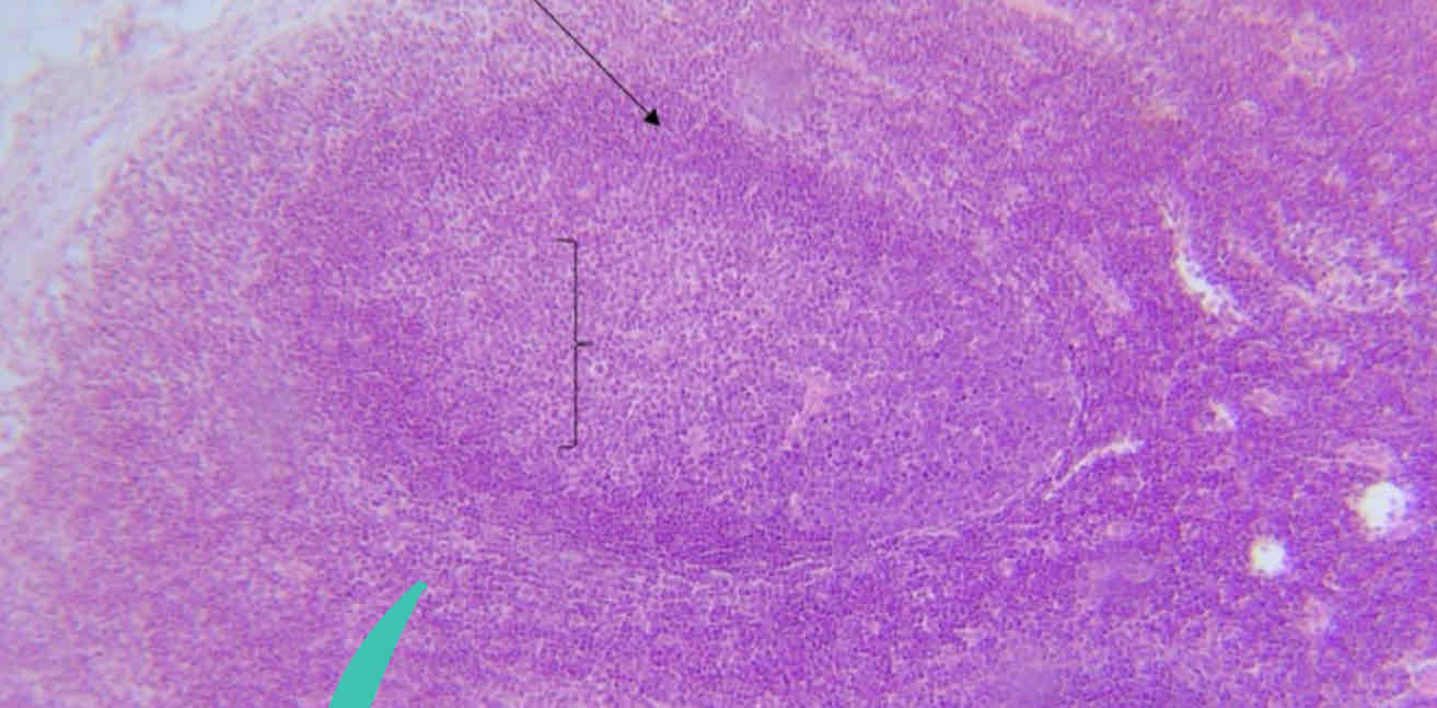
1.Lymph Nodules
a. Corona
b. Germinal Center
2.Lymphocytes
3.Reticular Fibers
Organ
a.
b.
Preponderant Cell:
Preponderant Fibers:
Ligamentum Nuchae
Dense Elastic Connective Tissue
Organ:
Tissue:
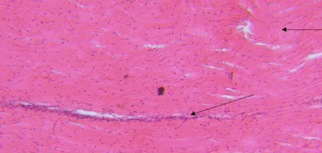
1) Dense Elastic Connective Tissue
a. Fibroblasts
b. Elastic Fibers
c.Areolar Tissue
2)Fibroblasts
3)Elastic Fibers
1.Tissue
a.
b.
c.
2.PC
3.PF
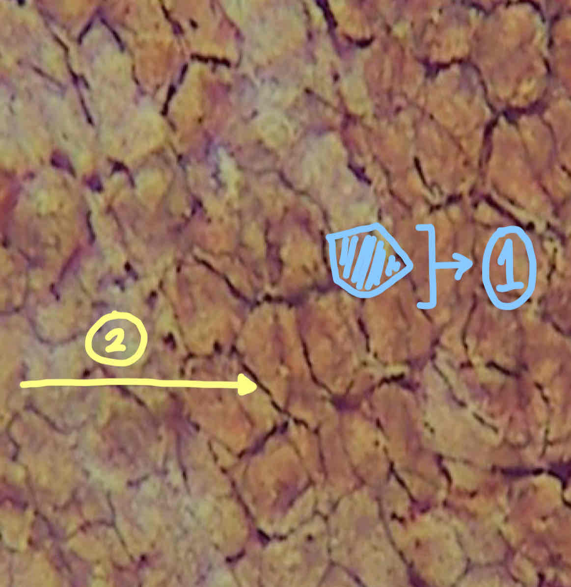
Organ: Mesentery
Tissue: Mesothelium
Organ
Tissue
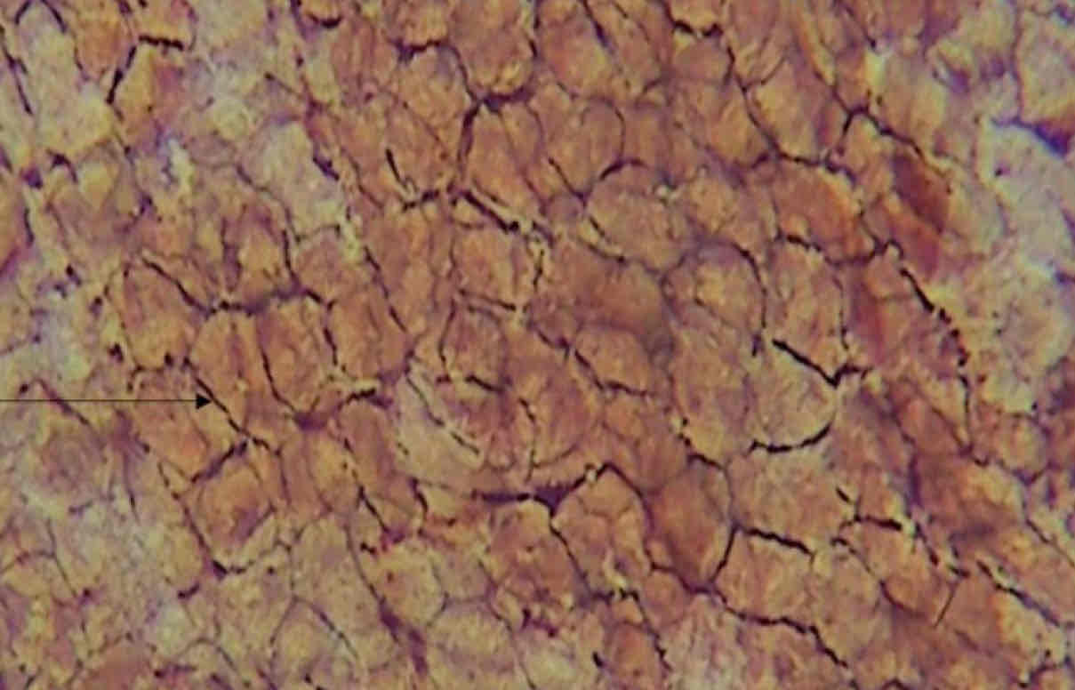
1) Hexagonal Cells with Wavy Outline
2) Lateral Surface Specialization: Serrated Border
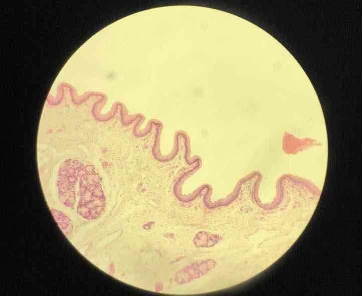
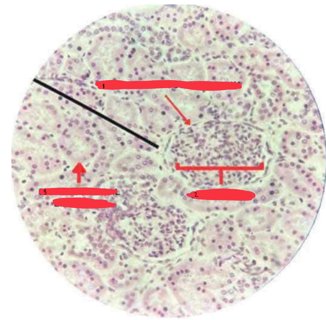
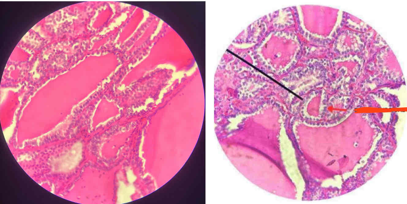
Kidney
Simple Squamous Epithelium
Organ
Tissue
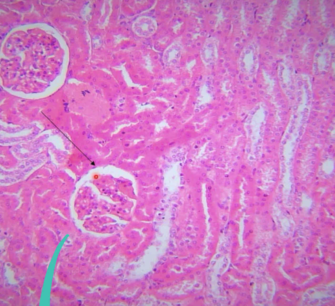
Simple Squamous Epithelium
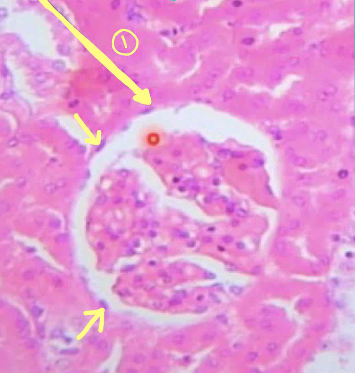
Organ: Thyroid Gland
Tissue: Simple Cuboidal Epithelium
Organ:
Tissue:
Simple Cuboidal Epithelium
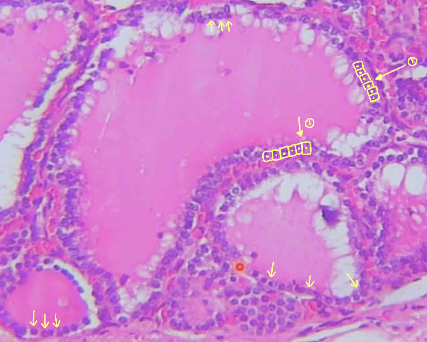
Ileum
Simple Columnar Epithelium
Organ:
Tissue:
Simple Columnar Epithelium
Striated Border
Goblet Cell
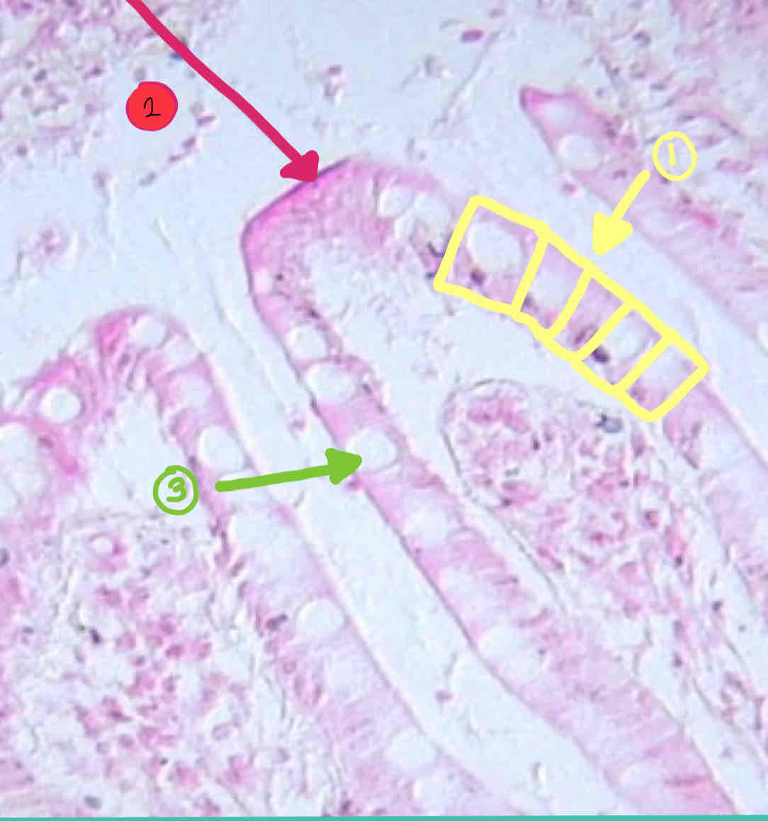
Gallbladder
Simple Columnar Epithelium
Organ
Tissue
1.Simple Columnar Epithelium
2.Microvilli
1) Tissue:
2) Free Surface Specialization:
Thick Skin
Stratified Squamous Epithelium (Keratinized)
1) Layer of Keratin
2) Tissue: Stratified Squamous Epithelium (Keratinized)
3) Connective Tissue Papilla
4) Areolar Tissue
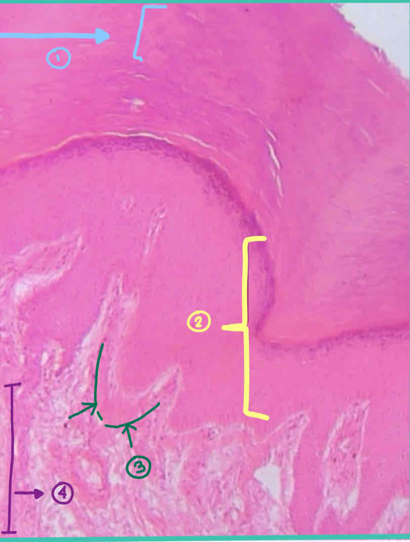
Esophagus
Stratified Squamous Epithelium (Non Keratinized)
Organ:
Tissue:
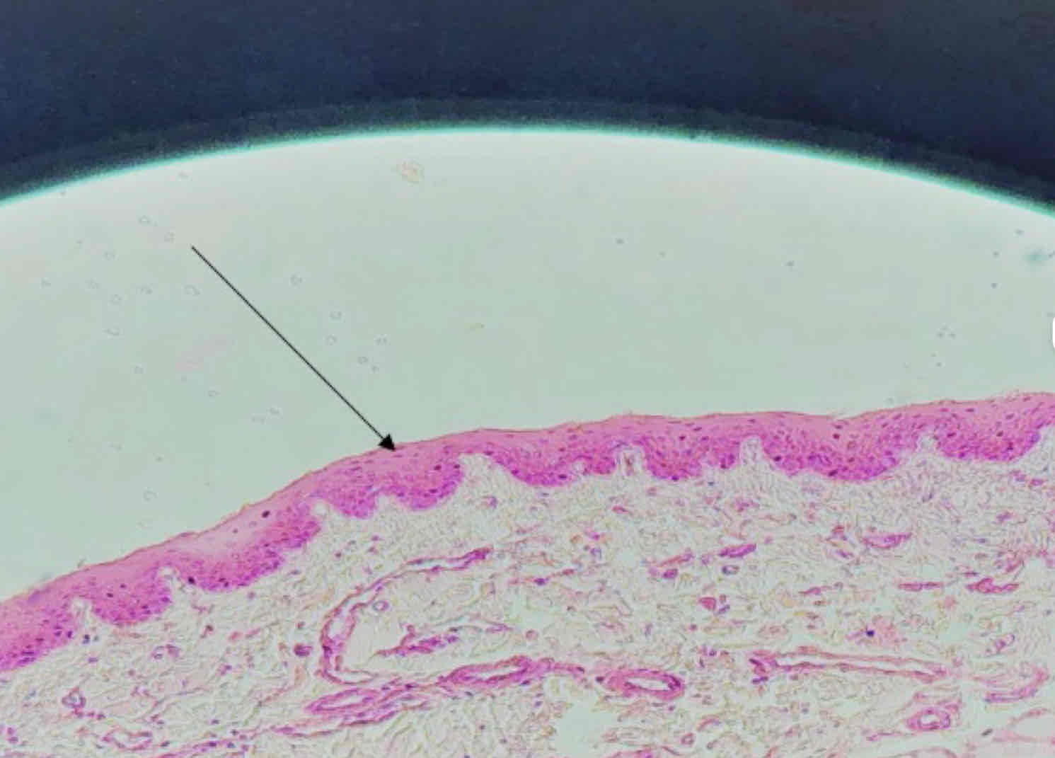
1) Stratified Squamous Epithelium (Non-Keratinized)
2) Connective Tissue Papilla
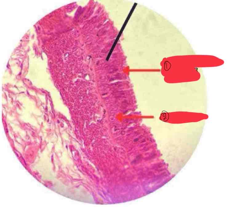
Urinary Bladder
Transitional Epithelium
Organ
Tissue
1) Transitional Epithelium
2) Condensed Border
1) Tissue:
2) Free Surface Specialization:
Larynx
Pseudostratified Columnar Epithelium
Organ
Tissue
1.Pseudostratified Columnar Epithelium
2.Goblet Cells
3.Cilia
Epididymis
Pseudostratified Columnar Epithelium
Organ
Tissue
1) Pseudostratified Columnar Epithelium
2) Stereocilia
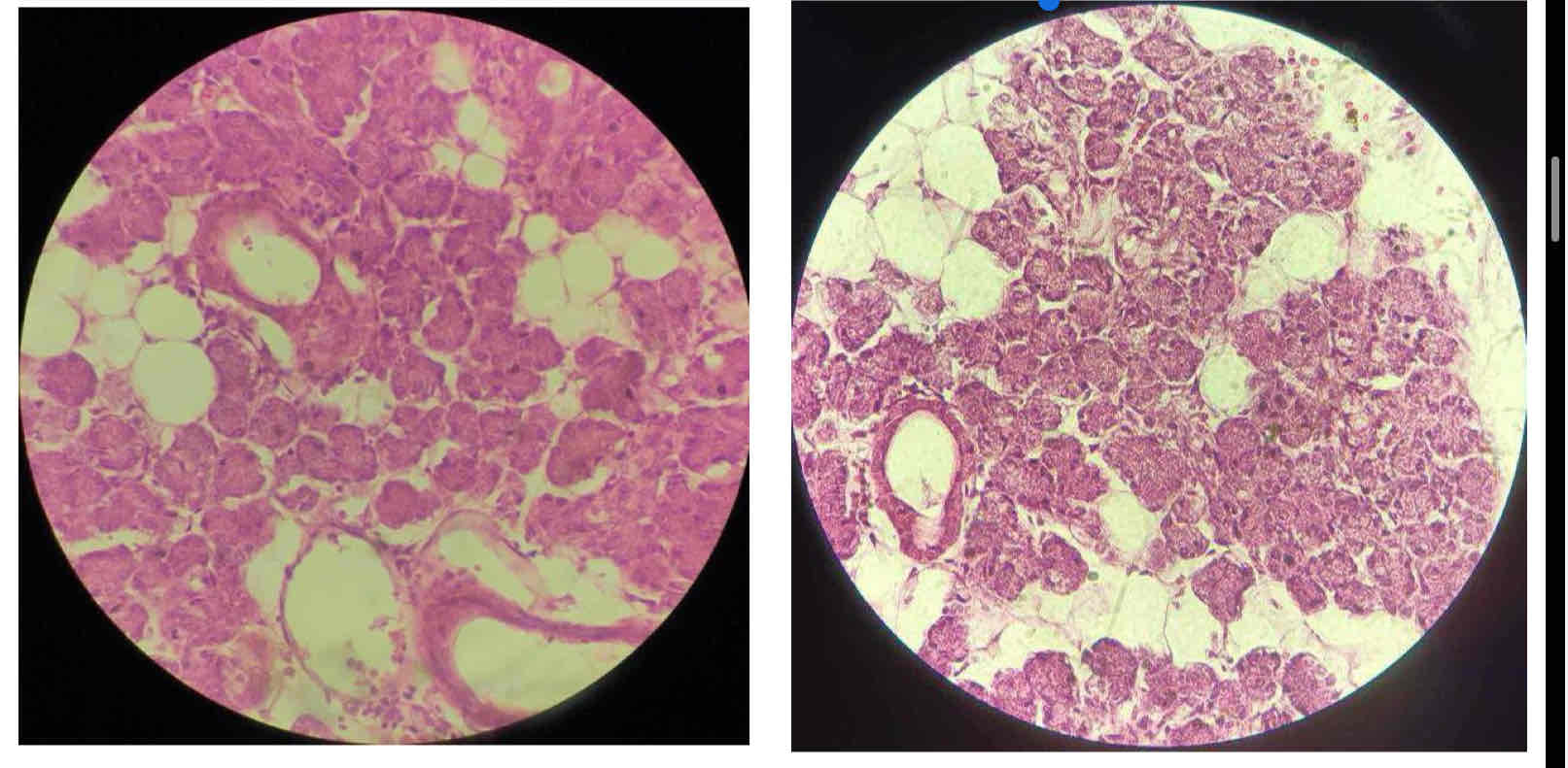
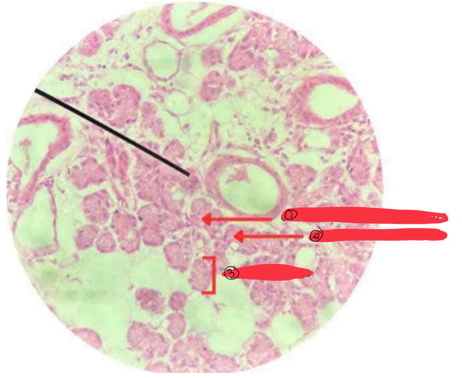
Parotid Gland
Serous Glandular Epithelium
Organ
Tissue
1) Serous Glandular Epithelium
2) Serous Acinus
Tongue
Mucous Glandular Epithelium
Organ:
Tissue:
1) Mucous Glandular Epithelium
2) Mucous Acinus
Sublingual Gland
Sero-Mucous Gland
Organ:
Tissue:
1) Sero-Mucous Gland
2) Mucous Glandular Epithelium
3) Serous Glandular Epithelium
4) Serous Demilune
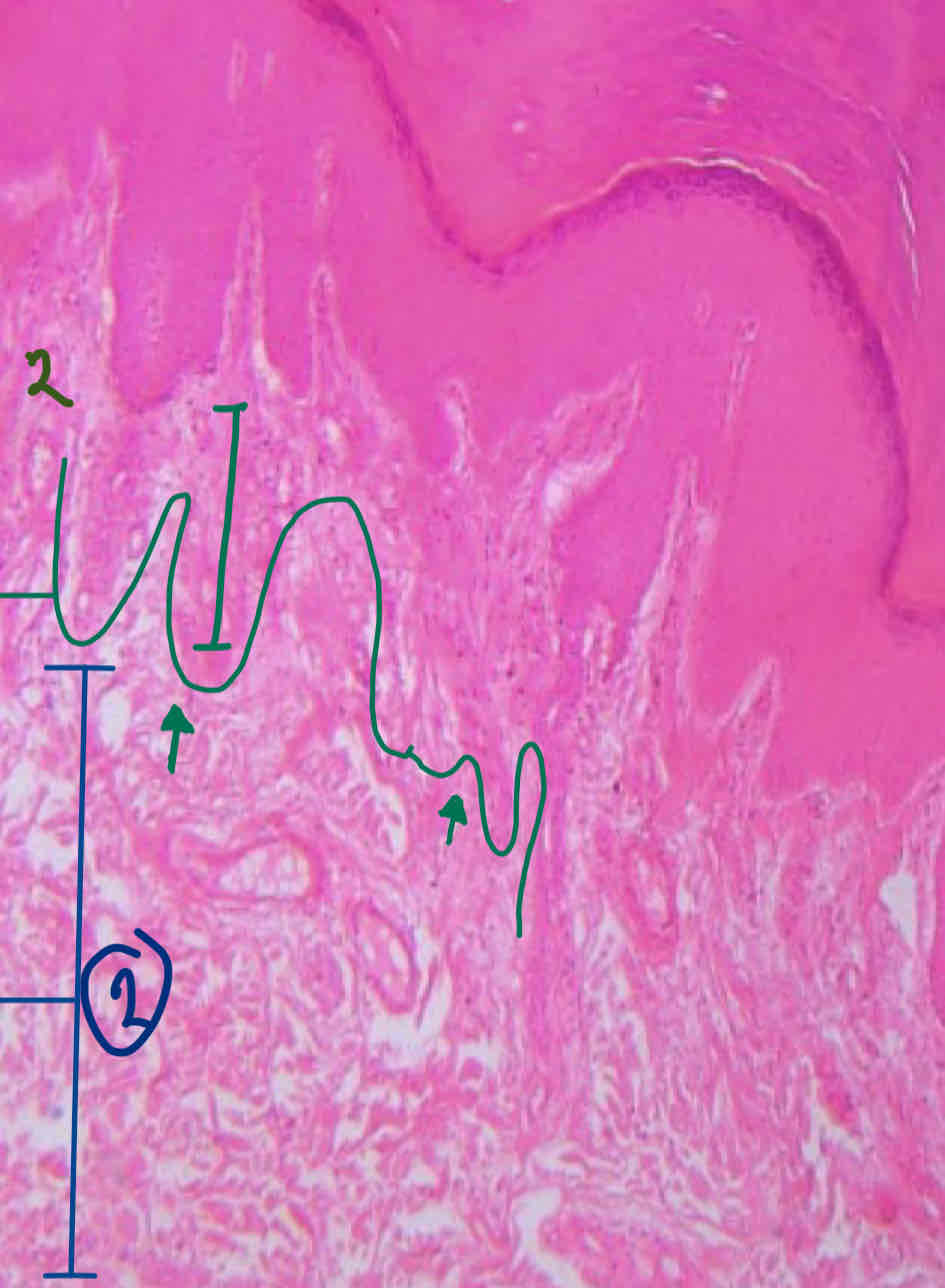
1) Epidermis
2) Dermis
Stratum corneum
Stratum lucidum
Stratum granulosum
Stratum spinosum
Stratum basale
Layers of the Epidermis
Papillary Layer
Reticular Layer
Layers of the Dermis
Stratum corneum
Stratum granulosum
Stratum spinosum
Stratum basale
Papillary Layer
Reticular Layer
Epidermis
Dermis
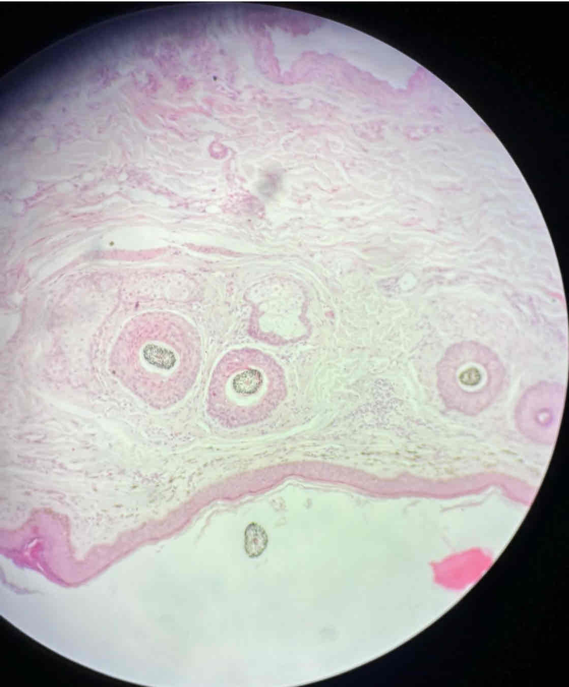
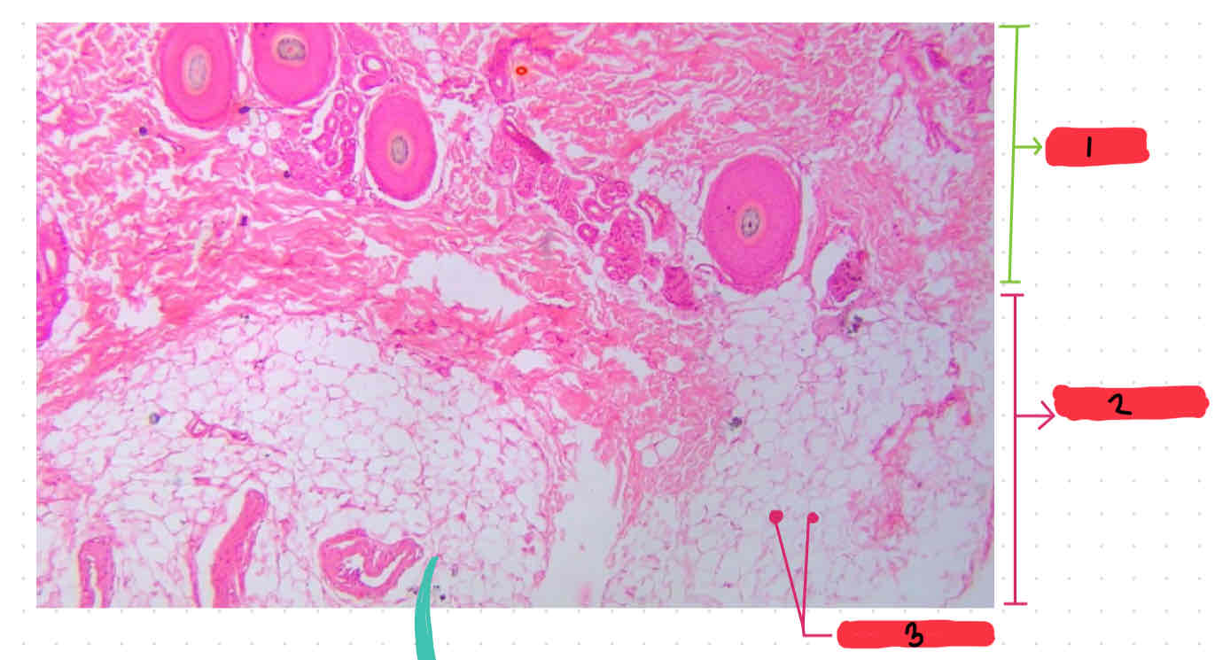
Hair Follicle
Sebaceous Glands
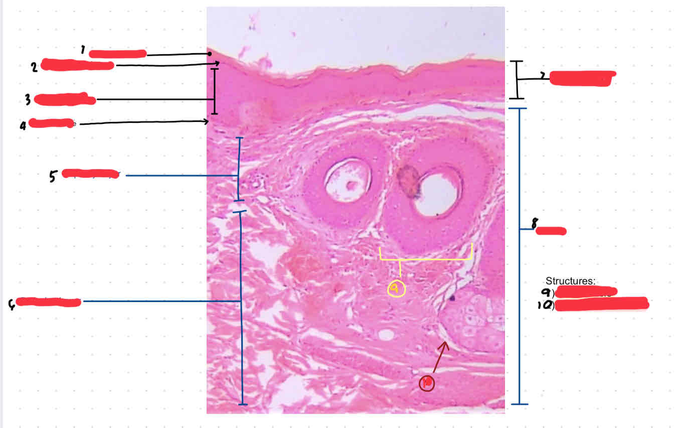
1.Dermis
2.Hypodermis
3.adipose tissue
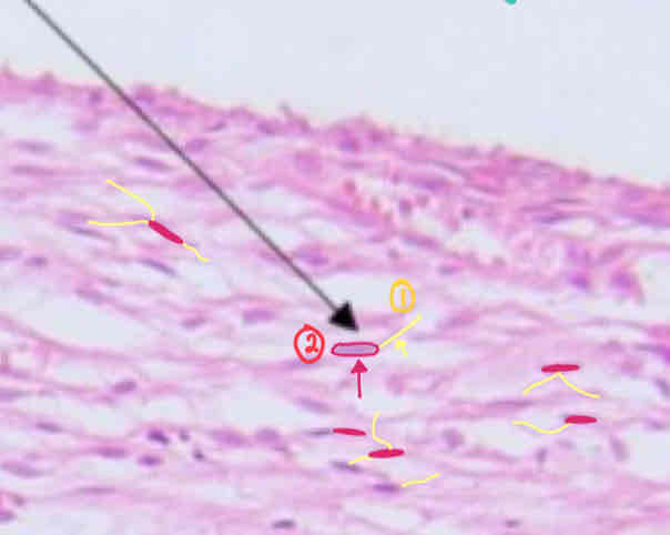
1) Hair Follicle
a.Hair Matrix
b.Hair Root
c.Epidermal Root Sheath
d.Dermal Root Sheet
2) Sweat Glands / Tubular Glands
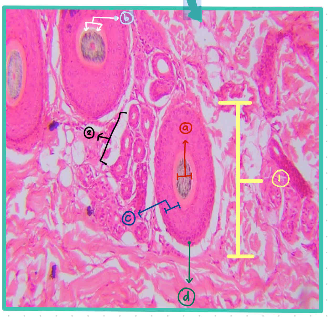
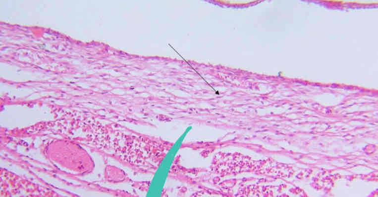
Hair Follicle
Sebaceous Gland
Arrector pili muscle
Hair Bulb
Hair Papilla
Hair Matrix
Hair Root
Epidermal Root Sheath
Dermal Root Sheath
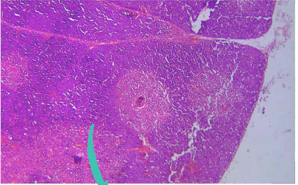
Spleen
Trabecula
White Pulp / Splenic Nodule
Red Pulp
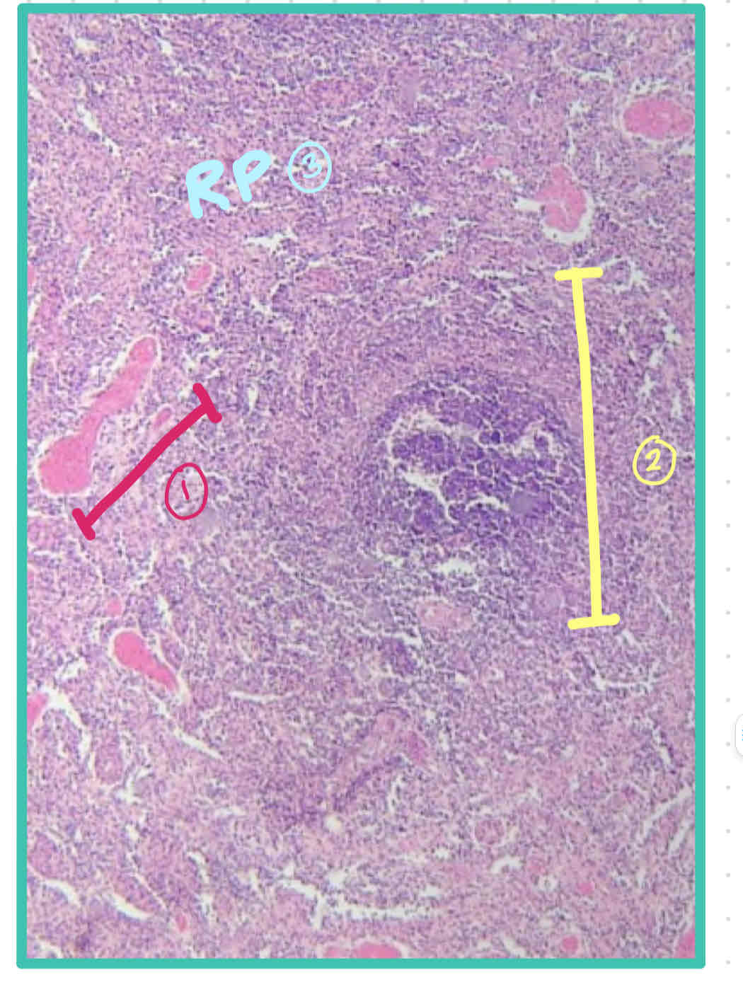
Bilroth's Cord/ Splenic Cord
Sheathed Artery
Area: White Pulp / Splenic Nodule
Central Artery
Capsule
Trabecula
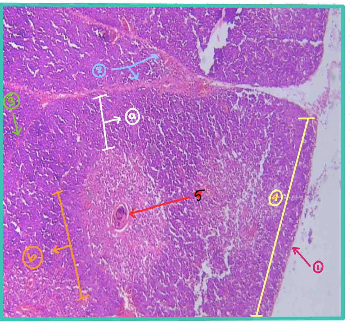
Thymus
Capsule
Septa
Trabecula
Thymic Lobule
a. Cortex
b.Medulla
Hassall's Corpuscle
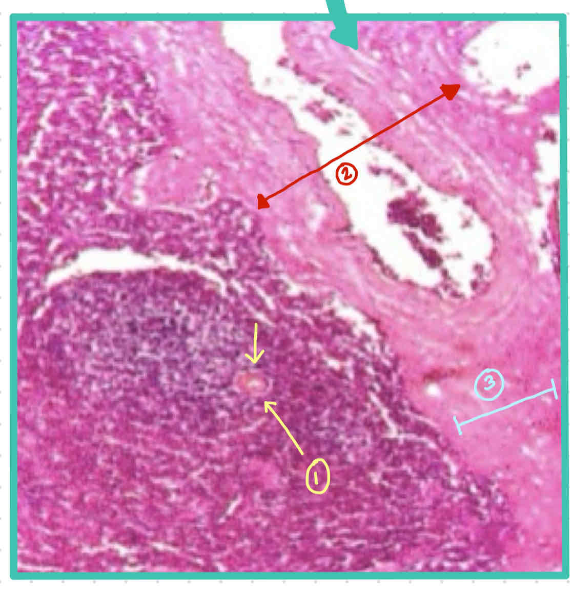
Lymph Node
Fibrous Capsule
Hilus
Trabecula
Lymph Nodule
a.Corona
b.Germinal Center
Subcapsular Lymphatic Sinus
Lymph Node