Brainstem and blood supply to the brain
1/139
There's no tags or description
Looks like no tags are added yet.
Name | Mastery | Learn | Test | Matching | Spaced | Call with Kai | Chat |
|---|
No analytics yet
Send a link to your students to track their progress
140 Terms
Describe the location of the three components of the brainstem and their anatomical relations.
Locate and identify the key structures that are visible on the midbrain (including in cross-section), pons and medulla oblongata.
Outline the course and appreciate the functional significance of some of the major ascending and descending neural pathways that pass through the brainstem, linking the cerebral hemispheres and the spinal cord.
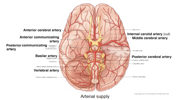
Describe the formation of the Circle of Willis, naming its major contributing arteries and identifying all major communicating and cerebral artery components.
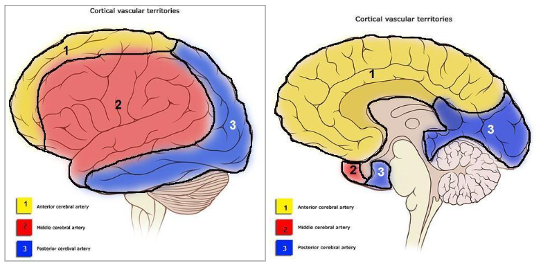
Describe the origin, course, and recall the specific cortical supply territories of the anterior, middle and posterior cerebral arteries.
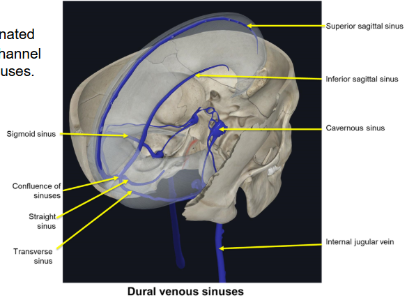
Describe the venous drainage of the brain and locate the major venous sinuses.
What are the 3 components of the brainstem ?
midbrain, pons and medulla oblongata
What is the bulb another term for?
medulla oblongata
What are some functions of the brainstem?
Condit for tracts (sensory and motor)
Head and neck sensory, motor and autonomic (CN III-XII)
Consciousness, cardiovascular system, respiratory system, pain perception (reticular formation)
Damage is life-threatening
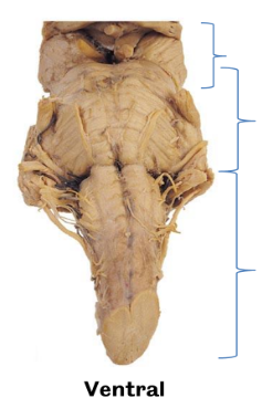
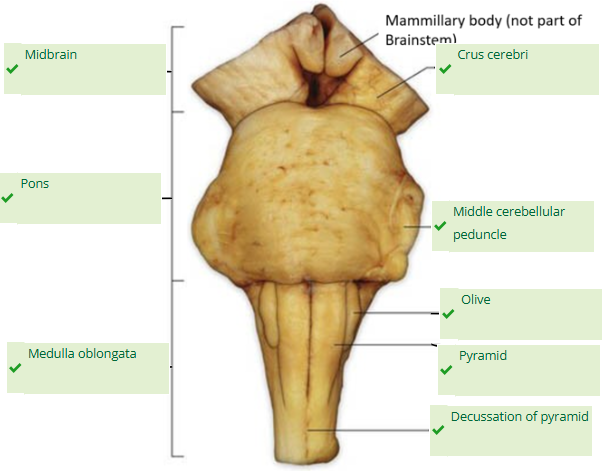
What structure and view is this? Which landmarks are visible?
ventral view of the brainstem
crus cerebri, olive, pyramid visible, transverse fibres of pons
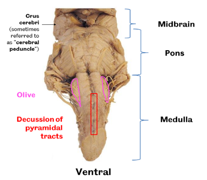
Function of crus cerebri
voluntary motor control, coordination because carries crucial motor fibers from the cerebral cortex to the brainstem and spinal cord
Function of brainstem pyramids
carries corticospinal tract fibres
Function of olive
coordination of movement, link to cerebellum
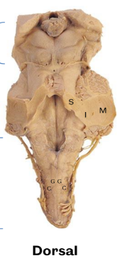
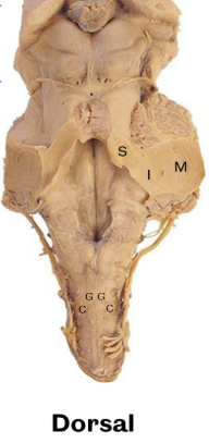
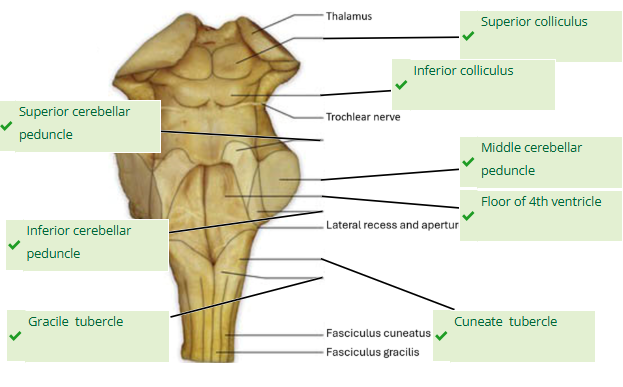
What structure and view is this? Which landmarks are visible?
dorsal view of the brainstem
cerebellum mostly removed here, Thalamus, pineal gland, colliculi sup and inf, trochlear nerve, dorsal columns sensory from spine terminate in the gracile and cuneate nuclei seen as tubercles
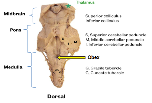
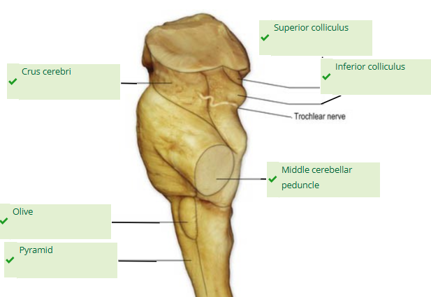
Function of superior vs inferior colliculus
reflexes linked to auditory vs sensory - very old response eg i see lion, i must run away from lion, together coordinate sensory-motor responses
Function of gracile vs cuneate nuclei
process information from lower limb vs upper body (c for side and cephalic so lateral to gracile and for top of body)
What is the obex?
(Latin for "barrier") is the point in the caudal medulla of the human brain where the fourth ventricle narrows to become the central canal of the spinal cord
Label these
superior, middle and inferior cerebellar peduncle
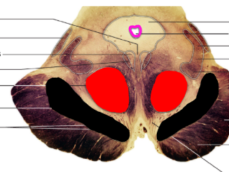
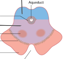
What view is this ? Label structures
horizontal section of the midbrain
Describe the volume and pathways of the brainstem’s connections to surrounding structures
numerous and bidirectional, links between the brain and spinal cord, brainstem and cerebellum and cerebellum and spinal cord
Some pass through e.g., corticospinal, spinothalamic, Some have origins in, or terminate in, the brainstem nuclei
What are 2 motor pathways spanning through the midbrain?
corticospinal and corticonuclear
What is the function of the corticospinal and corticonuclear pathways ?
voluntary/conscious control over skeletal muscles
What area does the corticospinal motor pathway control?
below the head
What area does the corticonuclear motor pathway control?
head and neck
What are 2 sensory pathways spanning through the midbrain?
reactive spinothalamic and discriminative dorsal column-medial lemniscus (DCML)
What are the 2 systems through which the brain is irrigated?
internal carotid system and vertebral-basilar system (2 internal carotid arteries and 2 vertebral arteries)
What is the function of the spinothalamic pathway?
reactive sensory pathway ie pain, temperature, “crude” touch and pressure
What is the function of the DCML pathway?
discriminative sensory pathway ie proprioceptive (limb position), fine touch and vibration
Are spinothalamic and DCML ascending or descending?
ascending/afferent tract
How do the internal carotid arteries enter the skull?
through carotid canal
How do the vertebral arteries enter the skull? Where do they arise?
through foramen magnum
from subclavian artery
Branches of the internal carotid and vertebral-basilar systems interconnect to form which structure?
form the cerebral arterial circle (The Circle of Willis)
Which short blood vessels complete the anterior and posterior portions of the circle of Willis respectively?
anterior and posterior communicating arteries - connect left and right anterior cerebral arteries and the posterior and anterior circulation respectively (vertebrobasilar and internal carotid artery systems)
Which main blood vessels are involved in supplying the brain with blood?
cerebral arteries (A, M and P), internal carotid arteries, vertebral artery, communicating arteries
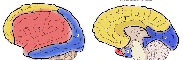
Label the following cortical vascular territories (areas of blood supply)
Where do all venous sinuses drain into?
internal jugular vein
What are the major dural venous sinuses?
Superior sagittal sinus
Inferior sagittal sinus
Straight sinus
Transverse sinus
Confluence of sinuses
Cavernous sinus
Sigmoid sinus
What carries out venous drainage in the brain?
Cerebral veins that collect deoxygenated blood and cerebrospinal fluid and channel it into a network of dural venous sinuses, which all drain into internal jugular vein
Which pathways spanning the brainstem are descending vs ascending ?
Descending (motor) pathways:
Corticospinal tract
Corticobulbar tract
Ascending (sensory) pathways:
Spinothalamic tract
Dorsal column medial lemniscus (DCML)
What is the relationship between the brainstem and the motor and sensory pathways?
Brainstem acts as conduit for the tracts
What splits the midbrain down its centre?
cerebral aqueduct - channel for CSF
What does peduncle mean?
foot/stalk
In which view of the midbrain is the olive visible?
ventral view
What forms the floor of the 4th ventricle?
rhomboid fossa
What is the function and make-up of the red nucleus?
grey matter, involuntary movement, arm swinging during walking, crawling in infants
What is the function and make-up of the substantia nigra?
grey matter, controls voluntary movement, motor planning, and reward-based learning by producing dopamine + regulates muscle tone and movement coordination via the nigrostriatal pathway
Where does the corticonuclear tract originate?
pre-central gyrus ie primary motor cortex
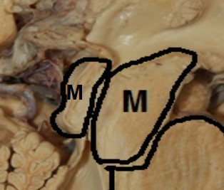
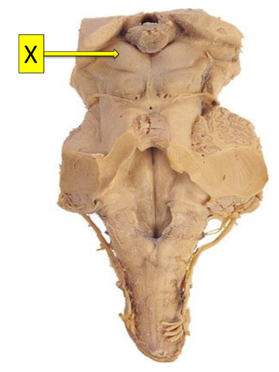
Which one statement BEST describes the function of structure X?
Centres within it control planning and reward.
It controls fine tuning of voluntary movement.
Centres within it control the autonomic functions of respiration, blood pressure, vomiting and heart rate.
It is critical for visual processing.
The area of the brain where memories are formed.
Centres within it control the autonomic functions of respiration, blood pressure, vomiting and heart rate.
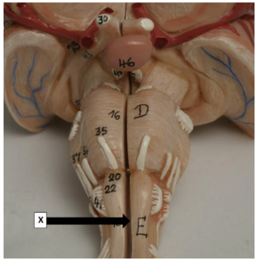
Which one statement is correct concerning point X?
It is the superior cerebellar peduncle which carries information from the cortex to spinal cord.
It is the gracile fasciculus and receives information from the upper limbs.
It is the gracile fasciculus and receives information from the lower limbs.
It is the point where pyramidal fibres of the corticospinal tract may cross over.
Is where the olivary nucleus is contained
It is the point where pyramidal fibres of the corticospinal tract may cross over.
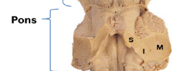
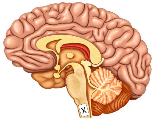
Which structure rests at point X on the occipital bone?
the pons
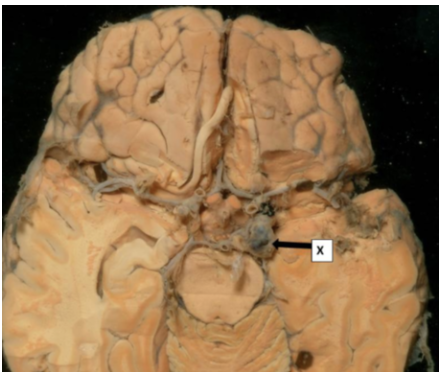
Which statement(s) are correct concerning structure X?
i. This is a tumour of the Circle of Willis
ii. This is an aneurysm of the Circle of Willis
iii. Risk factors for this include genetics and alcohol abuse.
iv. Risk factors for this include high blood pressure, genetics and head injury
ii and iv
classic saccular or berry aneurysm
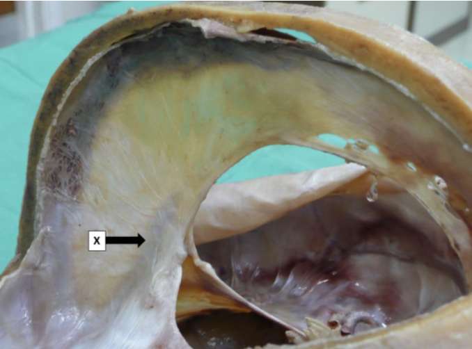
Which statement(s) concerning structure X is/are true?
i. This is the transverse sinus.
ii. This is the straight sinus.
iii. It lies mostly within the falx cerebri.
iv. It lies mostly within the falx cerebelli.
ii and iii
this is the straight sinus lying in the dural reflection of the falx cerebri
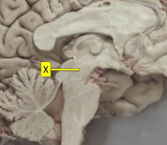
Structure X is
tegmentum of midbrain
Which ONE statement is true concerning structure X:
It is the superior colliculus of the tectum and it functions as a visual reflex centre.
It is the superior colliculus of the tegmentum and it functions as an auditory reflex centre.
It is the superior colliculus of the tegmentum and it functions as a visual reflex centre.
It is the superior colliculus of the tectum and it functions as an auditory reflex centre.
Is the middle cerebellar peduncle connecting the cerebellum to the pons.
It is the superior colliculus of the tectum and it functions as a visual reflex centre.
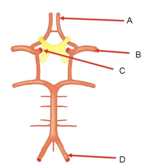
Which two structures provide the main blood supply to the brain?
C and D - one internal carotid artery and one vertebral artery, they provide main blood supply to the brain through circle of willis
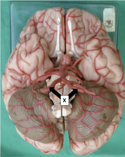
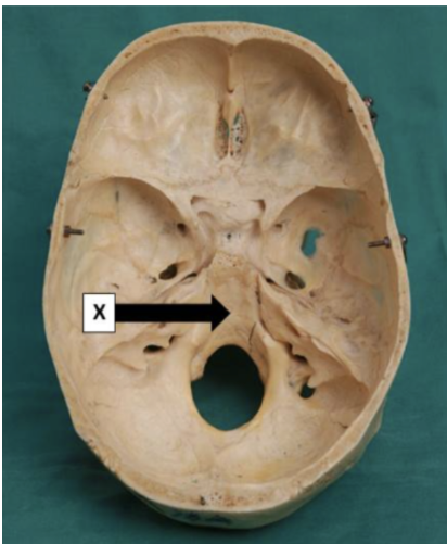
The vessels labelled X enter the cranial cavity through the:
foramen magnum
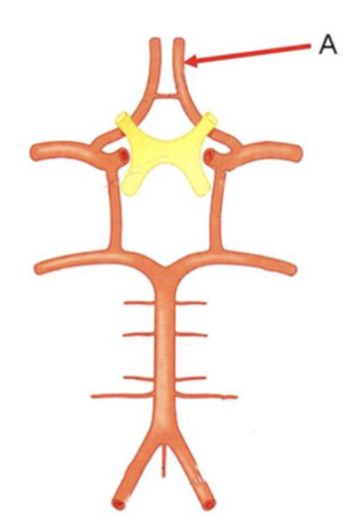
The Vessel A generally supplies:
medial territory of cerebral hemispheres
this is the anterior cerebral artery that mostly supplies medial portions of the cerebral cortex (frontal and parietal lobes mainly)
What does the brainstem connect the cerebrum to?
to the spinal cord and cerebellum
Which structures make up the brainstem and which critical functions do they allow?
midbrain, pons, medulla oblongata
coordinating critical functions like breathing, heart rate and consciousness
Which cranial nerves does the brainstem house?
CNIII through XII (10 of 12 CNs)
What forms the anterior vs posterior circulation of the brain?
paired internal carotid arteries vs paired vertebral arteries
What does the convergence of the anterior and posterior circulations of the brain form?
create an essential protective anastomosis called circle of willis
Which arteries branch from the circle of willis to supply the cerebral hemispheres?
anterior, middle, posterior cerebral arteries
What are the brainstem and cerebellum primarily supplied by?
vertebrobasilar system and its branches
How is the venous drainage of the brain unique? How does it function?
because does not follow the arterial pattern
drain into a complex network of valve-less channels embedded within the dura mater, known as the dural venous sinuses then internal jugular veins
Where do the dural venous sinuses ultimately empty into to return blood to the systemic circulation?
ultimately empty into the internal jugular veins to return blood to the systemic circulation
What is the brainstem a conduit for?
for all major ascending (sensory) and descending (motor) nerve tracts traveling between the cerebrum and the body
nuclei for ten of the twelve cranial nerves (CN III through CN XII)
What functions are CNs III through XII essential for?
motor and sensory functions of the face, head, and neck
Is the mammillary body part of the brainstem?
no!
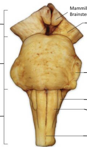
Label this ventral view of the brainstem
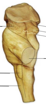
Label this lateral view of the brainstem
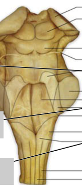
Label this dorsal view of the brainstem
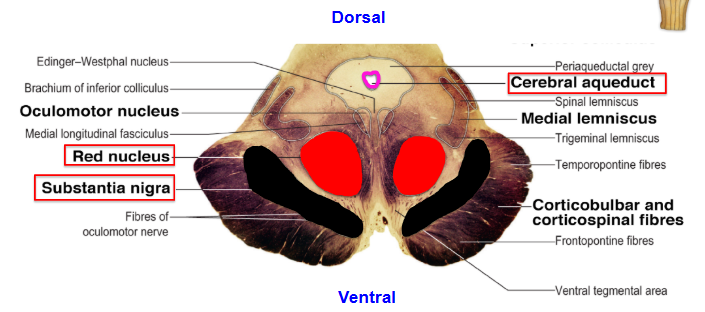
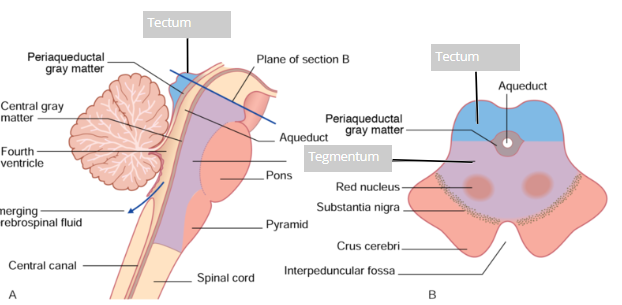
Which 3 main regions are made visible by a cross section of the midbrain?
tectum, tegmentum and crura cerebri/cerebral peduncles
What serves as the roof of the midbrain
tectum
What serves as the floor of the midbrain
tegmentum
Which passage for CSF traverses the midbrain? What structures does it connect?
cerebral aqueduct
third and fourth ventricles
What surrounds the cerebral aqueduct? What is this area vital for?
periaqueductgray (PAG also known as central gray matter)
pain modulation
What is the tectum dorsal to?
to the cerebral aqueduct
What structures does the tectum contain?
superior and inferior colliculi (collectively corpora quadrigemina)
What is the role of the structures making up the corpora quadrigemina?
superior colliculus - visual reflexes
inferior colliculus - auditory reflexes
What is the tegmentum ventral to?
to the aqueduct
Which key structures does the tegmentum house?
red nucleus and substantia nigra
Function of red nucleus
motor coordination
Function of substantia nigra
dopamine production for motor control, degeneration linked to Parkinson’s disease
Where are the crura cerebri/cerebral peduncles located and what do they carry?
massive fiber tracts most ventral to the tegmentum
carry corticospinal, corticopontine, and corticobulbar fibers descending from the cortex
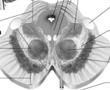
Label the following cross section of the midbrain
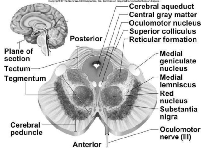
Describe the anatomical relations of the tectum, tegmentum and crura cerebri?
tectum is dorsal to cerebral aqueduct, tegmentum is ventral to aqueduct and crura cerebri most ventral to tegmentum
Which longitudinal zones is the brainstem organised into? What system are they related to?
tectum and tegmentum
to the ventricular system
What does tectum vs tegmentum mean in latin?
roof vs covering
What forms the central core of the brainstem, ventral to the cerebral aqueduct and fourth ventricle?
tegmentum
Are the tectum and tegmentum restricted to the midbrain?
tectum is, tegmentum extends through entire length of midbrain, pons, medulla
What does the medulla house?
reticular formation, cranial nerve nuclei and major ascending sensory pathways
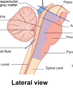
Identify the tectum and tegmentum in this lateral view of the brainstem

Identify the tectum and tegmentum in this transverse view of the brainstem
Which 2 motor pathways originate in the cerebral cortex? What are they responsible for?
Corticospinal (Pyramidal) Tract and the Corticonuclear (Corticobulbar) Tract
voluntary movement
What does the corticospinal tract control specifically? Where does its fibers descend from and synapse onto?
trunk and limbs
descend from motor cortex and synapse onto lower motor neurons (LMNs) in ventral horn of spinal cord
What from the corticospinal tract forms the lateral corticospinal tract and at what point does this happen? What does this allow?
At the medulla, approximately 85-90% of the fibers cross (decussate) to form the lateral corticospinal tract, enabling contralateral (opposite side) control of the body.

Does the corticospinal or corticonuclear tract synapse onto lower motor neurons?
both do! former synapses onto ones in the ventral horn of the spinal cord = contralateral control of the body
latter synapses onto ones within the motor nuclei of the cranial nerves = bilateral input to most cranial nerves

Which muscles are controlled contralaterally by cranial nerves ?
lower facial and hypoglossal muscles
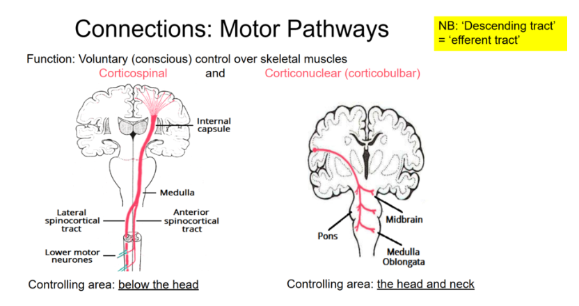
Which muscles does the corticonuclear tract control? Where do its fibers descend and where do they synapse?
head and face eg chewing, facial expression, swallowing
descend to brainstem where they synapse on LMNs in motor nuclei of cranial nerves
