EKG Quiz 1
1/48
There's no tags or description
Looks like no tags are added yet.
Name | Mastery | Learn | Test | Matching | Spaced | Call with Kai |
|---|
No analytics yet
Send a link to your students to track their progress
49 Terms
What is an electrocardiogram (ECG)
The electrical measurement of cardiac function
In the depolarization and repolarization cycle, what is the state of a resting cardiac cell?
Polarized
Where is the aortic valve located?
Between the left ventricle and the aorta
Where is the tricuspid valve located?
Between the right atrium and right ventricle
What is another name for the mitral valve?
Bicuspid valve
What is the innermost layer of the heart?
Endocardium
What is the primary pacemaker of the heart?
SA node
On a graph sheet, what does the horizontal axis represent?
Time
What happens during diastole?
Ventricles relax and fill with blood
What happens during systole?
Ventricles contract and eject blood
What is the correct order of the cardiac conduction?
SA node -> interatrial pathways -> AV node -> Bundle of His -> left and right bundle branches -> Purkinje fibers
What is the normal rate range for the SA node?
60-100 bpm
What is the normal rate range for the AV junction?
40-60 bpm
What is the normal rate range for ventricular/Purkinje fibers?
20-40 bpm
What happens when the right ventricle contracts?
Blood is sent to the lungs via the pulmonary artery
What type of blood does the pulmonary artery carry away from the heart?
Deoxygenated blood
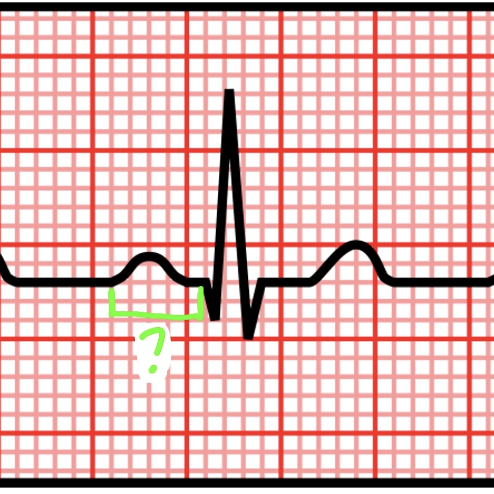
On ECG graph paper, what does one large box represent?
0.20 seconds
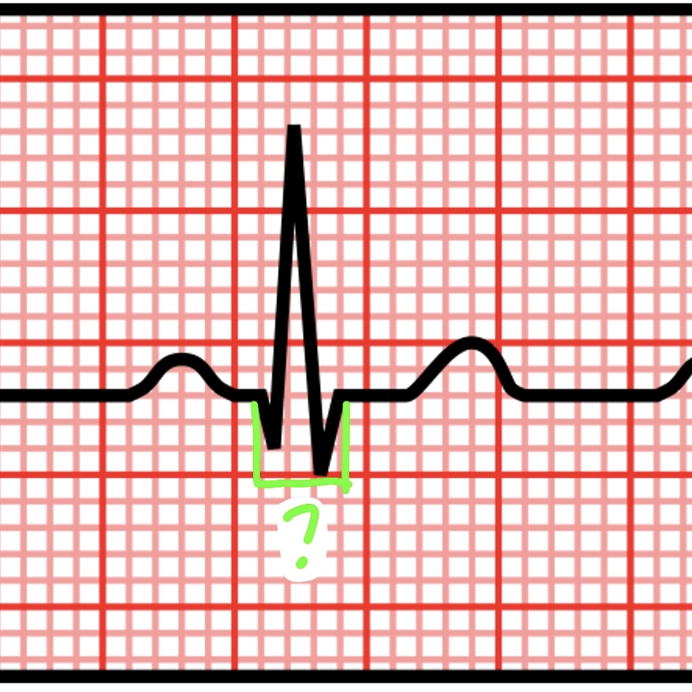
On ECG graph paper, what does one small box represent?
0.04 seconds
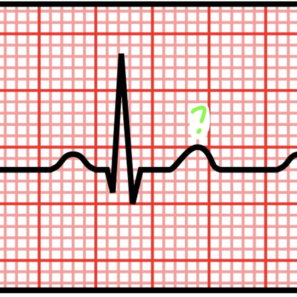
What do P waves represent?
Atrial depolarization
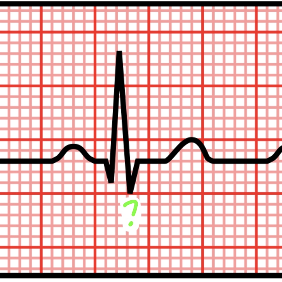
What does the QRS complex represent?
Ventricular depolarization
What is coronary circulation?
Circulation of blood within the heart
What is systemic circulation?
Circulation of blood throughout the entire body
What does a 12 lead EKG show?
Multiple views and all waves of the heart’s electrical activity
What is a single rhythm strip used for?
Observing one lead over an extended period
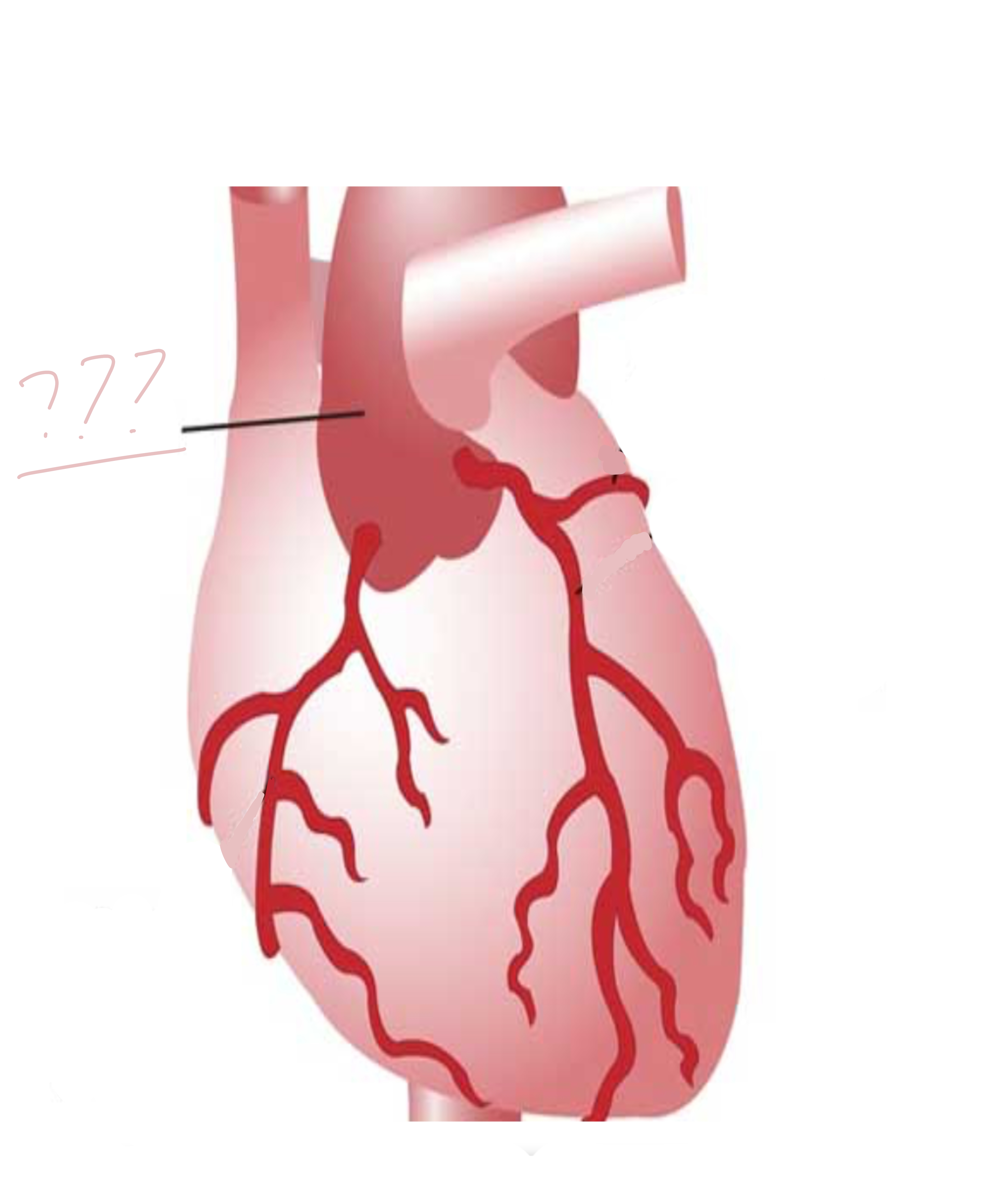
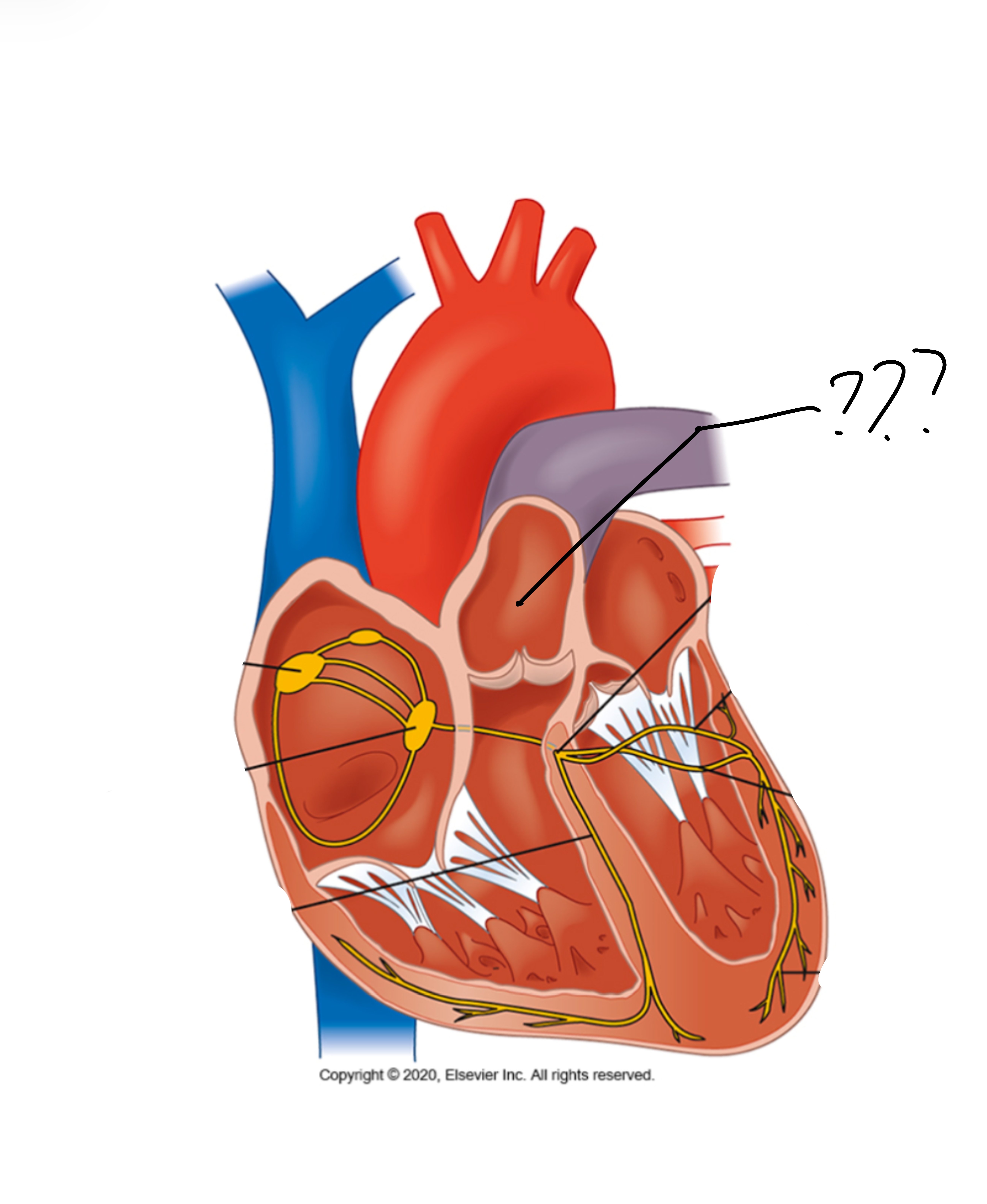
Aorta
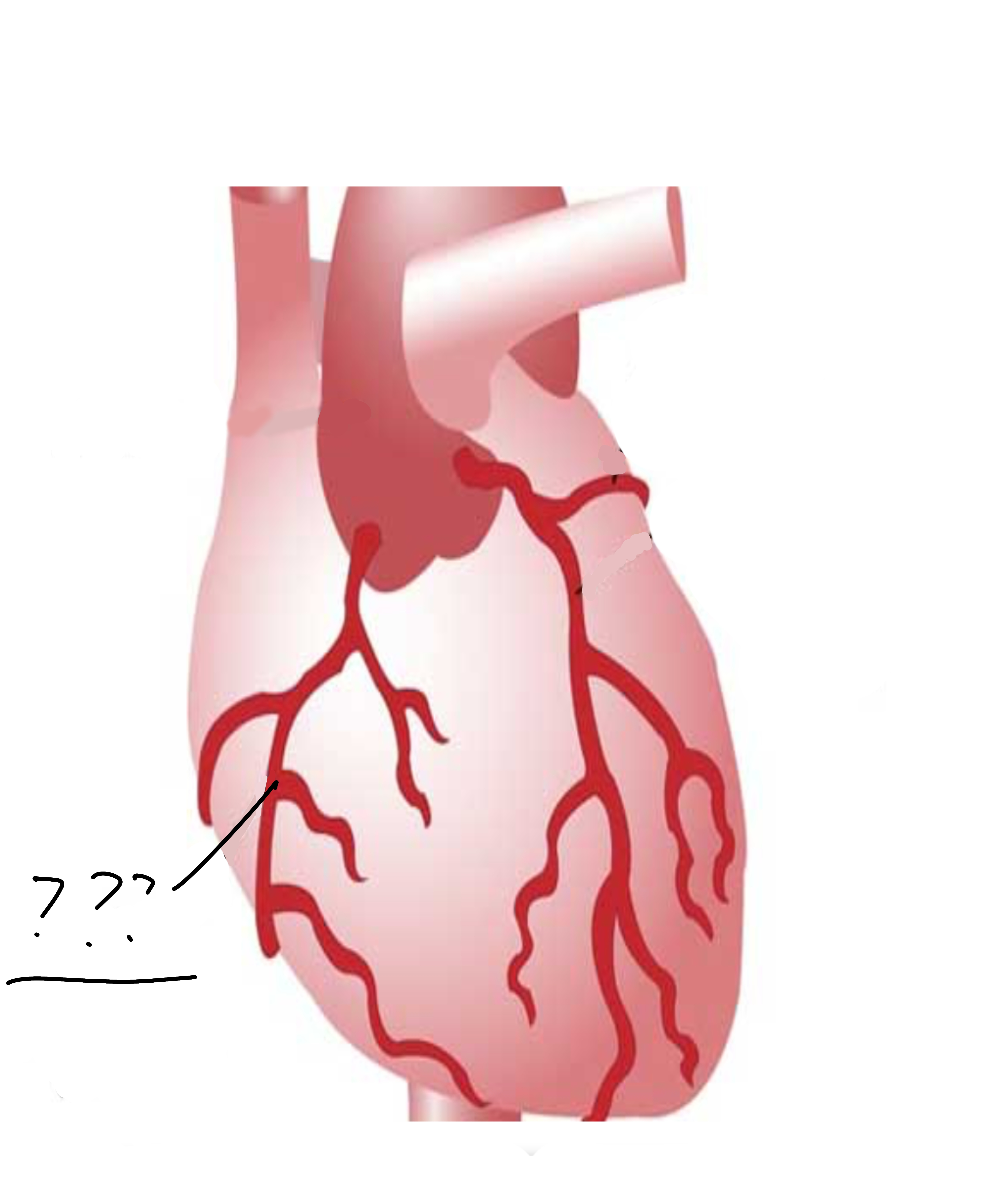
Right coronary artery
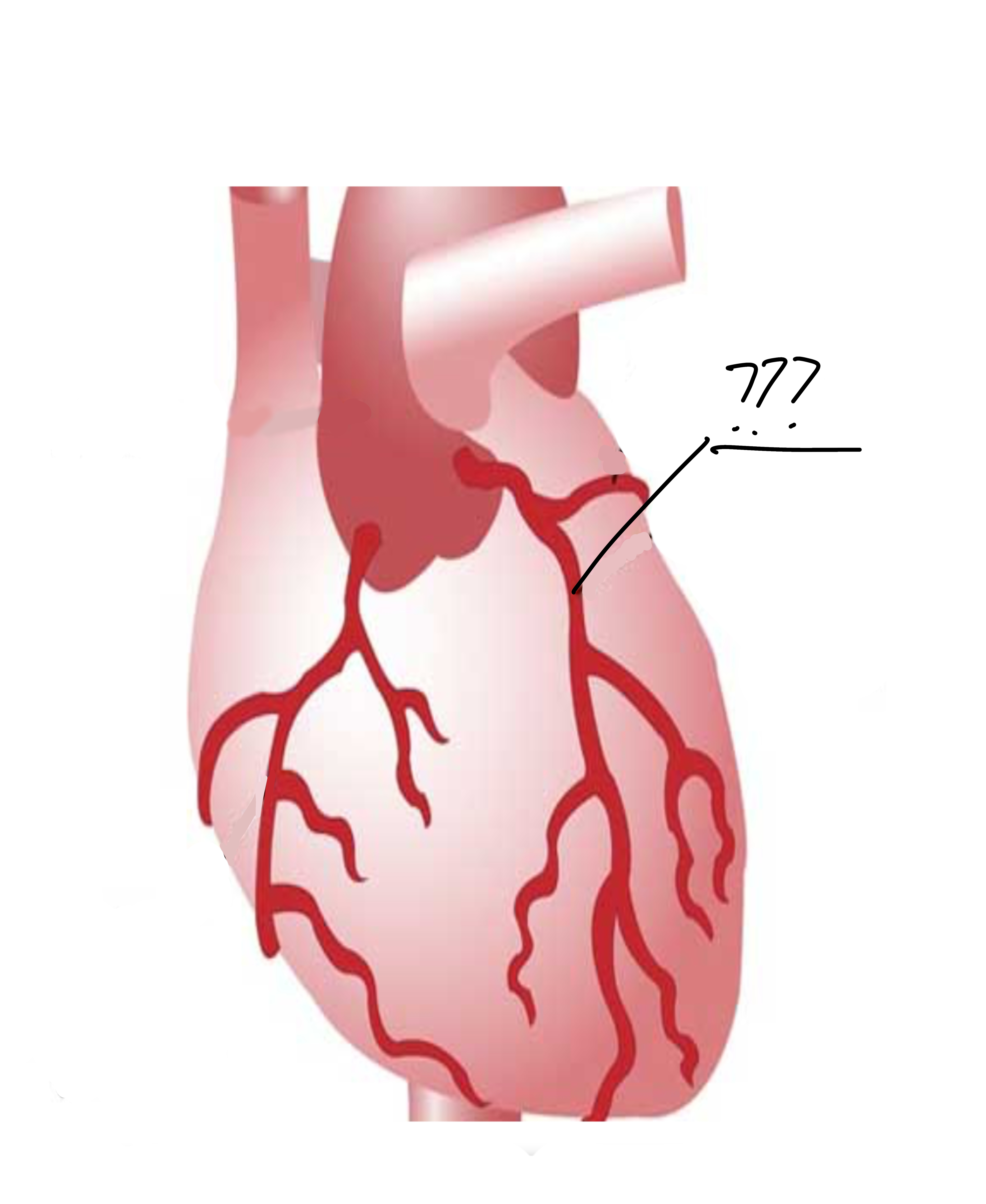
Left coronary artery
Interarterial pathways

SA node

AV node

Right bundle branches
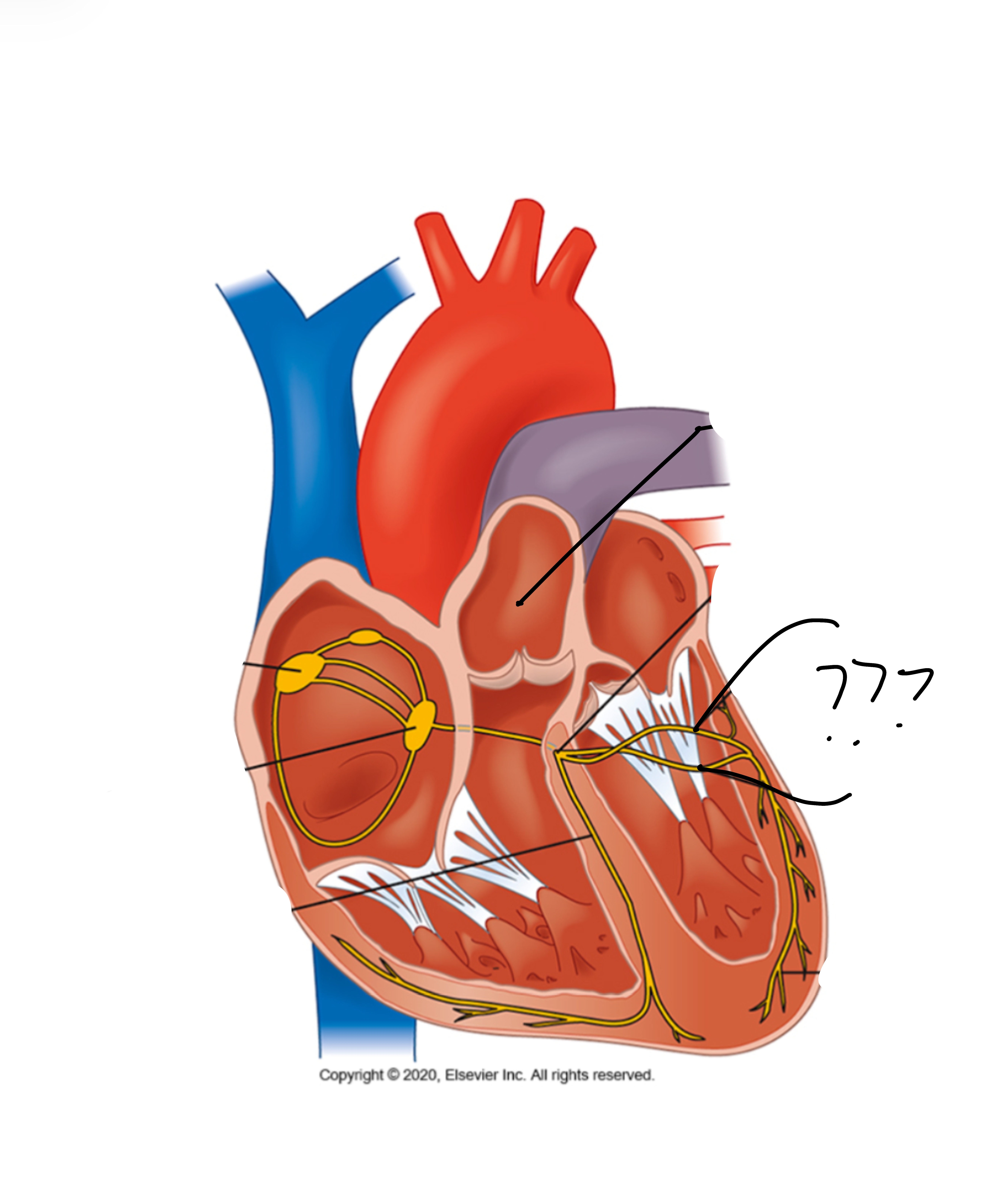
Left bundle branches
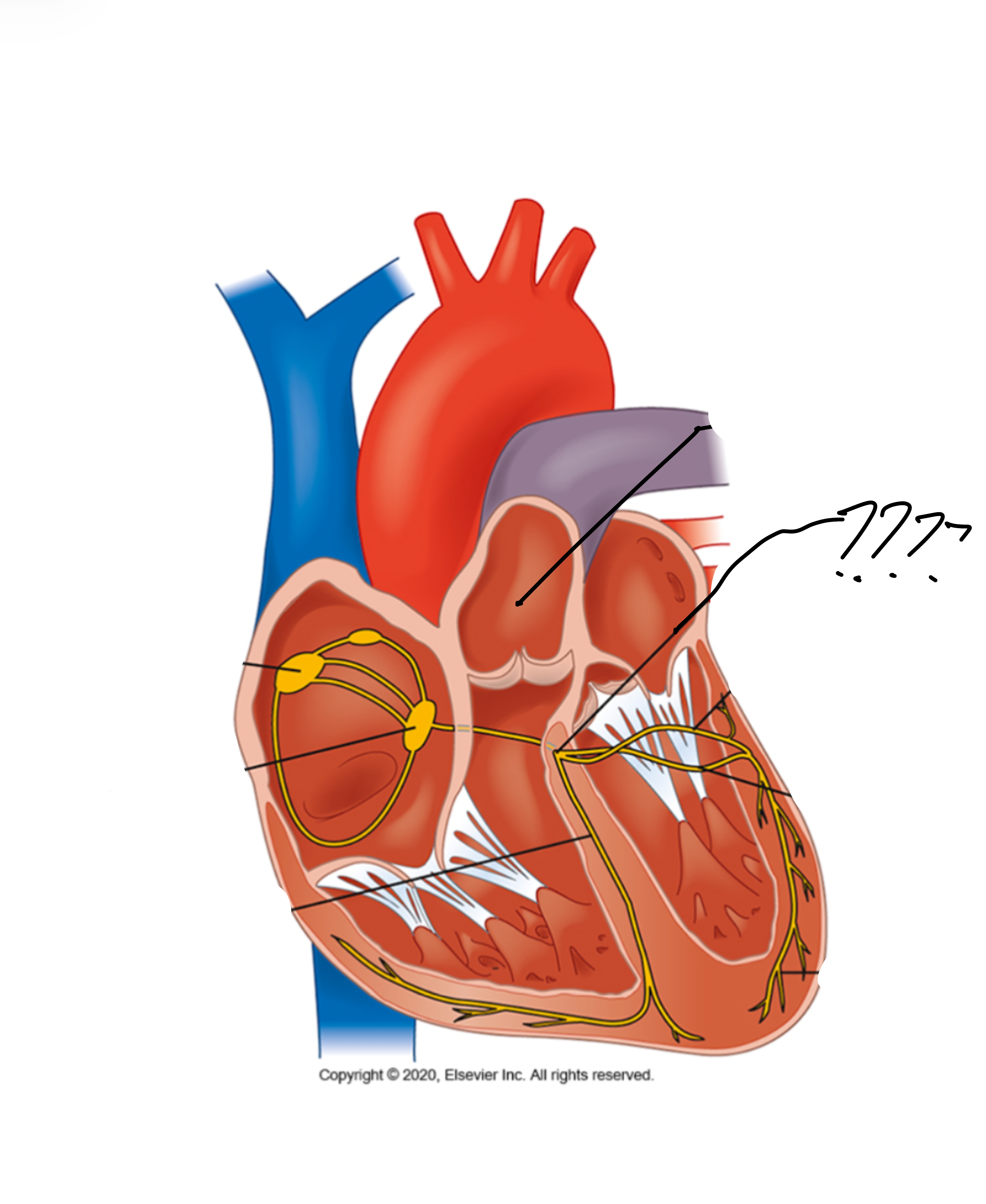
Bundle of His

Purkinje fibers
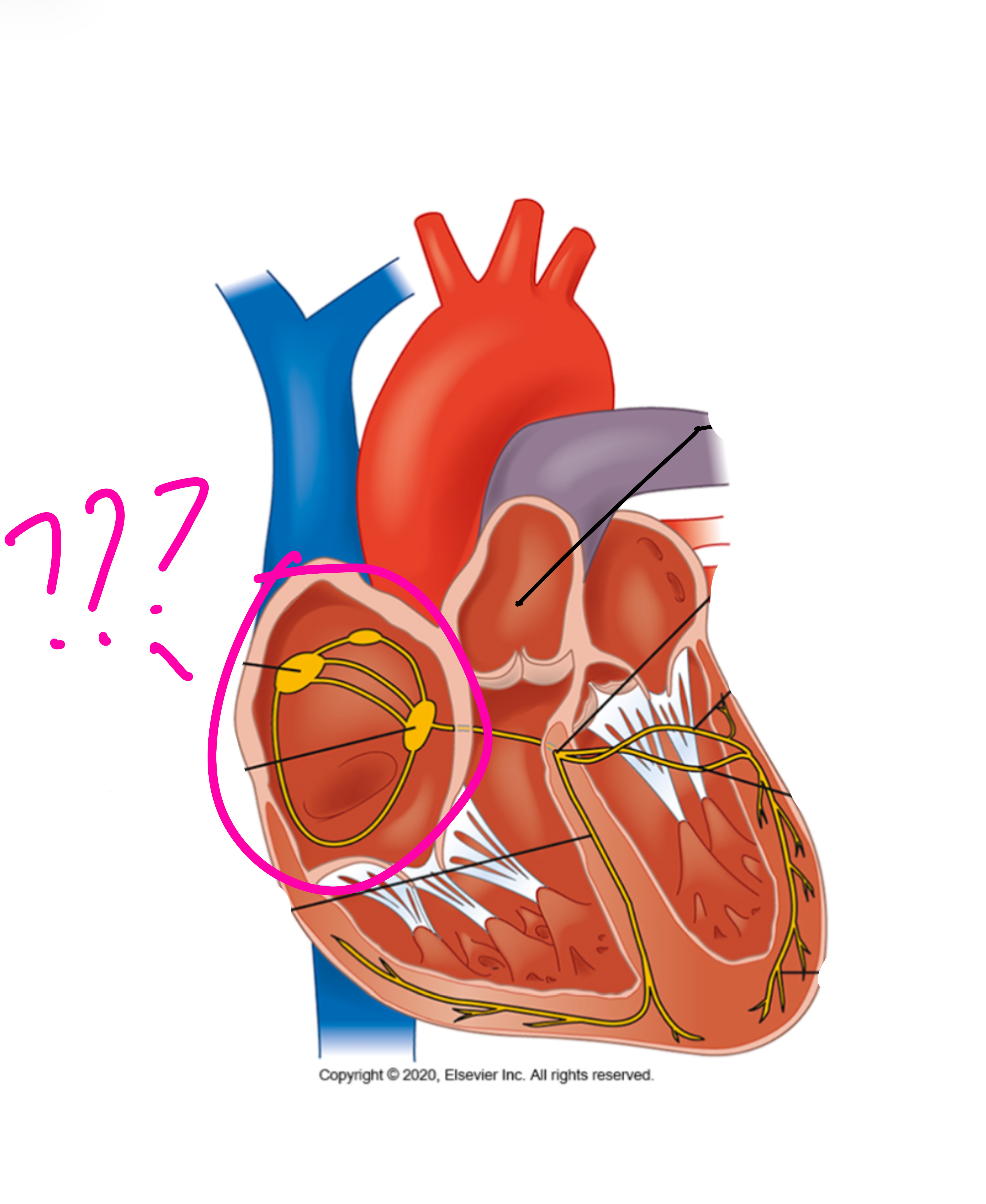
Right atrium
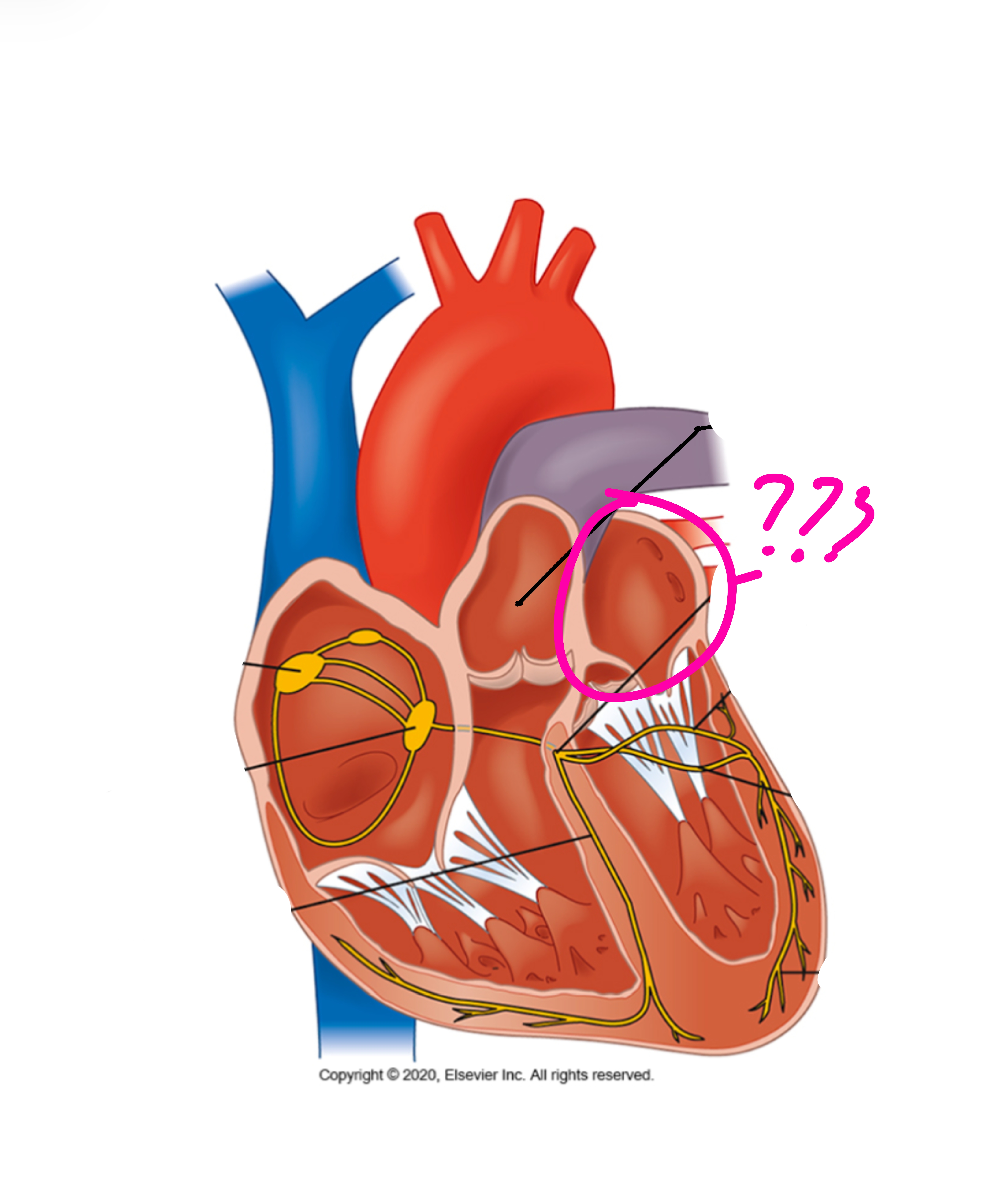
Left atrium
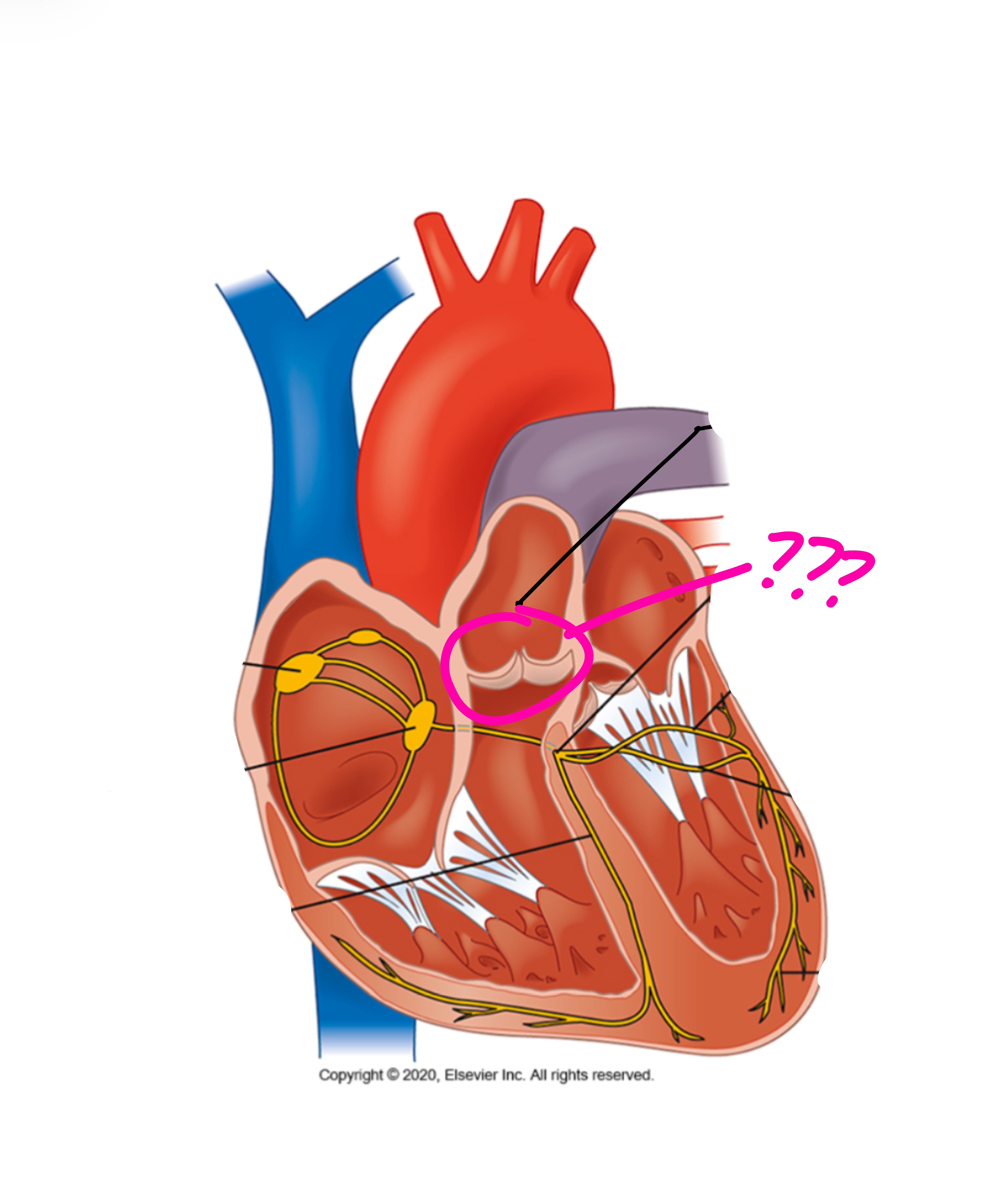
Pulmonary valve

Myocardium
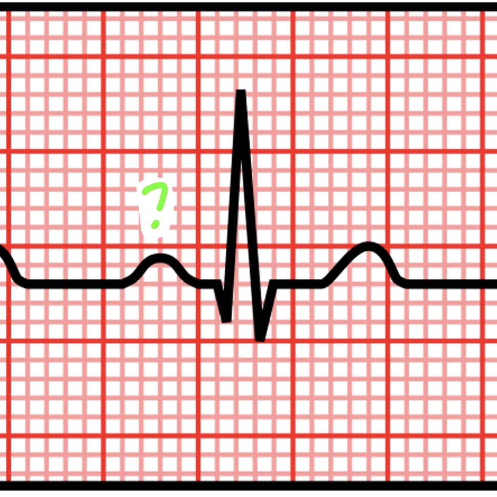
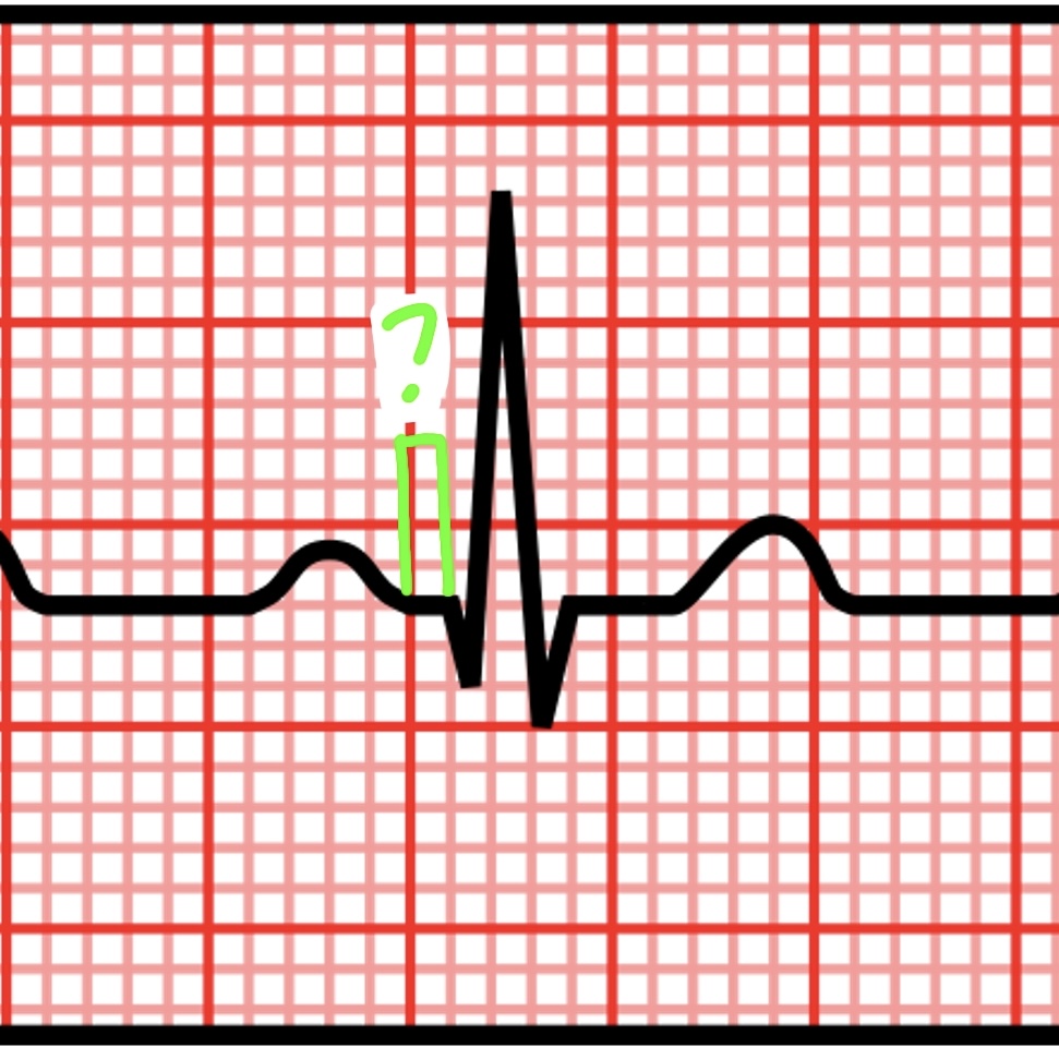
P wave
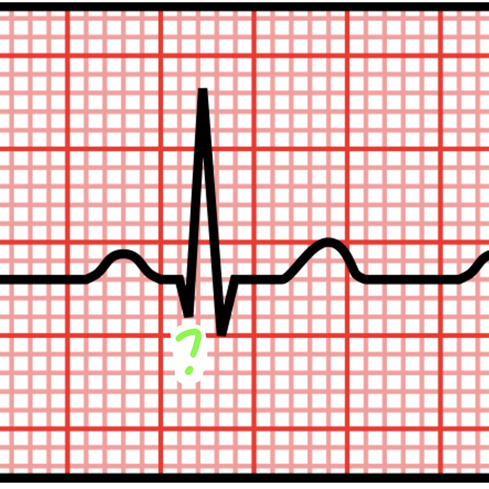
Q wave
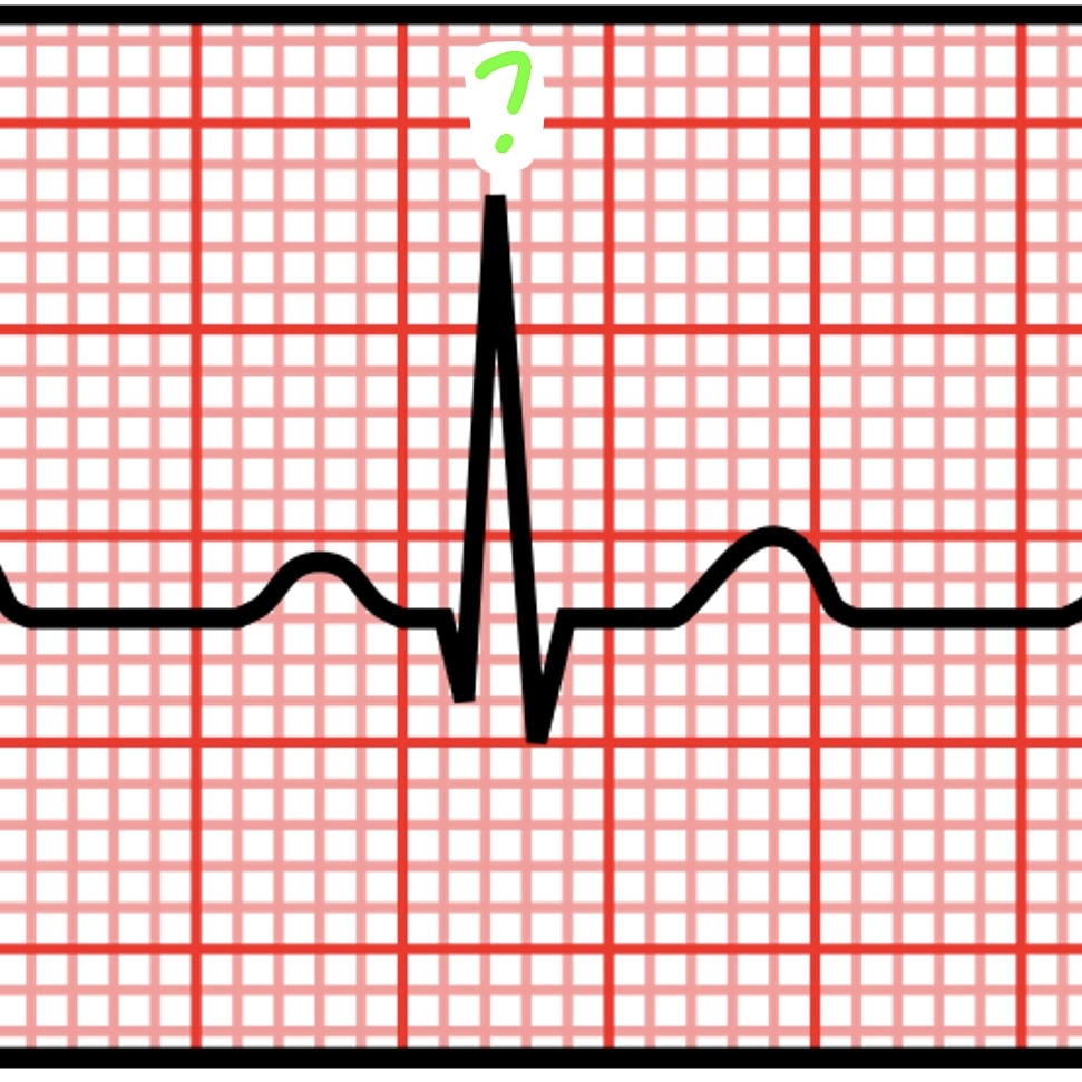
R wave
S wave
T wave
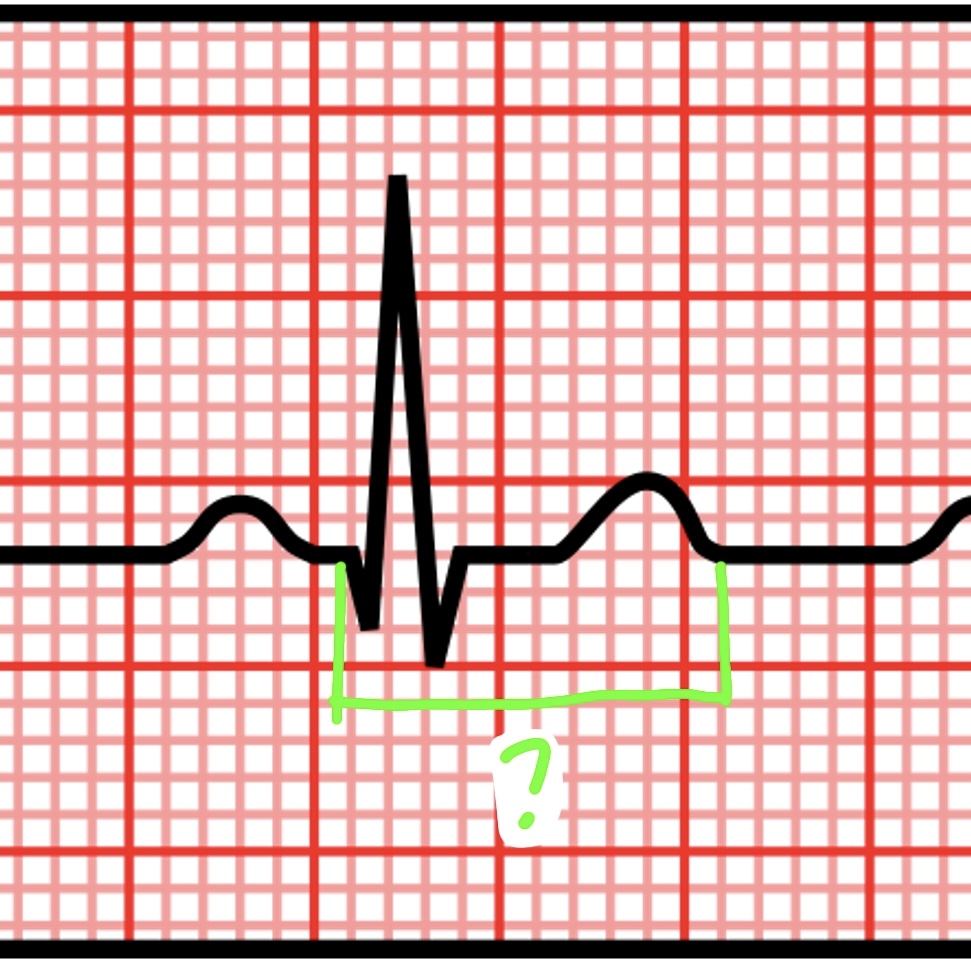
PR interval
QRS complex
ST interval
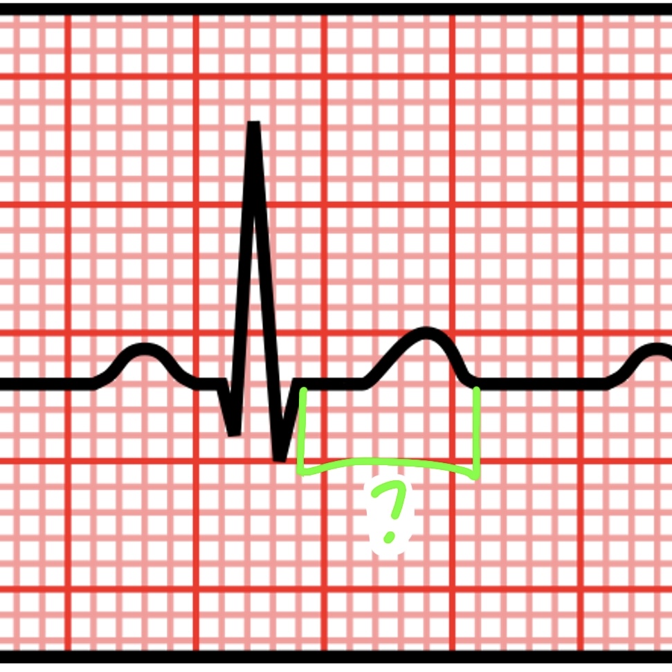
ST segment
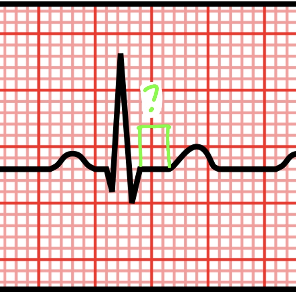
PR segment
QT interval