Sensory Systems Cue Cards
1/71
There's no tags or description
Looks like no tags are added yet.
Name | Mastery | Learn | Test | Matching | Spaced |
|---|
No study sessions yet.
72 Terms
What is sensation and what is the mechanism?
Transduction of a physical stimulus into a biological neuron (changes in membrane potential), which involves a sensor leading to activation of:
Chemically gated ion channel
Mechanically gated ion channel GPCR – secondary messenger system (or a variation of one of the above)
Sensor activity → change in membrane potential
Δ membrane potential → neurotransmitter release
What is the difference between olfactory and gustatory sensation?
Olfactory = smell, gustatory = taste
Olfactory receptor neurons are neurons, thus the sensory receptors are found on the dendrites of the neuron
Gustatory sensation is with a sensory receptor cell, which synapses with a neuron
What are olfactory receptors detecting and where are these receptors located and what is the pathway?
Detection of chemicals in the air we inhale
Receptors for olfaction are sensory receptor neurons
In the roof of the nasal cavity we have a layer of dendrites of olfactory neurons, this essentially connects to the ganglia brain cells the olfactory bulb which connects to the brain stem
The cribiform plate is a bone with little holes (perforations), that allow for axons to transit from the olfactory cavity to the olfactory bulb
The cranial nerve is the olfactory nerve
What is the pathway of receptor activation in olfaction?
Activates G-Protein coupled receptor, creating cyclic nucleotides
Chemical binds to the receptor, activates the G protein
cAMP builds up inside the olfactory receptor neuron
Activates cyclic nucleotide gated channel (non-specific cation channel), allowing for the movement of cations (calcium and sodium entering the cell and potassium leaving)
This changes the membrane potential, depolarising the cell and generating action potentials
Those action potentials project to the olfactory bulb, sending information to the brain
Increase of calcium within the cell also increases calcium sensitive processes within the cell
What allows for variation in an olfactory receptor?
High sequence variation along the 7 transmembrane spanning domains (amino acid sequences within the domains) allows for receptors for a wide variety of odorants
How can odorants bind to olfactory receptors if they are often hydrophobic but the receptors are embedded into a mucosa?
The cilia of the dendrites of the olfactory receptor neurons are forming a carpet within the mucosa (lining the olfactory cavity)
There is an interface between the air and the mucus, the odorant must dissolve into the mucus before it can bind to the olfactory receptor
The odorant is often hydrophobic meaning it doesn't dissolve well, so it must bind to a binding protein:
OBP – Odor binding protein
Binds to the hydrophobic odorant and allows it dissolve into the mucosal layer to diffuse towards the sensory receptor
How do odorant systems allow you to become partially desensitised to a smell?
Secondary messenger systems from g protein activation are relatively slow and their effects last for minutes:
The secondary messenger systems are the reason why you can become partially desensitised to a smell after being in a smelly room for a minute
What is the ORN and what does it allow for?
ORN – discharge rate of ions is odorant concentration dependent
The activity of olfactory sensory neurons is dependent on the concentration of the odorant in the environment
The nose doesn't just detect the presence, it detects the concentration
More concentration = greater response to stimuli (up to a max point)
How does the olfactory system distinguish different odorants?
Odorants can bind to multiple receptors with different affinities.
The sum of odorant sensory neuron activity helps us identify different kinds of odorants
What is the structure of the olfactory bulb and it’s projections
The olfactory bulbs have balls called glomeruli which have the dendrites of the post-synaptic neurons
Axons do not project to the closest glomeruli
Olfactory sensory neurons of the same family project to the same glomeruli
Glomeruli are coded by the odorants they are sensitive to
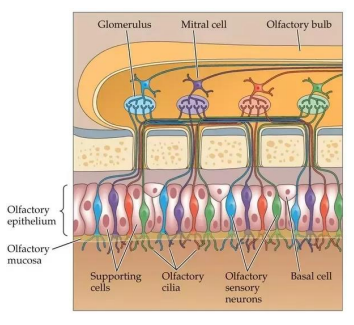
What are the 6 layers of the olfactory bulb?
Olfactory nerve layer (ONL)
Glomerular layer (GL)
most superficial
Contains synapses between the olfactory sensory neurons and the mitral or tufted cells
External plexiform layer (EPL)
fibre layer
Mitral cell layer (MCL)
Contains cell bodies of the mitral cells
mitral cells form synapses with granule cells (creating circuits)
Internal plexiform layer (IPL)
Fibre layer
Where axons and dendrites of the mitral cells and glomerular cells are forming small circuits
Granule Cell layer (GCL)
What are the three cell types within the olfactory bulb and what is the connection between them?
Mitral Cell
Second order sensory neurons
Axons form olfactory tract
Tufted cells
Granular cells
Inhibitory interneuron, within the olfactory bulb
Granule cells dendro-dendritic synapse with secondary dendrites of mitral cells.
Lateral inhibition (GABAergic) of neighbouring Mitral cells
Suppression of noise – sharpen the output of the glomerulus.
Mitral cells activate granule cells
Via glutmate
How does lateral inhibition occur which underlies odour discrimination?
It occurs in the dendro-dendritic synapse between mitral and granule cells
High magnification of the synapse between the granule and mitral cell, two way communication between cells
Activity in the mitral cell will release glutamate onto the granule cell which is excitatory
This drives GABA release from the granule cell onto the mitral cell
This is an autoinhibitory circuit
The granule cell also has synapses on the adjacent mitral cells, inhibiting multiple mitral cells
What are the five distinct taste sensations?
Bitter
Sour
Sweet
Savoury/umami
Salty
What is the structure of taste buds, papillae and taste receptor cells?
Taste receptor cells (TRC) are located within taste buds that line the crevice of papillae
Three different shapes of papillae, located in distinct locations of the tongue.
Taste buds are composed of 10- 150 TRC
TRC project microvillae to the apical surfaces of the taste bud
forming the taste pore
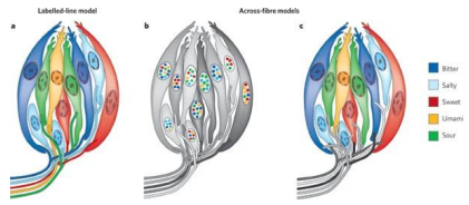
What are the two theories in how taste qualities are encoded in taste receptor cells?
Labelled-line model
each TRC is programmed to discriminate one flavour sensation
Across-fibre models
Two versions
each TRC can discriminate multiple flavours, but are tuned for different preferences.
Each TRC can only discriminate a distinct flavour, but afferent axons connect to multiple TBC and code for flavour in mixed modalities.
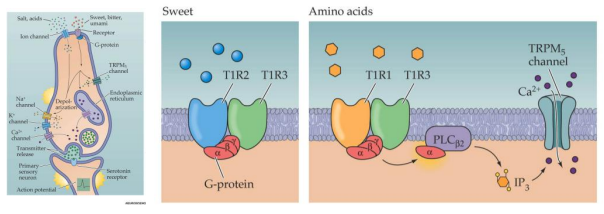
What is the structure of taste receptors?
Taste receptors are heterodimers
Taste receptors are either G-coupled protein receptors or chemically gated ion channels that detect specific chemicals in food, contributing to the sensation of taste.
How does taste receptor signal transduction work?
The taste receptor is a sensory cell not a neuron
Forms synapses with the first order sensory neuron
Either a GCPR or a chemically gated ion channel
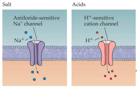
Salt/acids:
Activate ligand gated ion channel
Sweet, bitter, umami:
Activate second messenger system (GCPR)
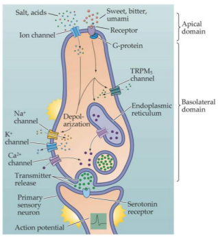
How does signal transduction in salt and acid flavours occur?
Chemically gated ion channels
How does signal transduction in sweet, bitter and umami flavours occur?
G-coupled protein receptors that activate second messenger systems. Umami and sweet are dependent on the presence of certain G-protein dimers
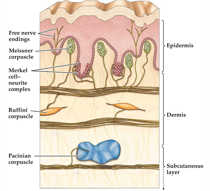
What kind of skin is best for the somatosensory system of the skin?
Most of the body is covered by hairy skin. The palmar surface of the hands and the soles of the feet are covered by glabrous skin, with skin ridges a prominent feature.
What are the four types of mechanoreceptors in glabrous skin?
There are four types of mechanoreceptors in glabrous skin.
Meissner corpuscles and Merkel complexes are close to the surface
Ruffini organs and Pacinian corpuscles are deeper in the skin.
These receptors are innervated by large myelinated axons with cell bodies in the dorsal root ganglia.
Transmission of this information to the brain generates our conscious experience of touch.
What is the role of Pacinian corpuscles and how do they work?
Corpuscles detect force and have nerve structures inside:
Force comes through the skin and finds it's way to the receptors
This mechanically opens the receptors, enabling sodium ions to enter the cell, depolarising the cell and generating an action potential
What are skin receptors detecting?
Skin receptors are detecting a change of shape in the skin
Detect until it is damaging
What is PIEZO-2 and why is it important in somatosensory transduction?
The molecular machine that transduces force into changes in membrane voltage
Forms a channel in the membrane and responds to force by a subtle conformational change that opens the receptor
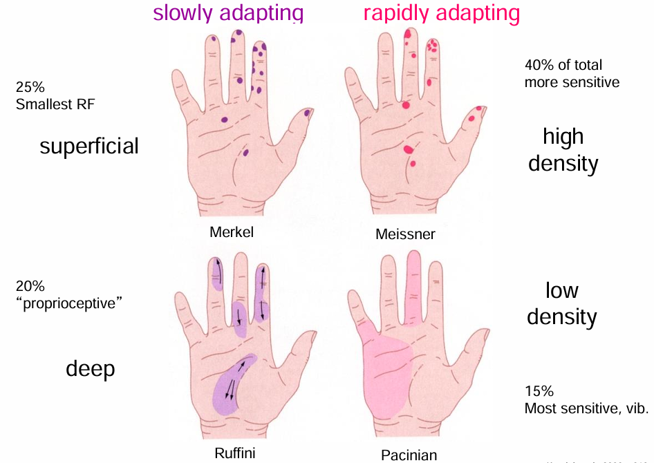
What are the slowly adapting mechanoreceptors and what is their effect?
Merkel complexes are found at the tips of the epidermal ridges where they respond to indentation (makes up 25% of mechanoreceptors)
Ruffini endings are found in the upper dermis, have a sustained response to skin movement and stretch (important for grip) (makes up 20% of mechanoreceptors)
What are the rapidly adapting mechanoreceptors and what is their effect?
Meissner receptors are found near the skin surface and have a transient response to skin movement (makes up 40% of mechanoreceptors)
Pacinian receptors are located deep in the dermis and hypodermis and have a transient response to vibration (makes up 15% of mechanoreceptors)
What is the relationship between the mechanoreceptors and their receptor fields?
Deep receptors have larger receptive fields (with Merkel cells having the smallest receptor field)
Describe the different types of peripheral nerve axons?
Peripheral nerve axons can be either:
efferent (carrying signals from the CNS)
afferent (carrying signals to the CNS)
Although mixed in the peripheral nerves, they have separate pathways (the roots - either motor or sensory) connecting to the CNS.
The cell bodies for the sensory nerves sit in the dorsal root ganglion
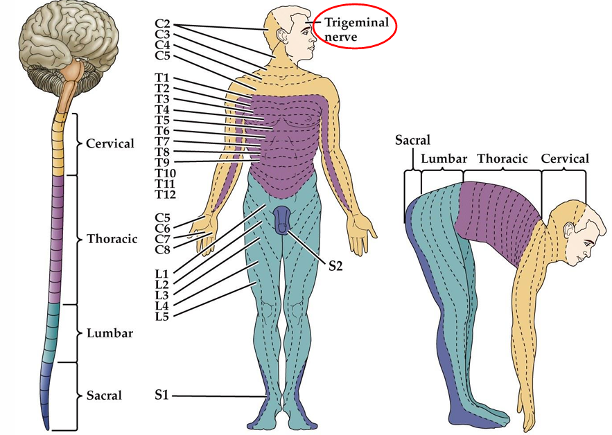
What are dermatomes?
Where spinal nerves project to
How does information from mechanoreceptors convey information to the brain?
Somatosensory afferents convey information from the skin surface to central circuits (via a single neuron), then using two more neurons, up to the brain:
First neuron covers most of the distance until the brainstem
Then a new neuron in the dorsal column nucleus crosses over and goes up the brain
the third neuron in the thalamus which takes the information into the cerebral cortex
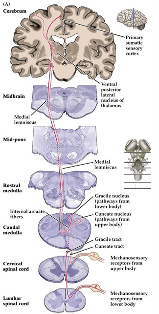
What do the dory primary somatosensory cortex regions each contain?
All four regions have a complete map of the body circuit (four separate maps of the same thing)
Can cortical territory change or is it fixed?
Cortical territory is plastic
Functional changes can occur in the somatic sensory cortex following amputation of a digit (boundary shifts in the representations of the cortex)
Functional expansion of a cortical representation can also occur from undertaking a repetitive behavioural task
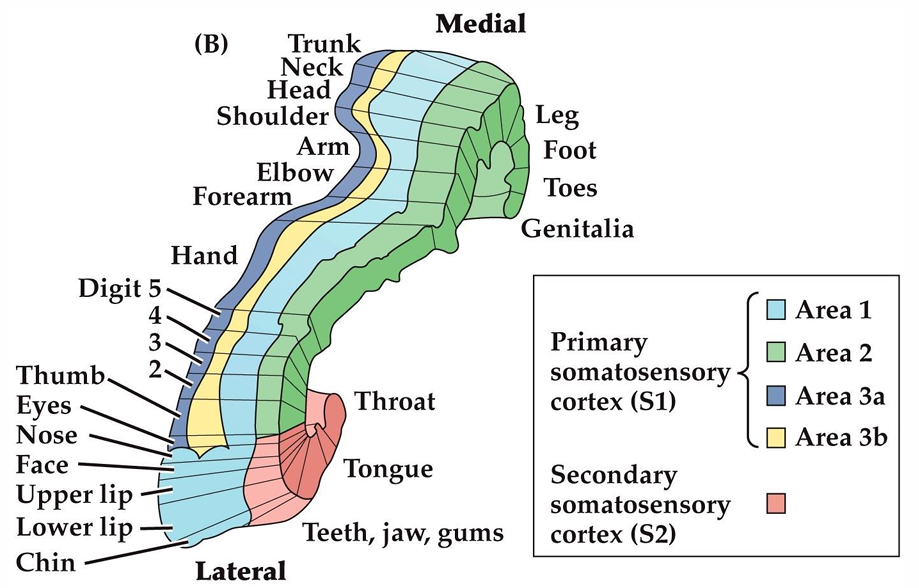
What is the structure of the eyeball?
Protective layer:
Cornea (transparent) and sclera
Vascular layer (nutritious layer providing blood supply to eye):
Choroid
Ciliary body
Iris
Inner layer (nerves):
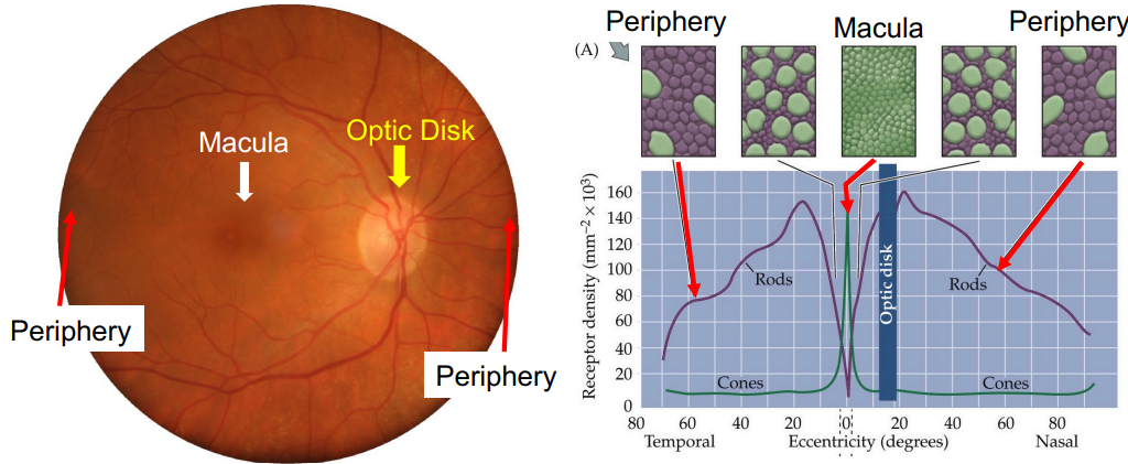
Retina (inner surface of the eyeball (5/6 of the eye)
The macular is a very important region of the retina that allows you to see centrally
Optic nerve contains the axons of the output neurons of the retina (retinal ganglia cells)
What limits visual acuity?
Optical factors
Pupil Size
Clarity of optical media (cornea must be clear)
Cataracts, Corneal Opacities
Refractive errors -> blur (size problems in the eyeball)
Myopia, Hypermetropia, Astigmatism, Presbyopia
Neural factors
Ability of the neurons to connect and process information
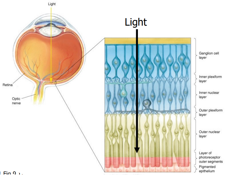
What is the structure of the retina
Retina has many layers
Light passes through all of the layers of the retina before it reaches the photoreceptors
This is because of the cells that are underneath the retina keep the photoreceptors alive and nourished
The retina is made up of many types of neurons that form many different layers, allowing for neural processing of information
What are the two types of photoreceptor cells that detect light and what is their function?
The cells that detect light: rods and cones
Rods
night vision
very sensitive
one type
no colour vision
100 million
are not in the macula (fovea)
Cones
day vision
less sensitive
three types
colour vision
5 million
density of cones in the retina determines how well you can see
How do photoreceptors enable vision?
Contain photopigments that are activated by light.
Rods contain Rhodopsin
Light sensitive protein
Cones contain one of three different cone-opsins
Opsins bind to vitamin A (11-cis Retinal - kinked) inside of the rhodopsin (capturing the light)
causes a conformational change in the cis-vitamin A (9changes to trans)
This activates the light sensitive protein and leads to a cascade that enables vision
How do photoreceptors release neurotransmitters?
In the retina, neurons do not fire action potentials
Neurotransmission occurs purely based on the membrane potential
Photoreceptors are hyperpolarized by light
Use glutamate as their neurotransmitter which are being released continuously
Depolarisation = more neurotransmitter release
Hyperpolarisation = less neurotransmitter release
Respond to light with graded changes in membrane potential (not Action Potentials)
What is the mechanism by which photoreceptors effect membrane potential?
Photoreceptors have cyclic GMP gated sodium channels on their membrane
In the dark:
cGMP gates a sodium channel causing continuous influx of sodium ions.
Causes depolarization of the cell.
In the light:
cGMP breaks down to GMP
cGMP no longer gates the sodium channels
Flow of Na ions ceases
Cell is hyperpolarized
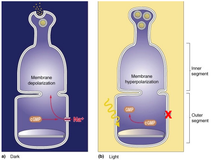
Specifically how does light lead to phototransduction?
Light activates rhodopsin (by changing the shape of the vitamin A)
Initiates a G protein cascade that ultimately leads to the closure of cGMP gated sodium channels.
Rhodopsin gets activated by light -> Activates G protein called Transducin -> PDEnzyme is activated ->breaks down cGMP -> Closure of sodium channels -> hyperpolarization
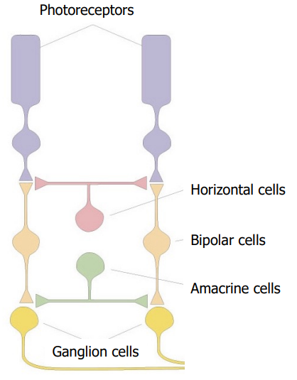
How is information about light passed through the retina?
Retinal “Through” pathway:
Photoreceptors synapse onto bipolar cells which synapse onto the output neurons the ganglion cells
Modulated by Lateral interactions:
Horizontal cells
Amacrine cells
The modulation enables us to see colour, flickering lights, motion etc
What are the major features of bipolar cells?
10 different types:
1x Rod bipolar cell
9x cone-bipolar cells.
Important for spatial vision, & colour vision.
Use glutamate as their neurotransmitter
Bipolar cells receive input from photoreceptors and form output synapses with retinal ganglion cells
What are ganglion cells?
Ganglion cells are the output neurons of the retina
Ganglion cells respond to light by either increasing or decreasing their action potential firing rate
Many different types (at least 40): ON, OFF, M and P
Each ganglion cell tells the brain something different
Release Glutamate
Ganglion cell responses:
Increase or decrease in firing
Transient or sustained response
Fire action potentials to communicate with higher brain centres
Their axons form the optic nerve
Visual information is passed to higher cortical centres in parallel.
What is the receptive field of a ganglion cell?
the area of retina that when stimulated with light changes the cell’s membrane potential.
The response of the ganglion cells to light depends on where in the receptive field the light falls
This allows us to see edges exceptionally well
What are horizontal cells?
Lateral inhibition cells that modulate
Input from photoreceptors.
Provide output onto photoreceptors.
Use inhibitory neurotransmitter GABA.
Respond to light by hyperpolarizingW
What are amacrine cells?
Axon less cells of which there are many different types
Important for lateral inhibition
For the most part ACs are considered inhibitory cells (release inhibitory NTs: glycine, GABA)
What are the output neurons and output regions of ganglion cells?
Output neurons of the retina:
M (parasol) ganglion cells
Magnocellular = large
Large receptive fields
Motion detection, flicker and analysis of gross features.
P (midget) ganglion cells:
Parvocellular = small
More numerous
Visual acuity and colour vision
Output targets of ganglion cells:
Many brain regions
Majority target the lateral geniculate nucleus (thalamus)
Which sends it to the correct region of the primary visual cortex
What is the overall visual pathway?
Retina
Optic nerve
Lateral geniculate nucleus
Optic radiations (white matter tract)
Primary Visual cortex (V1)
What is the optic chiasm and what is its significance?
The fibres from right and left optic nerves combine to form the Optic chiasm.
Lies at the base of the brain, anterior to the pituitary
Fibres cross at Optic chiasm.
Right visual hemifield is “viewed” by the left hemisphere.
Left visual hemifield is “viewed” by the right hemisphere.
Note: visual hemifield is not the same as that eye, it's the side of the eye
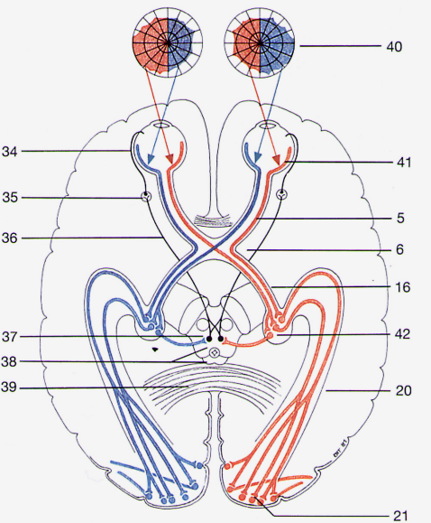
What is the lateral geniculate nucleus?
LGN is part of the thalamus
Functional streams of information from both eyes passed to LGN
Six layers (numbered 1-6)
Contain 2 types of cells
Magnocellular cells (Layers 1,2)
Receive input from M (parasol) ganglion cells, one from each eye
Parvocellular layers (layers 3-6)
Receive input from P (midget) ganglion cells, two from each eye
What are optic radiations?
White matter tract that goes from the thalamus to the occipital lobe
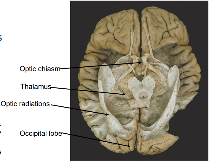
What is the primary visual cortex and what part of it encodes information from where?
The primary visual cortex is a section of the occipital lobe, located on either side of the calcarine fissure
Specific information coming from the retina targets a very specific area of the primary visual cortex
The posterior part of the PVC encodes information from the macula, so thus is the most important part of the PVC
The further anterior you get within the PVC, the more peripheral the vision it encodes
How is input from the LGN received and integrated in the primary visual cortex?
Input from the LGN to the primary visual cortex is inputted into layer 4C
Function organization of V1 (high order)
Arranged in Columns:
Cells arranged vertically comprise a functional unit.
Orientation columns
Neurons respond best to a bar of light at a specific orientation
Ocular dominance columns.
Refers to function of neurons in Layer 4C
Two LGNs-receive input from each eye (but segregated into different layers)
M type GC/LGN input to Layer 4Ca
P-type GC/LGN input to Layer 4Cb
Mixing of information from each eyes occurs in layers IVB and Layer III.
What are the two large cortical streams of visual processing?
dorsal pathway (Where?)
area MT
ventral pathway (What?)
area V4
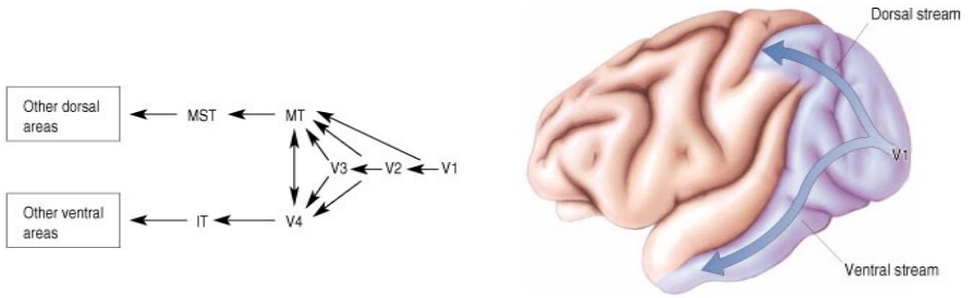
What is area MT and what is it important for?
Area MT: middle temporal lobe is an area in the dorsal pathway that is specialized for processing object motion.
Receives retinotopic information from a number of cortical areas including V2 & V3
Receives input from cells in Layer IVB of the primary visual cortex. (ie M-type GCs/LGN)
What is area V4 and what is it important for?
Area V4 is located in the ventral pathway and is important for processing color and form. It plays a critical role in visual perception by integrating color information with shape recognition.
Area V4:
Receives input from the blob and interblob regions of the primary visual cortex via V2.
Neurons in V4 have large receptive fields that are both orientation selective and colour selective.
What is area IT and what is it important for?
Area IT (inferotemporal cortex) is located in the ventral pathway and is crucial for processing visual object recognition and complex visual stimuli, including faces and other intricate shapes. It integrates information from earlier visual areas to contribute to high-level visual perception.
A major output of Area V4.
Neurons respond to a wide variety of abstract shapes and colours.
Important for visual memory and perception.
Important for perception of faces.
What is being detected in hearing?
Wavelength (pitch)
Amplitude (loudness)
Waveform (tone / timbre)
What is the structure of the middle ear and what is its purpose?
Tympanic membrane absorbs the sound waves coming into the ear
Three middle ear bones are a system of levers that put force on the fluid filled inner ear
to solve the impedance mismatch (sound energy is absorbed by water)
What is the structure of the inner ear and what is its purpose?
Three fluid filled chambers: scala tympani, scala vestibuli and scala media
Basler membrane is made of collagen fibres
Inner hair cells are sensory cells that get activated when sound waves are travelling through the inner ear
Convert sound waves into electrical signals
Outer hair cells are receiving motor and effector information instead of sensory information that can modulate, and stiffen the basal membrane
Hair cells are not hair, they are cilia and are only called hair cells because they protrude from the cell membrane
The hairs stick into the tectorial membrane
How do waves travel along the cochlea?
Stapes produces a pressure wave that travels along and causes the round winder to bounce around. The scala media and basilar membrane responds to that movement and gets deflected
How much deflection depends on the frequency of the sound and where you are on the membrane
What is spectral decomposition?
Different parts of the basilar membrane resonate with different frequencies, so the sound travels along until it reaches the region it resonates with
Different hair cells get activated differently by different frequencies
Where you activate the basilar membrane will depend on the frequency of the sound
Very broad, with broad peaks
How does the fact that the basilar membrane is moving activate the hair cells?
System of two levers, basilar membrane and tectorial membrane
Because the levers have different points of rotation, when they move they cause the hair cells to bend
Thus, sound detection is mechanotransduction (it's mechanical)
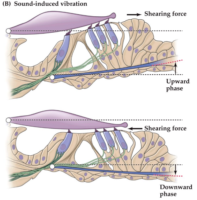
How does mechanoelectrical transduction occur via hair cells?
Cilia have tip links which attach them to each other
When the tip links move, it mechanically opens K+ channels allowing K+ to flow into the cell
Potassium ions entering the cell causes depolarisation
This leads to release of neurotransmitter that activates the auditory nerve which goes to the brainstem for information processing
Why does K+ go into the cell in mechanoelectrical transduction?
Because it is an exception, potassium has a higher concentration outside the cell
Potassium has a high concentration because there is a stria vascularis membrane that pumps potassium in and takes sodium out
Uses a lot of ATP
This pump not working causes deafness as this concentration gradient and channel is vital for hearing
At 100 hertz, potassium enters the cell and releases neurotransmitters (probably glutamate) from the synaptic vesicles, this would probably occur at 100 times per millisecond. What happens for a 20,000 hertz sound?
Instead, the brain interprets the location on the basilar membrane to detect the frequency
Although at a low frequency, these mechanisms will be occurring less rapidly, it is purely the location on the basilar membrane that detects the frequency of the sound
What is the receptor field of auditory nerve fibres?
The receptor field for a single hair cell is quite broad, but they do have a peak of maximal activation
What is the central auditory pathway dorm the cochlea to the brain?
Sound travels from the cochlea through the auditory (cranial) nerve
Goes to the brainstem targets, then to the medial genicular thalamus
Projects from here to the auditory cortex which is in the temporal lobe
There is a lot of crossing over and sharing information in the auditory pathway.
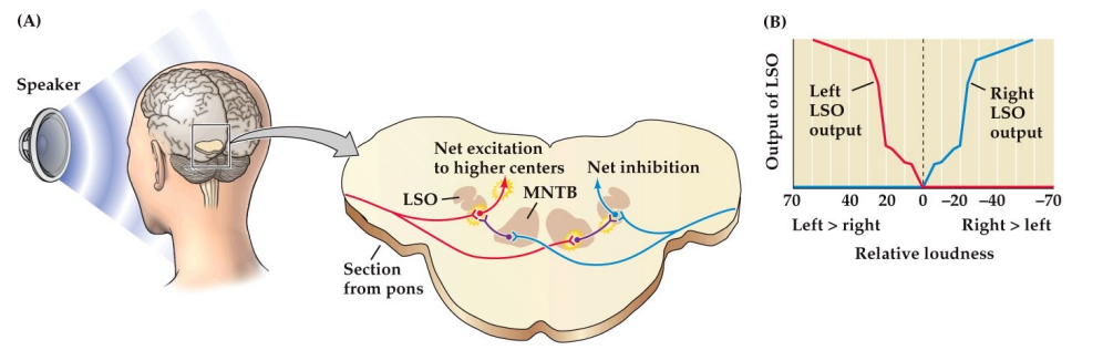
How does the LSO compute the location of a sound?
By inter-aural time differences
Detects and exaggerates sound loudness to judge where it is from
Gets exaggerated through an inhibitory pathway where the other side then gets inhibited
What is the purpose of the brainstem and where is the vestibular cochlear nerve?
All tracts (bundles of axons in the CNS) that travel between the brain and the spinal cord must pass through the brainstem
The brainstem contains vital integrative centres for vital functions (cardiovascular and respiratory control)
The brainstem contains the central nuclei for the cranial nerves 3 to 12.
Vestibular cohlear nerve is the 8th nerve
The brainstem contains several small nuclei with hugely divergent projections to the rest of the brain that determine conscious state
The cerebellum joins the brainstem by the cerebellar peduncles
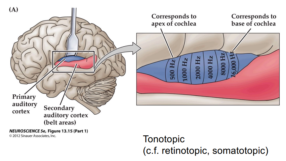
How is the auditory cortex mapped?
The auditory cortex has different regions for different frequencies (mapped in a tonotopic way)
anterior = low (apex of cochlea)
posterior = high (base of cochlea
Cortical representation of auditory input is a little asymmetric when comparing left and right
Could be because the left hemisphere is so dominant in the processing of language
The cortical territory is plastic.