Bio1110 Human Cardiovascular system
1/67
There's no tags or description
Looks like no tags are added yet.
Name | Mastery | Learn | Test | Matching | Spaced | Call with Kai |
|---|
No analytics yet
Send a link to your students to track their progress
68 Terms
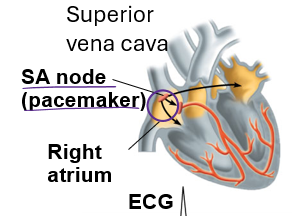
Right atria
located at the top right of the heart (looks like its on the left) receives blood from the upper and lower body and the heart muscle itself
Left atria
located in the top left of the heart, receives blood from the lungs
right ventricle
Located in the bottom right of the heart, sends blood to the pulmonary
Two atrioventricular valves
connects the left atria and ventricle together, and another on the right that connects the right atria and ventricle together, the right AV valve allows deoxygenated blood to flow from the atria to the ventricle, same for the left but its oxygenated blood
Semilunar valves
valves that allow blood to enter or leave the heart, the right one connects to the lungs and left one connects to the rest of the body
What side is the blood deoxygenated?
the right side
What side is the blood oxygenated?
the left side
1st order of blood flow for the heart
deoxygenated blood enters the heart through superior vena cava (upper body) and inferior vena cava (lower body), and the coronary sinus (the hearts) and drains into the right atrium
2nd order of blood flow for the heart
deoxygenated blood is sent through the AV valve into the right ventricle and then pumped out to the lungs through the semilunar valve
3rd order of blood flow for the heart
now oxygenated blood returns to the heart through pulmonary veins to the left atrium
4th order of blood flow for the heart
AV valve opens and lets in oxygenated blood into the left ventricle, and is then pumped out through the semilunar valve to the rest of the body, and the cycle repeats
What happens when blood tries to go back into the atrium from the ventricle?
The AV valve snaps shut
Diastole means …
whatever its describing is relaxed
Systole means …
whatever its describing is contracting
1st step of the cardiac cycle
Atrial and ventricular are diastole, so both are relaxed, ventricles begin to fill and the semilunar valve is closed and AV valves are open, lasts 0.4 seconds
During the first step of the cardiac cycle when everything is diastole the heart is using …
negative pressure
2nd step of the cardiac cycle
Atrial systole(contracts) and ventricular diastole, fills the ventricles completely, lasts 0.1 seconds
During the second step of the cardiac cycle when the atria contracts the heart is using …
positive pressure
3rd step of the cardiac cycle
Atrial diastole and ventricular systole, ventricles contracts which produces positive pressure, semilunar valves are open and blood is leaving the system, lasts 0.3 seconds then returns to the heart and step 1
Heart rate =
cardiac cycles/min
Cardiac output =
volume/min
How does the heart pump blood in only one direction?
the AV and semilunar valves prevent blood from being pumped the wrong direction, they act as one way gates
Do the atria and ventricles contract at the same time?
No, there is one step where neither of them are contracting and during the other two stages its either one or the other
How does the heart produce its familiar “lub-dub” heart sounds
lub sound happens = when the AV valves close and Dub sound = when the semilunar valves close, the lub/the first beat is louder because the semilunar valves is thinner and snaps closed quickly
Electrical impulse travels through …
gap junctions in heart tissue
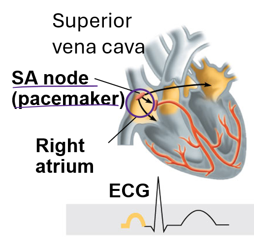
1st step of setting the heart rhythm
signals from sinoatrial node spread through the atria and they contract
2nd step of setting the heart rhythm
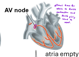
Signals are passed to the Atrioventricular nodes which act as a relay after a small delay (allows time for atria to finish contraction and delivering all the blood)
3rd step of setting the heart rhythm
Bundle branches pass signals to the heart apex
4th step of setting the heart rhythm
Signals spread throughout the ventricles
What is the heart apex?
the bottom of the heart, so the contraction comes from the bottom of the ventricle, away from the semilunar valve to make sure we empty out ALL the blood
Where are the Sinoatrial (SA) nodes located?
Where are the Atrioventricular (AV) nodes located?
The SA node is referred to as the …
pacemaker, because it generates the first electrical impulse of every heartbeat, sets the heart rate
Anatomy of arteries and function
three layers, walls are thick, strong, and elastic, endothelium = simple squamous epithelium, smooth muscle controls diameter and path of blood flow
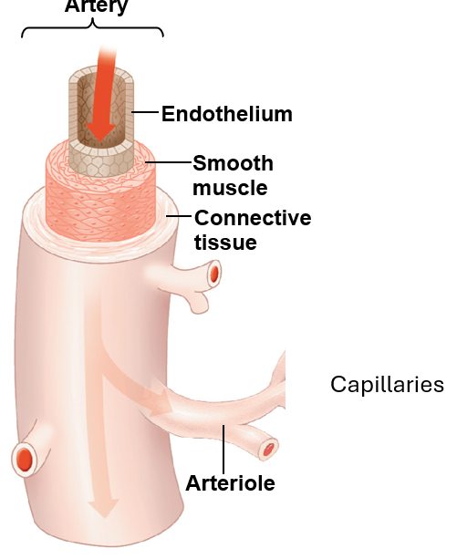
Arterioles control pressure in …
arteries (vasoconstriction, vasolidation
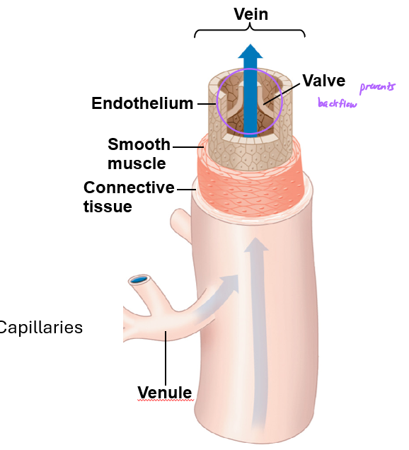
Anatomy of veins and function
three layers, walls are thinner than arteries (about a third), smooth muscle, endothelium, have valves
What do coronary blood vessels do?
supply the hear with blood, keeps the cardiac tissue alive, the heart takes pressure so its not a good exchange area so it has its own capillary beds
What is a portal system?
a special circulatory pathway where blood passes through two capillary beds before returning to the heart
Example of portal system in humans
The hepatic portal vein takes blood from the small intestines to the liver, takes O2 but it also takes things like sugars, vitamins, amino acids, minerals, or anything else absorbed from the small intestines to the liver
What happens at the first little hump of the ECG
Signals from SA node spread through the atria and they contract
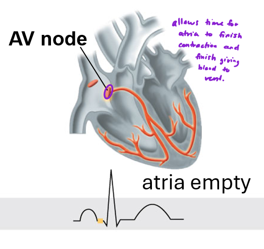
What happens at the first flat line of the ECG
signals are passed to the AV node which acts as a relay after a small delay
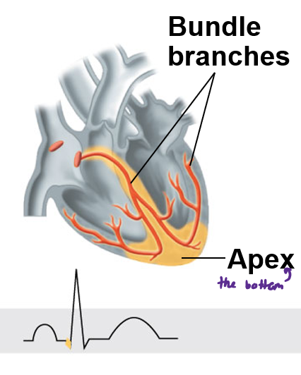
What happens at the first drop line of the ECG
bundle branches pass signals to the heart apex
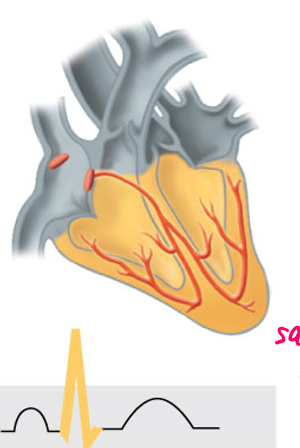
What happens at the sharp point of the ECG
signals spread throughout ventricles and it contracts
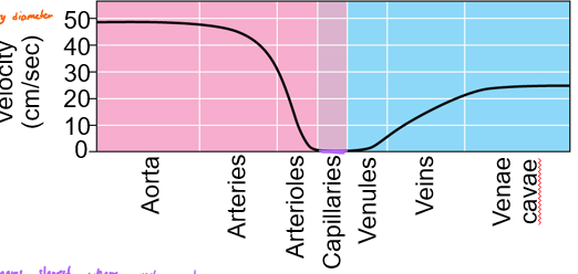
How does blood pressure change throughout the circulatory system?
it starts high and as it travels away from the heart it decreases, is really low once it passes through the capillary beds
How does velocity change throughout the circulatory system?
highest after leaving the heart, drops as it reaches the capillary beds to zero then rises as it leaves (not as high as when it first leaves the heart)
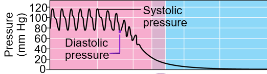
What is systolic blood pressure?
pressure that stretches artery walls, represents the pressure in arteries when the heart contracts (usually greater than 120 mm Hg)
What is diastolic blood pressure?
when the arteries aren’t being stretched, they relax, represents the pressure in your arteries when your heart is relaxed between beats (usually less than 80)
How does blood pressure read?
systolic / diastolic so ideal is 120/80
Does pressure of the blood ever reach zero?
No, smooth muscle squeezes to cause systolic pressure
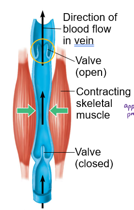
What mechanisms assist veins with the return of blood to the heart?
an open valve, and contracting skeletal muscle
How is distribution of blood regulated by arterioles?
by dilation of arterioles, when they dilate resistance drops and blood flow increases; when they constrict resistance rises and blood flow decreases
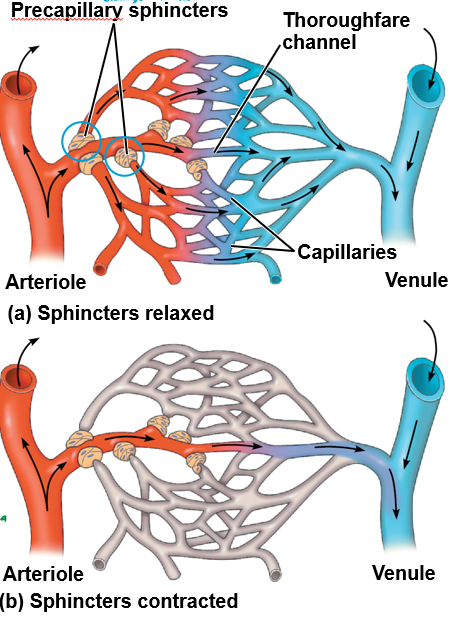
How is the distribution of blood regulated by precapillary sphincters?
they are rings of smooth muscle located right where a capillary branches off an arteriole, when they dilate, capillary opens and blood flows in, when they constrict the capillary is temporarily closed
Overall, precapillary sphincters and arterioles distribute blood based on …
metabolic need
What organs always have blood supplied heavily to their capillary beds?
The brain, heart, kidneys and liver
What percent of capillaries are used at a given time?
5-10%
Examples of things that don’t get constant blood flow
the digestive system (we aren’t eating all the time so nothing to digest) and skeletal muscles ( we aren’t using every muscle all the time)
How does fluid move between a capillary and the interstitial fluid?
pressure pushes the water out of the vessel, blood pressure is greater than osmotic pressure in the capillaries
What two factors are important to understanding which direction the fluid will move between a capillary and the interstitial fluid?
Hydrostatic pressure (the pushing force from the blood pressure) and Osmotic pressure (the pulling force that brings water back into the vessels)
What happens to the extra water that is lost?
it gets pushed into the interstitial fluid, it’ll go back and forth, which is bad because it will eventually become less liquidy
How does water get back in the bloodstream?
not through the interstitial fluid because it will be less liquidy, instead lymphatic vessels pick it up and return it to the blood as lymph, creates a one-way loop for water
What else do Lymphatic vessels do?
collect fluid from body tissues and transport lipoproteins from the intestine to the bloodstream
What are the components of blood?
Cellular elements (Leukocytes, Platelets, and Erythrocytes) and Plasma
What are the major functions of plasma?
water = a solvent, ions = osmotic balance, pH buffering, and regulation of membrane permeability, plasma proteins
What do Leukocytes do?
white blood cells, they are for defense and immunity, we have the least amount of these
What do Platelets do?
blood clotting, we have a general amount of these
What do Erythrocytes do?
red blood cells, have hemoglobin, transport O2 and CO2, we have the most of these
What plasma proteins do we have?
Albumin = osmotic balance, pH buffering, Immunoglobulins (antibodies) = defense and immunity, Apolipoproteins = lipid transport, and Fibrinogen = blood clotting
Leukocytes include …
Basophils, Lymphocytes, Eosinophils, Neutrophils, and Monocytes