unit 2 exam practice: lymphatic, digestive, metabolism
1/63
There's no tags or description
Looks like no tags are added yet.
Name | Mastery | Learn | Test | Matching | Spaced |
|---|
No study sessions yet.
64 Terms
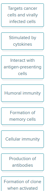
Drop each label into the appropriate box(es), indicating whether the label is associated with B cells or T cells (or both). Labels can be used more than once.
B: humoral immunity, prod of antibodies, stimulated by cytokines, formation of clone and memory cells
T: cellular immunity, interact with antigen-presenting cells (APC), target cancer cells and virally infected cells, stimulated by cytokines, formation of clone and memory cells
Lymph drainage is important for what functions?
Check All That Apply
Absorption of dietary fats.
Return of small proteins from tissue fluid to blood.
Regulation of osmotic pressure of blood.
Provision of nutrients to tissue cells.
Transport of foreign particles from tissue fluid to lymph nodes.
Absorption of dietary fats.
Return of small proteins from tissue fluid to blood.
Transport of foreign particles from tissue fluid to lymph nodes.
Class I MHC antigens are found in the membranes of (Click to select) all body cells except RBCs/ antigen-presenting cells, thymus cells, and activated T cells .
Class II MHC antigens are found in the membranes of (Click to select) antigen-presenting cells, thymus cells, and activated T cells/ all body cells except RBCs .
class i: all body cells except RBC
class ii: antigen-presenting cells, thymus cells and activated T cells
Antibodies binding to their antigen can activate complement. This, in turn, leads to opsonization of the antigen-antibody complexes. What is meant by opsonization?
Multiple Choice
Lysis of the cells bearing the antibody-antigen complexes.
Coating antibody-antigen complexes, making them more easily phagocytized by macrophages and neutrophils.
Clumping together of the antibody-antigen complexes.
Clumping of antibody-antigen complexes into insoluble substances.
Coating antibody-antigen complexes, making them more easily phagocytized by macrophages and neutrophils.
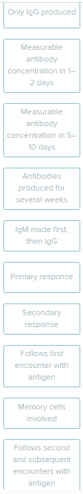
The labels describe either a primary or secondary immune response. Drop each label into the appropriate category.
Primary response: follows first encounter with antigen, IgM made first, then IgG, measurable anti-body concentration in 5-10 days
Secondary response: follows second and subsequent encounters with antigen, IgG produced, antibodies produced for several weeks, memory cells involved, measurable antibody concentration in 1-2 days
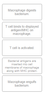
Place the events leading to T cell activation in the correct order
macrophage engulf bacterium
macrophage digest bacterium
bacterial antigens are inserted into cell membrane of macrophage along with MHC protein
t cell binds to displaced antigen/MHC on macrophage
t cell activated
A molecule called a(n) antigen/antigen/hapten/immunoglobulin is typically a large, complex molecule that stimulates an immune response.
A small molecule that can not stimulate an immune response on its own, but may cause an immune response when it binds to a larger molecule is called a(n) antigen/antigen/hapten/immunoglobulin
a. antigen
b. hapten
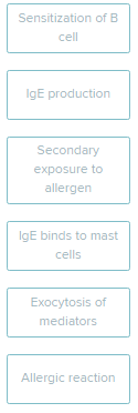
Put the steps that occur during an immediate-reaction allergic response in the correct order.
sensitization of B cell
IgE production
IgE binds to mast cells
secondary exposure to allergen
exocytosis of mediators
allergic reaction
The production of antibodies is the result of a ___ immune response, involving ____ lymphocytes.
An antibody is a_____ molecule produced by cells called _____ cells. These cells result from the proliferation of activated B cells.
Each antibody targets a specific _____, with the intention of destroying or neutralizing it.
Antibodies are Y-shaped molecules. The "stem" of the Y is part of the _______ region of the molecule. At the tips of the "arms" of the Y are the_____ regions that act as antigen-binding sites.
humoral, b
protein, plasma
antigen
constant, variable
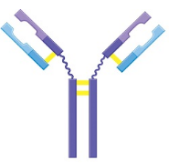
Fill in the blanks for the figure legend, indicating the identity of the different colored segments of the antibody molecule.
label each color
light blue: variable of light chain
dark blue: constant of light chain
light purple: variable of heavy chain
dark purple: constant region of heavy chain
For each description, indicate the correct group of lymph nodes.
Filter lymph from lower limbs, lower abdominal wall, external genitalia
pelvic lymph node
cervical lymph node
inguinal lymph node
supratrochlear lymph node
inguinal lymph node
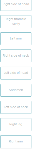
Consider each area of the body and the destination of its lymphatic drainage. Drop each label into the appropriate box. (right lymphatic duct or thoracic duct)
and left thoracic cavity
right lymphatic duct :
right side of head
right side of neck
right arm
right thoracic cavity
thoracic duct:
left arm
left side of head
abdomen
left side of neck
right leg
left thoracic cavity
List the function(s) of lymph nodes.
Check All That Apply
Filter lymph
Immune surveillance
Produce macrophages
Filter blood
Collect tissue fluid
filter lymph
immune surveillance
What is the role of skeletal muscle contractions in the flow of lymph?
Multiple Choice
It causes lymph fluid in nearby lymphatic vessels to be pushed further away from the upper thorax.
It pushes tissue fluid into the lymph capillaries due to increased interstitial fluid pressure.
It stops the flow of lymph as the lymphatic vessels are compressed.
It compresses lymphatic vessels, encouraging the lymph to flow toward the upper thorax.
It compresses lymphatic vessels, encouraging the lymph to flow toward the upper thorax.
In a typical blood capillary bed, the balance of hydrostatic and colloid osmotic pressures results in _____ occurring at the arterial end of the capillaries and ______ occurring at the venous end.
Overall, _____ from the plasma normally exceeds _______ ,resulting in the net formation of tissue fluid.
filtration
reabsorption
filtration
reabsorption
Delayed-reaction allergic responses are mediated by B cells.
True or False
false
explanation:
Delayed-reaction allergies require time to develop (approximately 48 hours) and involve the release of chemicals factors from T cells and macrophages.
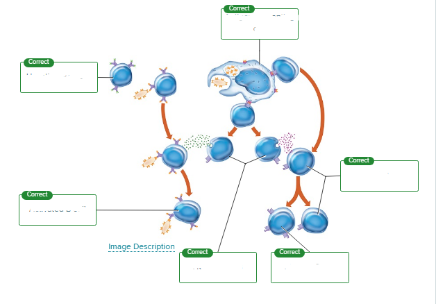
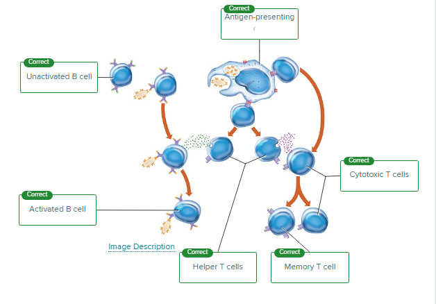
This figure illustrates the activation of B and T cells. Label the cells that are involved in the process.
unactivated B cell
activated B cell
antigen presenting cell
cytotoxic cell
helper T cell
memory T cell
What are lymphatic nodules?
Multiple Choice
Compact masses of lymphatic tissue
Encapsulated lymphatic organs
Regions of bone marrow that produce lymphocytes
Compact masses of lymphatic tissue
What term refers to any molecule that elicits an immune response?
Multiple Choice
Clone
Antigen
Cytokine
Antibody
Antigen
Match the description with the correct type of cytokine.
A table presents information under the following column headers: function and cytokines.
Block viral replication, stimulate macrophages to engulf viruses, stimulate B cells to produce antibodies, attack cancer cells |
interferons
Match the description with the correct type of cytokine.
A table presents information under the following column headers: function and cytokines.
Stops tumor growth, releases growth factors, causes fever (with bacterial infection), stimulates T and B cell differentiation |
tumor necrosis factor
Match the description with the correct type of cytokine.
A table presents information under the following column headers: function and cytokines.
Stimulate bone marrow to produce lymphocytes |
colony-stimulating factors
Match the description with the correct type of cytokine.
A table presents information under the following column headers: function and cytokines.
Control lymphocyte differentiation and proliferation |
interleukins
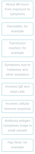
Click and drag each characteristic into the appropriate box, describing each type of hypersensitivity. (type 1-4)
autoimmune reactions
lysis of antigen due to compliment
hay fever, involves IgE and mast cells, symptoms due to histamine and other mediators
transfusion reaction, lysis of antigen due to complement
autoimmune reaction, antibody-antigen complexes lodge in small vessels
dermatitis, involves cellular immune response, 48 hrs from exposure
Name the cells included in the mononuclear phagocytic system.
Check All That Apply
Eosinophils
Macrophages
Lymphocytes
Monocytes
Neutrophils
macrophage
monocytes
neutrophils
(Click to select) IgA IgD IgE IgG IgM antibodies are the first type of antibody produced in response to an infection. They activate the complement system.
(Click to select) IgA IgD IgE IgG IgM antibodies are found in exocrine gland secretions, such as within the nose, breathing passages, digestive tract, ears, eyes, and vagina. They defend against bacteria and viruses.
(Click to select) IgA IgD IgE IgG IgM antibodies are found in all body fluids and are the smallest but most abundant of the antibodies. They activate the complement system.
(Click to select) IgA IgE IgD IgG IgM antibodies are found as surface receptors on most B-cells.
(Click to select) IgA IgD IgE IgG IgM antibodies are found in the lungs, skin, and mucous membranes. This type of antibody is involved in
m
a
g
d
e
Natural killer (NK) cells are what type of cell?
Lymphocyte
Helper cell
Monocyte
Phagocyte
Lymphocyte
The (Click to select) innate adaptive defenses of the body protect against many different types of pathogens. For this reason, they are also referred to as the (Click to select) specific nonspecific defenses.
These defenses include mechanical and chemical barriers, (Click to select) macrophage natural killer lymphocytic cells that are able to lyse cell membranes of pathogens, and the process of (Click to select) phagocytosis inflammation antibody production during which redness and swelling can occur as the tissue responds to injury or invading pathogens.
The defensive mechanisms that target specific pathogens are called (Click to select) adaptive innate defenses. These responses involve the white blood cells called (Click to select) lymphocytes erythrocytes monocytes .
B cells are involved in the process called (Click to select) humoral cellular immunity. Activated B cells become (Click to select) natural killer phagocytic plasma cells which produce antibodies. T cells are involved in the (Click to select) cellular humoral immune response, directly interacting with their target cells.
innate, nonspecific
natural killer, inflammation
adaptive, lymphocytes
humoral, plasma, cellular
Indicate where in the lymph nodes the listed cell types are found by dragging labels to the appropriate boxes. Some labels are used more than once.
Lymphatic nodules vs medulla vs germinal centers
b cells
t cells
macrophages
lymphatic nodules: b cells macrophages
medulla: t cells
germinal centers: b cells
Complete the sentences describing the process of inflammation.
Stimulation of specific receptors in the area of inflammation results in _____ pain/redness . Stimulation is due to pressure associated with swelling and to the presence of inflammatory chemicals that irritate receptors.
In addition, the _____ number/permeability of capillaries is increased, causing the leakage of fluid into tissue spaces. This leakage results in ____ pain/swelling , also called edema.
pain
permeability, swelling
Antibodies binding to an antigen can lead to a variety of mechanisms that destroy or inactivate the (Click to select) antigen antibody .
One direct mechanism used by antibodies during an immune response is the clumping of antigen-bearing cells, a process called (Click to select) agglutination neutralization precipitation opsonization . Another mechanism is used if antigens are soluble. Binding of antibodies causes these soluble antigens to undergo (Click to select) precipitation agglutination neutralization opsonization , forming insoluble substances.
The process of (Click to select) neutralization precipitation opsonization agglutination occurs when antibodies cover toxic portions of antigens, preventing their effects.
Formation of antibody-antigen complexes can activate (Click to select) T cells macrophages complement , which in turn leads to (1) (Click to select) coagulation opsonization chemotaxis agglutination to increase phagocytosis of antigen; (2) (Click to select) precipitation opsonization lysis chemotaxis , attracting more leukocytes to the area; and (3) (Click to select) neutralization coagulation precipitation lysis , or rupture of membranes of foreign cells.
antigen
agglutination, precipitation
neutralization
complement, opsonization, chemotaxis, lysis
_____ _____ _____ immunity occurs when the person is exposed to a _____ pathogen, develops the disease, and becomes immune as a result of the _____ immune response.
_____ _____ _____ immunity can be induced by any substance that contains the antigen that is purposefully introduced into the body. For example, a vaccine stimulates a primary response against the antigen without causing symptoms of the disease.
_____ _____ _____ immunity is a short-term immunization due to the injection of _____ that are not produced by the recipient's immune system.
_____ _____ _____ immunity occurs in the fetus during pregnancy, as certain antibodies are passed from the maternal blood into the fetal bloodstream.
naturally acquired active, live, primary
artificially acquired active
artificially acquired passive, antibodies
naturally acquired passive
Pressures occurring at capillary beds lead to filtration and reabsorption of fluid between the tissue fluid and blood in the capillaries. A result of these pressures is an increase in (Click to select) osmotic pressure tissue fluid blood pressure because filtration exceeds reabsorption.
The increase in tissue fluid leads to an increase in the (Click to select) hydrostatic pressure osmotic pressure of tissue fluid, forcing fluid into the (Click to select) lymph capillaries blood capillaries . Fluid then flows through lymphatic vessels toward the lymphatic trunks.
The lymphatic vessels structurally resemble small (Click to select) arteries veins and have a low hydrostatic pressure. Other mechanisms are needed to aid in the flow of lymph back to the circulation.
The contraction of skeletal muscles provides a (Click to select) osmotic pump muscular pump respiratory pump which squeezes these vessels, increasing pressure in them. When the muscle relaxes there are (Click to select) valves channels gaps inside the vessels which prevent backflow.
This return of fluid is also assisted by the (Click to select) muscular pump respiratory pump osmotic pump which generates low pressure in the thoracic cavity and high pressure in the abdominal cavity. The lymph then is moved from the lymphatic vessels to the blood vessels.
tissue fluid
hydrostatic pressure, lymphatic capillaries
veins
muscular pump, valves
respiratory pump
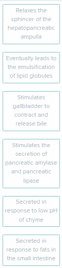
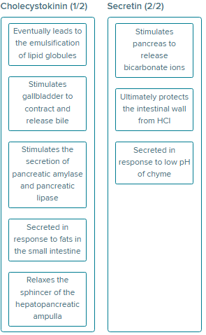
For each label, determine whether it describes the actions of secretin or the actions of cholecystokinin. Drag and drop each label into the correct category.
stimulate pancreas to release bicarbonate ions
ultimately protects the intestinal wall from HCl
CCK: eventually leads to the emulsification of lipid globules, stimulate gallbladder to contract and release bile, stimulate the secretion of pancreatic amylase and lipase, secreted in response to fats in the small intestine, relaxes the sphincter of the hepatopancreatic ampulla
secretin: stimulate pancreas to release bicarb, protect the intestinal wall from HCl, secreted in response to low pH of chyme
Complete each sentence describing the sphincters located throughout the digestive tract.
|
orbicularis oris
inferior
lower esophageal
pyloric
ileocecal
internal anal
external anal
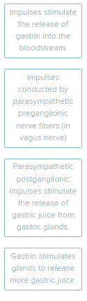
Put in correct order the events resulting in the stimulation of the gastric glands.
impulses conducted by parasympathetic preganglionic nerve fibers (in vagus nerve)
parasympathetic postganglionic impulses stimulate the release of gastric juice from gastric glands - hormone released
impulses stimulate the release of gastrin into the blood stream - hormone travels
gastrin simulates glands to release more gastric juice- hormone reacts
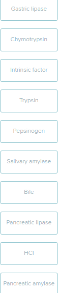
Drag each label into the appropriate position, identifying where each secretion enters the GI tract.
mouth: salivary amylase
stomach: gastric lipase, HCl, IF, pepsinogen
small intestine: chymotrypsin, trypsin, bile, pancreatic lipase, pancreatic amylase
Three main vessels make up the hepatic portal triad. These structures are found on the (Click to select) inferior left right superior side of the liver.
Blood arrives at the liver through two circulatory vessels. Blood from the digestive tract is diverted to the liver via the (Click to select) hepatic artery hepatic portal vein hepatic vein , which carries nutrient-rich, oxygen- (Click to select) poor rich blood to the liver.
A branch off the celiac trunk, the (Click to select) hepatic artery hepatic portal vein hepatic vein is responsible for delivering oxygen- (Click to select) poor rich blood to the liver. Thus, this vessel provides for the large metabolic demand of the liver.
Bile produced in the liver flows out of the liver in the (Click to select) bile common hepatic cystic duct. This duct merges with the (Click to select) bile common hepatic cystic duct from the gallbladder to form the (Click to select) bile common hepatic cystic duct, which leads to the small intestine.
inferior
hepatic portal vein, poor
hepatic artery, rich
common hepatic, cystic, bile
The act of protein digestion begins in the mouth with (Click to select) chemical mechanical digestion.
Once in the stomach, (Click to select) chemical mechanical digestion begins with the enzyme (Click to select) lipase pepsin trypsin , which hydrolyzes peptide bonds.
Chemical digestion is continued in the small intestine, where enzymes from the (Click to select) liver pancreas small intestine (trypsin, chymotrypsin, and carboxypeptidase) and from the (Click to select) liver pancreas small intestine (peptidase) continue the breakdown of proteins.
The final products are (Click to select) amino acids fatty acids monosaccharides that are absorbed through the intestinal wall into the (Click to select) lacteals bloodstream .
mechanical
chemical, pepsin
pancreas, small intestine
amino acids, bloodstream
The mesentery is the part of the (Click to select) omentum pericardium peritoneum pleural membranes that is responsible for connecting portions of the small intestine to the abdominal wall.
This double-layered membrane is located between the (Click to select) parietal visceral peritoneum that lines the abdominal cavity and the (Click to select) visceral parietal peritoneum that surrounds the digestive organs.
The mesentery is attached to the (Click to select) anterior lateral posterior part of the abdominal wall and allows the segments of the intestines to move against one another without friction.
The mesentery contains (Click to select) arteries and veins fibers omenta villi that branch to supply the small intestine.
peritoneum
parietal, visceral
posterior
arteries and veins
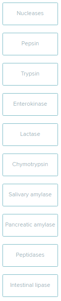
Indicate where each enzyme (or its inactive precursor) is produced.
mouth vs pancreas vs stomach vs small intestine
mouth: salivary amylase
pancreas: chymotrypsin, pancreatic amylase, nucleases, trypsin
stomach: pepsin
small intestine: enterokinase, peptidases, intestinal lipase, lactase
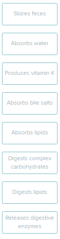
For each of the following labels, determine if they are a function of the large intestine. Then, drag the label to the appropriate location.
function of LI: store feces, absorb water, produce vitamin K, digest complex carbs
Name the steps of lipid absorption.
fats form chylomicrons
synthesis of fats in the ER
fatty acids enter the epithelial cell
chylomicrons enter lacteal
lymph transports chylomicrons
fatty acids enter the epithelial cell
synthesis of fats in the ER
fats form chylomicrons
chylomicrons enter lacteal
lymph transports chylomicrons
The inner lining of the stomach contains openings of the ducts from the (Click to select) gastric intestinal mucous parietal glands. These glands contain three types of cells, whose secretions together form (Click to select) duodenal gastric intestinal pancreatic juice.
The (Click to select) chief mucous parietal cells secrete pepsinogen and the (Click to select) chief mucous parietal cells secrete hydrochloric acid (HCl). The third cell type secretes mucus. These cells are the (Click to select) chief mucous parietal cells.
One function of HCl is to remove some amino acids from pepsinogen, converting it to (Click to select) intrinsic factor mucus pepsin trypsin , an active enzyme.
Both HCl and pepsin function to chemically breakdown and digest (Click to select) fats proteins starch .
Mucus is an (Click to select) acidic alkaline secretion that helps to protect the stomach lining.
gastric, gastric
chief, parietal, mucous
pepsin
proteins
alkaline
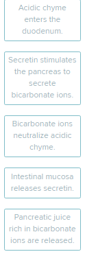
Place in correct order the events that maintain the pH of the duodenum.
acidic chyme enters the duodenum
intestinal mucosa release secretin
secretin stimulates the pancreas to secrete bicarb ions
pancreatic juice rich in bicarb ions are released
bicarb ions neutralize acidic chyme
What part of the tooth is composed of cellular tissue similar to bone, but harder?
Multiple Choice
Cementum
Dentin
Enamel
Pulp
Dentin
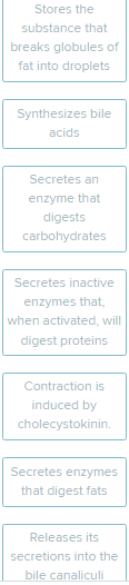
Identify the digestive organ shown in each image below. Then, for each label, decide which organ is being described. Drag and drop each description onto the appropriate image.
liver vs gall bladder vs pancreas
synthesize bile
liver: synthesizes bile acids, bile, and releases its secretions into the bile canaliculi
gall bladder: stores the substance that breaks globules of fat into droplets, contraction is induced by CCK
secretes enz that digests carbs, secretes inactive enzymes that, when activated, will digest proteins, secretes enz that digest fats
In order to use proteins as an energy source, they must first be digested into amino acids . These digestion products then must undergo deamination , removing the -NH2 groups. The remaining parts of these molecules can enter various points of the respiratory pathway, resulting in the production of ___.
ATP
For each scenario, place the label in the correct box, indicating whether it would raise or lower the total metabolic rate (TMR).
starvation
eating breakfast
aging
fighting bacterial infection
hypothyroidism
raise: eating breakfast,, fighting bacterial infection
lower: aging, hypothyroidism, starvation
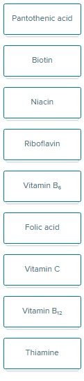
what vitamin/s function in normal RBC production
folic and B12

what vitamin/s function in the production of nucleic acids
folic, b12, niacin, biotin, b6

what vitamin/s function in normal collagen production
vitamin C

what vitamin/s function in protein metabolism
folic acid, c, biotin, b6

what vitamin/s function in energy metabolism
b12, thiamin, riboflavin, niacin, pantothenic acid
What is the common name for these chemicals
biotin
folic acid
ascorbic acid
cyanocobalamin
b7
b9
C
b12
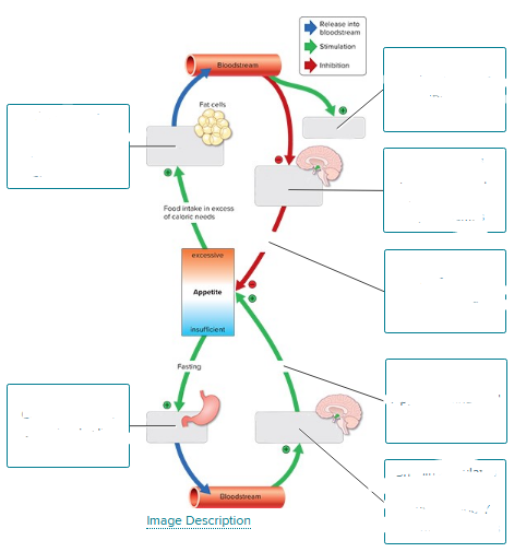
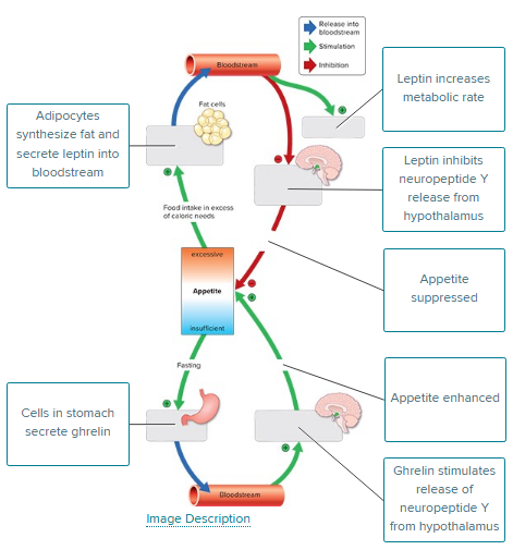
Control of appetite involves the hormones leptin and ghrelin. Complete the figure illustrating the control of appetite.
top
adipocytes synthesize fat and secrete leptin into bloodstream
leptin inc metabolic rate
leptin inhibits neuropeptide Y release from hypothalamus
appetite suppressed
bottom
cells in stomach secrete ghrelin
ghrelin stimulates release of neuropeptide Y from hypothalamus
appetite enhanced
For each kilogram of body weight, an individual requires __________Blank of energy to maintain their basal metabolic rate.
Multiple Choice
1 calorie per day
1 calorie per hour
1 calorie per minute
10 calories per hour
1 calorie per hour