IB Biology Gr. 11 Exam Review - Unit 1 – Cellular Biology.
1/22
There's no tags or description
Looks like no tags are added yet.
Name | Mastery | Learn | Test | Matching | Spaced | Call with Kai |
|---|
No analytics yet
Send a link to your students to track their progress
23 Terms
What does it mean for a cell to differentiate?
Cell differentiation refers to the process by which a less specialized cell becomes a more specialized cell type with specific functions. In multicellular organisms, cells undergo differentiation during development to give rise to various cell types that make up different tissues and organs in the body. This process allows cells to take on specific roles and functions, contributing to the overall complexity and functionality of an organism.
Vocabulary partaining to cell differentiation.
Stem Cells:
Definition: Stem cells are undifferentiated cells that have the unique ability to develop into various specialized cell types. They can divide and produce both identical stem cells (self-renewal) and differentiated cells with specific functions.
Types: There are two main types of stem cells:
Embryonic Stem Cells (ESCs): Derived from embryos, these cells have the potential to differentiate into any cell type in the body. BECOME ANY TYPE
Adult (or Somatic) Stem Cells: Found in various tissues and organs throughout the body, these cells can differentiate into specialized cell types specific to the tissue in which they are located. BECOME LIMITED TYPES
Pluripotent Cells:
Definition: Cells that have the ability to differentiate into many different cell types but not all cell types. Embryonic stem cells are an example of pluripotent cells.
Multipotent Cells:
Definition: Cells that can differentiate into a limited number of cell types. Adult stem cells are often multipotent, having the potential to give rise to specific cell lineages within a particular tissue.
Totipotent Cells:
Definition: Cells with the potential to differentiate into any cell type, as well as extraembryonic tissues. Totipotent cells exist in the very early stages of embryonic development.
Gene Expression:
Definition: The process by which information from a gene is used to synthesize a functional gene product, such as a protein or RNA molecule. Changes in gene expression drive cell differentiation.
How are genes involved in the process of cell differentiation?
Genes play a crucial role in the process of cell differentiation. The genetic information encoded in DNA is organized into units called genes, which serve as instructions for building and maintaining the structures and functions of cells. The differentiation of cells is regulated by the activation or repression of specific genes in response to various signals and cues from the cell's environment.
The key mechanisms by which genes are involved in cell differentiation include:
Gene Expression:Cells express specific genes by transcribing the DNA into messenger RNA (mRNA), which is then translated into proteins.The type and amount of proteins produced by a cell determine its structure and function.During differentiation, certain genes are activated, leading to the production of proteins characteristic of the specialized cell type.
Signal Transduction: External signals from the cellular environment, such as growth factors, hormones, and signaling molecules, can trigger changes in gene expression. These signals activate intracellular signaling pathways that ultimately influence the transcription of specific genes involved in differentiation.
Epigenetic Regulation: Epigenetic modifications, such as DNA methylation and histone modification, play a role in controlling gene expression patterns during differentiation. These modifications can turn genes on or off, influencing the fate of a cell as it differentiates.
Master Regulator Genes: Certain genes, known as master regulator genes or transcription factors, play a central role in controlling the differentiation of cells. These genes regulate the expression of other genes and guide the cell toward a specific lineage or fate.
Gene Expression. Expand upon how genes are packaged and how they are activated.
Genes are packaged in the form of chromatin within the cell nucleus. Chromatin consists of DNA wrapped around histone proteins, forming nucleosomes. This complex structure helps organize and condense the genetic material, allowing it to fit within the confines of the cell nucleus. The level of compaction of chromatin can influence gene expression.
The packaging of genes can be broadly categorized into two states: euchromatin and heterochromatin.
Euchromatin is a less condensed and more accessible form of chromatin, allowing for gene expression.
Heterochromatin is tightly packed and typically associated with inactive genes.
Transcription factors are proteins that bind to specific DNA sequences, and their binding can either promote or inhibit the transcription of a gene. Enhancers are regulatory DNA sequences that can enhance the activity of promoters, which are regions where RNA polymerase binds to initiate transcription. During gene activation, certain transcription factors bind to enhancers and promoters, facilitating the recruitment of RNA polymerase. This initiates the process of transcription, where the DNA is transcribed into mRNA. The mRNA then serves as a template for protein synthesis during translation.
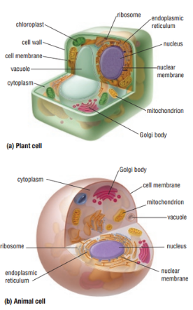
What structures are common to all cells?
will we need to draw/label a diagram?
Plasma Membrane: The plasma membrane, also known as the cell membrane, encloses the cell and separates its internal environment from the external surroundings. It regulates the passage of substances in and out of the cell, providing a selective barrier.
Cytoplasm: The cytoplasm is the gel-like substance inside the cell that surrounds organelles. It contains various cellular structures and provides a medium for cellular activities.
Nucleus: The nucleus is a membrane-bound organelle that houses the cell's genetic material (DNA). It serves as the control center for cellular activities and is involved in the regulation of gene expression.
Genetic Material (DNA/RNA): All cells contain genetic material, either in the form of DNA (deoxyribonucleic acid) or RNA (ribonucleic acid). DNA carries the genetic instructions for the cell's structure and function, while RNA is involved in protein synthesis.
Ribosomes: Ribosomes are cellular structures responsible for protein synthesis. They can be found free in the cytoplasm or attached to the endoplasmic reticulum.
Cytoskeleton: The cytoskeleton is a network of protein filaments and tubules that provides structural support to the cell, helps maintain its shape, and is involved in various cellular processes such as cell division and intracellular transport.
Endoplasmic Reticulum (ER): The endoplasmic reticulum is a membrane-bound organelle involved in the synthesis, folding, and transport of proteins and lipids. It can be rough (with ribosomes) or smooth (lacking ribosomes).
Golgi Apparatus: The Golgi apparatus is responsible for modifying, sorting, and packaging proteins and lipids for transport to their final destinations within or outside the cell.
Mitochondria: Mitochondria are the powerhouses of the cell, producing energy in the form of ATP through cellular respiration. They have their own DNA and are believed to have originated from an ancient symbiotic relationship between cells.
Vacuoles (in plant cells): Vacuoles are large membrane-bound sacs found in plant cells. They store nutrients, waste products, and help maintain turgor pressure, contributing to cell rigidity.
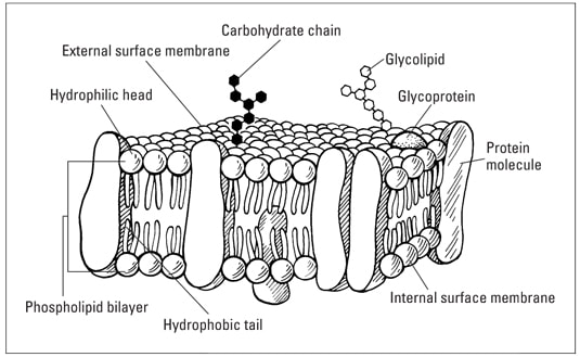
Label a diagram to show the structure of membranes.
Verify if this is in reference to the cell membrane.
Know the main properties of different forms of transport. (Passive Transport)
Passive Transport:
No Energy Expenditure:
Passive transport processes do not require the input of energy from the cell.
Movement occurs along the concentration gradient, from areas of higher concentration to lower concentration.
Diffusion:
Simple Diffusion: Small, non-polar molecules move directly through the lipid bilayer of the cell membrane.
Facilitated Diffusion: Larger or polar molecules move through channel or carrier proteins in the membrane.
Osmosis:
Osmosis is a specific form of passive transport involving the movement of water molecules across a selectively permeable membrane.
Filtration:
Filtration is the movement of water and solutes through a selectively permeable membrane due to pressure differences.
Know the main properties of different forms of transport. (Active Transport)
Active Transport:
Energy Expenditure:
Active transport processes require energy (usually in the form of ATP) to move substances against their concentration gradient.
Against Concentration Gradient:
Substances are moved from areas of lower concentration to areas of higher concentration.
Ion Pumps:
Ion pumps actively transport ions across the membrane, maintaining concentration gradients necessary for various cellular functions.
Endocytosis:
Endocytosis involves the uptake of large particles or fluids by the cell through the formation of vesicles.
- Phagocytosis: Cell engulfs solid particles.
- Pinocytosis: Cell engulfs liquids.
Exocytosis:
Exocytosis is the process by which large molecules are expelled from the cell. Secretory vesicles fuse with the cell membrane, releasing their contents.
Know the main properties of different forms of transport. (Other Forms)
Other Forms:
Bulk Flow:
Bulk flow involves the mass movement of fluids and the substances they contain.
Channel-Mediated vs. Carrier-Mediated Transport:
Channel-mediated transport involves the movement of ions or water through protein channels.
Carrier-mediated transport involves the binding of a substance to a carrier protein, which undergoes a conformational change to transport the substance across the membrane.
Selective Permeability:
Cellular membranes are selectively permeable, allowing certain substances to pass through while restricting others.
What is osmolarity? Relate this to hypertonic, hypotonic, isotonic, as well as solute concentrations.
Osmolarity is a measure of the total concentration of solute particles in a solution.
Relate Osmolarity to hypertonic, hypotonic, isotonic, as well as solute concentrations.
The terms hypertonic, hypotonic, and isotonic are used to describe the relative concentrations of solute particles in different solutions and how they affect the movement of water across a selectively permeable membrane, such as a cell membrane.
Hypertonic Solution:
A hypertonic solution has a higher osmolarity (concentration of solute particles) compared to another solution.
When a cell is placed in a hypertonic solution, water tends to move out of the cell, causing the cell to shrink or undergo crenation.
The high concentration of solutes in the external solution draws water from the less concentrated interior of the cell.
Hypotonic Solution:
A hypotonic solution has a lower osmolarity compared to another solution.
When a cell is placed in a hypotonic solution, water tends to move into the cell, causing the cell to swell or undergo cytolysis.
The lower concentration of solutes in the external solution allows water to move into the cell, where solute concentration is higher.
Isotonic Solution:
An isotonic solution has the same osmolarity as another solution.
When a cell is placed in an isotonic solution, there is no net movement of water. The concentration of solutes is balanced inside and outside the cell.
Cells maintain their shape and size in isotonic solutions.
In summary:
Hypertonic: Higher solute concentration outside the cell. Water moves out of the cell, causing it to shrink.
Hypotonic: Lower solute concentration outside the cell. Water moves into the cell, causing it to swell.
Isotonic: Equal solute concentrations inside and outside the cell. No net movement of water, and the cell maintains its shape.
Predict the movement of water for cells placed in different solutions.
Hypertonic Solution:
Prediction: Water will move out of the cell.
Result: The cell will shrink or undergo crenation.
Hypotonic Solution:
Prediction: Water will move into the cell.
Result: The cell will swell or undergo cytolysis.
Isotonic Solution:
Prediction: No net movement of water.
Result: The cell will maintain its shape and size.
What is the endosymbiotic theory?
The endosymbiotic theory is a widely accepted scientific concept that explains the origin of eukaryotic cells, which include all complex organisms, such as plants, animals, fungi, and protists. The theory proposes that certain organelles within eukaryotic cells, specifically mitochondria and chloroplasts, originated as free-living prokaryotic organisms that were engulfed by a host cell in a symbiotic relationship. Over time, these once-independent organisms evolved a mutually beneficial relationship with the host cell, leading to the development of the eukaryotic cell as we know it.
What is evidence of the endosymbiotic theory?
Mitochondria and Chloroplasts Have Their Own DNA:
Both mitochondria and chloroplasts possess their own DNA, separate from the nuclear DNA of the host cell. This DNA is circular, similar to that found in bacteria, and it encodes for some of the organelles' essential proteins.
Similarities to Bacteria:
Mitochondria and chloroplasts share similarities with bacteria in terms of size, shape, and structure. For example, mitochondria are similar in size and shape to certain bacteria, like those in the alpha-proteobacteria group, while chloroplasts resemble cyanobacteria.
Reproduction Similarities:
Both mitochondria and chloroplasts replicate independently of the cell by a process similar to binary fission, a form of asexual reproduction seen in bacteria.
Double Membrane Structure:
Mitochondria and chloroplasts have a double membrane structure. The outer membrane is similar to the host cell's membrane, while the inner membrane bears similarities to the membrane of free-living prokaryotes.
Endosymbiosis in Modern Organisms:
Endosymbiotic relationships still exist in modern organisms. Examples include certain species of amoebas that harbor intracellular bacteria and the relationships between certain insects and bacteria.
Genetic and Biochemical Evidence:
Comparison of the genetic material and biochemical processes in mitochondria and chloroplasts with those of free-living bacteria provides evidence supporting their shared evolutionary history.
How does mitosis differ from meiosis?
Mitosis:
Purpose:
Mitosis is a form of cell division that results in the production of two genetically identical daughter cells, each with the same number of chromosomes as the parent cell.
The primary purpose of mitosis is growth, tissue repair, and the maintenance of a constant number of chromosomes in somatic (body) cells.
Number of Divisions:
Mitosis involves one round of cell division.
The cell undergoes a single division, and the resulting daughter cells are identical to the parent cell.
Genetic Diversity:
Mitosis does not introduce genetic diversity because the daughter cells are genetically identical to the parent cell.
Chromosome Number:
The chromosome number in the daughter cells is the same as that of the parent cell (diploid).
Occurs in:
Mitosis occurs in somatic (non-reproductive) cells of multicellular organisms.
Meiosis:
Purpose:
Meiosis is a specialized form of cell division that occurs in germ cells (sperm and egg cells) and results in the production of four non-identical daughter cells, each with half the number of chromosomes as the parent cell.
The primary purpose of meiosis is to produce gametes (sperm and egg cells) for sexual reproduction.
Number of Divisions:
Meiosis involves two consecutive rounds of cell division: meiosis I and meiosis II.
The process reduces the chromosome number by half.
Genetic Diversity:
Meiosis introduces genetic diversity through processes such as crossing over (exchange of genetic material between homologous chromosomes) and independent assortment of chromosomes during meiosis I.
Chromosome Number:
The chromosome number in the daughter cells is half that of the parent cell (haploid).
Occurs in:
Meiosis occurs in germ cells (sperm and egg cells) and is involved in the formation of gametes for sexual reproduction.
What are their respective roles in multicellular organisms?
Roles in Multicellular Organisms:
Mitosis:
Mitosis plays a crucial role in growth, development, and tissue repair in multicellular organisms.
It ensures that the new cells formed are genetically identical to the original cell, maintaining the integrity and function of tissues.
Meiosis:
Meiosis is essential for sexual reproduction, as it produces gametes with half the chromosome number of somatic cells.
When gametes fuse during fertilization, the resulting zygote has a complete diploid set of chromosomes, contributing to genetic variability in offspring.
In summary, mitosis is involved in somatic cell division for growth and tissue maintenance, while meiosis is specific to germ cells and is crucial for the production of gametes and the introduction of genetic diversity in sexual reproduction.
Cancer.
Cancer is a complex and multifaceted group of diseases characterized by uncontrolled cell growth and the ability of cells to invade other tissues. The development and progression of cancer involve a series of events, including initiation, promotion, and progression. Additionally, the body has mechanisms to suppress the development of cancer, and the failure of these mechanisms can contribute to the emergence of cancer.
Describe the development of cancer.
Initiation:
Mutation: The process usually begins with a genetic mutation that occurs in a single cell's DNA. This mutation can be caused by various factors such as exposure to carcinogens (cancer-causing substances), radiation, or spontaneous errors during DNA replication.
Initiated Cell: The cell with the mutation is termed an initiated cell. At this stage, the mutation may not cause noticeable changes.
Promotion:
Promotion of Growth: Certain factors, known as promoters, encourage the initiated cell to undergo rapid and uncontrolled cell division. These factors may include chronic irritation, inflammation, or exposure to additional carcinogens.
Formation of Preneoplastic Lesions: The initiated cells form a group of abnormal cells, known as preneoplastic lesions or benign tumors.
Progression:
Malignant Transformation: Over time, additional genetic mutations may occur in the preneoplastic cells, leading to malignant transformation. Malignant cells acquire the ability to invade surrounding tissues and may eventually enter the bloodstream or lymphatic system, allowing them to spread to other parts of the body (metastasis).
Cancer development reviewing oncogene, proto-oncogene and tumour suppression.
The development of cancer is a complex process involving the accumulation of genetic and epigenetic alterations that disrupt normal cellular functions, leading to uncontrolled cell growth and the formation of a tumor. Key players in this process include oncogenes, proto-oncogenes, and tumor suppressor genes.
1. Proto-oncogenes:
- Proto-oncogenes are normal cellular genes that play essential roles in regulating cell growth, differentiation, and survival.
- These genes can be mutated or overexpressed to become oncogenes, which promote uncontrolled cell proliferation.
- Proto-oncogenes code for proteins involved in signal transduction pathways, cell cycle regulation, and apoptosis.
- Mutations such as point mutations, gene amplification, or chromosomal rearrangements can lead to the activation of proto-oncogenes.
2. Oncogenes:
- Oncogenes are mutated or overactive forms of proto-oncogenes that contribute to the development of cancer.
- Activated oncogenes promote abnormal cell growth and survival by bypassing normal regulatory mechanisms.
- Examples of oncogenes include the Ras gene family, which is involved in cell signaling, and the MYC gene, which regulates cell cycle progression.
3. Tumor Suppressor Genes:
- Tumor suppressor genes code for proteins that normally inhibit cell growth and prevent the formation of tumors.
- Loss-of-function mutations or inactivation of tumor suppressor genes contribute to uncontrolled cell division.
- Examples of tumor suppressor genes include TP53 (p53), which regulates the cell cycle and apoptosis, and RB1 (retinoblastoma), which controls cell cycle progression.
- The "two-hit hypothesis" suggests that both alleles of a tumor suppressor gene must be mutated or inactivated to eliminate its function, increasing the risk of cancer.
4. Tumor Suppressor Pathways:
- Tumor suppressor genes often function in pathways that regulate the cell cycle, DNA repair, and apoptosis.
- For example, the p53 pathway is crucial for detecting DNA damage and initiating cell cycle arrest or apoptosis to prevent the propagation of damaged cells.
- Dysfunction in these pathways can lead to the survival and proliferation of cells with genomic instability, contributing to cancer development.
5. Genomic Instability:
- The accumulation of genetic mutations and chromosomal abnormalities, known as genomic instability, is a hallmark of cancer.
- Genomic instability can result from defects in DNA repair mechanisms, making cells more susceptible to acquiring additional mutations over time.
In summary, cancer development involves the activation of oncogenes that promote uncontrolled cell growth and the inactivation of tumor suppressor genes that normally prevent such growth. The intricate balance between proto-oncogenes and tumor suppressor genes is crucial for maintaining normal cellular functions, and disruptions in these regulatory mechanisms contribute to the initiation and progression of cancer. Understanding these molecular mechanisms is essential for developing targeted therapies and strategies for cancer prevention and treatment.
Describe the suppression of cancer.
Tumor Suppressor Genes:
Normal Function: Tumor suppressor genes encode proteins that regulate cell division, repair DNA damage, and induce cell death (apoptosis).
Suppression Failure: Mutations or inactivation of tumor suppressor genes can lead to a loss of their normal function, allowing uncontrolled cell growth.
DNA Repair Mechanisms:
Normal Function: Cells have DNA repair mechanisms that correct errors and mutations in the DNA sequence.
Suppression Failure: Deficiencies in these repair mechanisms can lead to the accumulation of genetic mutations, contributing to the development of cancer.
Cell Cycle Checkpoints:
Normal Function: Cells have checkpoints in the cell cycle where the process is halted to ensure that DNA is intact and cell conditions are suitable for division.
Suppression Failure: Disruption of cell cycle checkpoints can result in the proliferation of damaged cells.
Apoptosis (Programmed Cell Death):
Normal Function: Cells that are irreparably damaged or have undergone genetic mutations may undergo apoptosis, preventing the survival and growth of abnormal cells.
Suppression Failure: Resistance to apoptosis allows damaged cells to survive and contribute to the formation of tumors.
Immune System Surveillance:
Immune System Recognition:
The immune system can recognize and eliminate abnormal cells, including those with cancerous characteristics.
Suppression Failure:
Immune suppression or evasion mechanisms developed by cancer cells can allow them to escape detection and destruction by the immune system.
Calculate length.
To calculate length, you need the size of the object and a scale bar.

Calculate magnification.
Magnification is the factor by which an image is enlarged. If you know the actual size of an object and its size on the image, you can calculate magnification using the formula:

Calculate mitotic index.
The mitotic index is a measure of the percentage of cells undergoing mitosis in a population.
