Week 3 & 4 - Brain Imaging
1/82
There's no tags or description
Looks like no tags are added yet.
Name | Mastery | Learn | Test | Matching | Spaced | Call with Kai |
|---|
No analytics yet
Send a link to your students to track their progress
83 Terms
COW
Circle of Willis
MCA
Middle cerebral artery
EAM
External acoustic meatus
GCS
Glasgow coma scale
SOL
Space occupying lesion (abnormal mass: tumour, cyst, haematoma)
OML
Orbitomeatal line
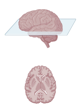
Name this plane
Axial
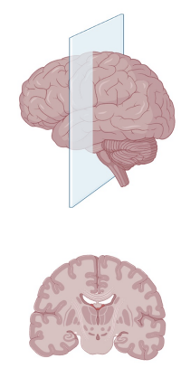
Name this plane
Coronal
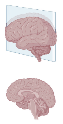
Name this plane
Sagittal
What happens at gestational week 3 in regards to brain development?
Brain development begins
Ectoderm thickens to form the neural plate
Neural plate folds inwards to form neural groove
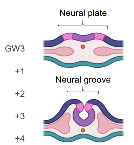
What happens at gestational week 4 in regards to brain development?
Neural groove fuses together creating the neural tube
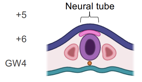
When does the neural tube differentiate into primary and secondary vesicles?
GW4 - 5
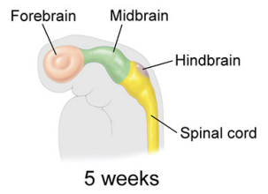
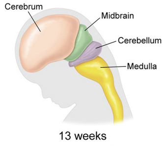
What happens at gestational week 7 - 13 in regards to brain development?
The vesicles fold and the brain begins to take shape
What is the size of a new born brain?
About 25% of the adult brain size
Why is the skull unfused at birth?
To allow for the delivery through the birth canal
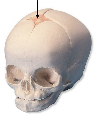
Name this section
Fontanelle
What is an epoch?
Stage of brain development
What are the 5 epochs of brain growth?
Birth - 9
Adolescence (9 - 32)
Adulthood (32 - 66)
Early aging (66 - 83)
Late aging (83+)
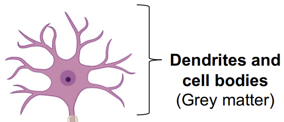
What does the grey matter in brain consist of?
Dendrites and cell bodies
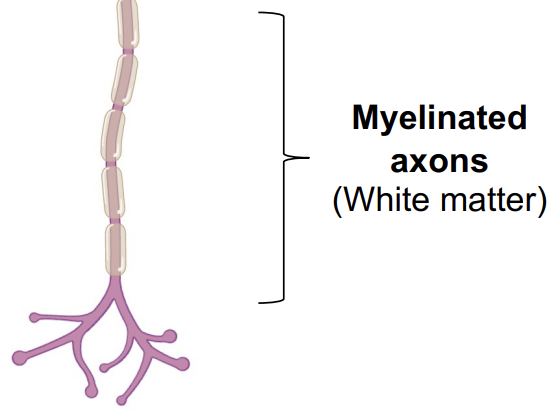
What does the white matter in brain consist of?
Myelinated axons
What are the 3 main parts of the brain and their main functions
Cerebrum (Higher functions and cognition)
Cerebellum (Coordination, balance, fine motor skills)
Brain stem (life support functions, PNS, CNS)

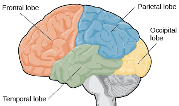
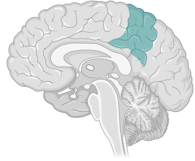
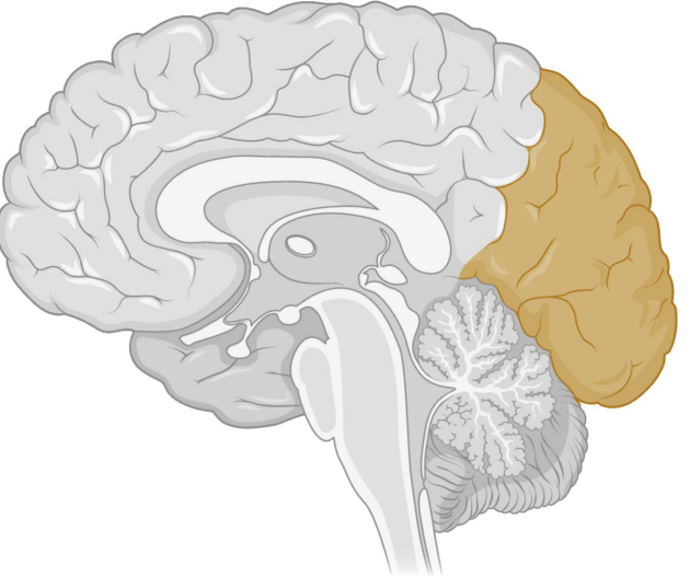
What are the lobes of the brain?
Frontal
Parietal
Occipital
Temporal
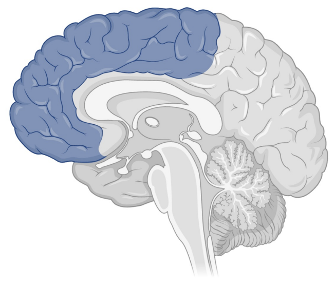
Function of frontal lobe
Decision making
Judgement
Voluntary movement (primary motor cortex)
Speech production (Broca’s area)
Function of parietal lobe
Processing and integrating somatosensory information
Perceive touch, pressure, pain, temperature
Spatial awareness
Proprioception (knowing where your body parts are)
Coordinating visual motor skills
Function of occipital lobe
Visual processing
Receiving and decoding raw signals from the eyes
Spatial processing
Depth perception
Colour determination
Recognition or objects and faces
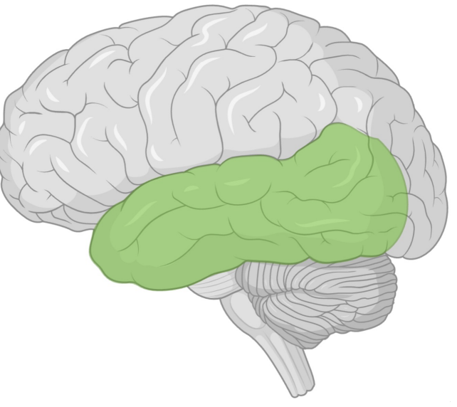
Function of temporal lobe
Processing sound
Language comprehension
Forming and retrieving memories
Linking sensory input with emotion and meaning
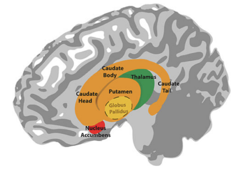
Function of the basal ganglia
Smooths involuntary movement
Cortical functions
Habit formation
Limbic system
Dysfunction = parkinson’s
Describe the function of the limbic system
Emotional regulation and memory
Amygdala: source of fight or flight
Hippocampus: converts short term memories into long term, memory storage, effected by Alzheimer’s
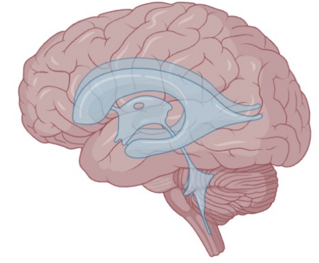
Describe the function of ventricles
4 ventricles that synthesise and circulate CSF through the nervous system
Shock absorbency
Delivers nutrients and flushes cellular waste
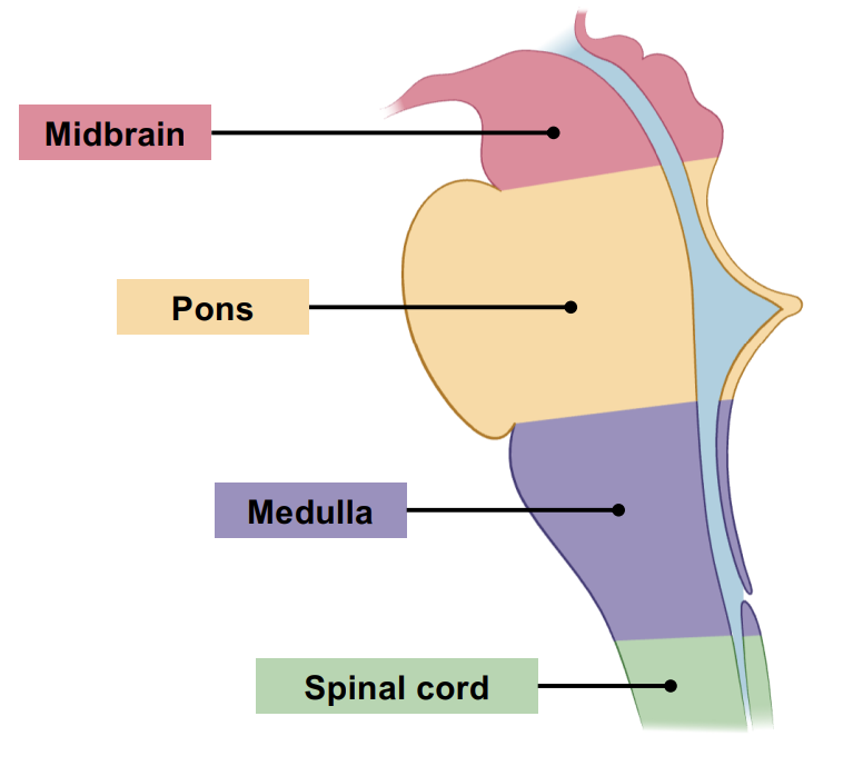
What’re the sections of the brain stem?
Midbrain
Pons
Medulla oblongata (point of left-right crossover)
Spinal cord
Describe the role of the blood brain barrier
Protects brain from systematic risks
Capillary endothelial cells = selectively permeable membrane
Prevents passage of toxins into the brain but allows oxygen and nutrients by active transport
What contrast is administered in patients with suspected BBB failure?
BBB failure = no ICM!!!
The carotid arteries
Arises from aortic arch
CCA branches into ECA and ICA
ICA branches into MCA - supplies oxygenated blood to cerebrum
The carotid siphon
S shape bend at the end of the ICA
Dampens pulsation (reduces changes in blood flow pressure)
Prevents turbulent blood flow arriving to the COW
Vertebral arteries
Arise from the subclavian arteries and travel through the transverse foramina of the cervical vertebrae
20% of the brains blood supply, supplies the brainstem and cerebellum
Susceptible to trauma
Meninges of the brain
Dura mater
Arachnoid mater
Pia mater
List some indications for brain imaging
Trauma
Neurological changes
Seizure
Stroke
SOL
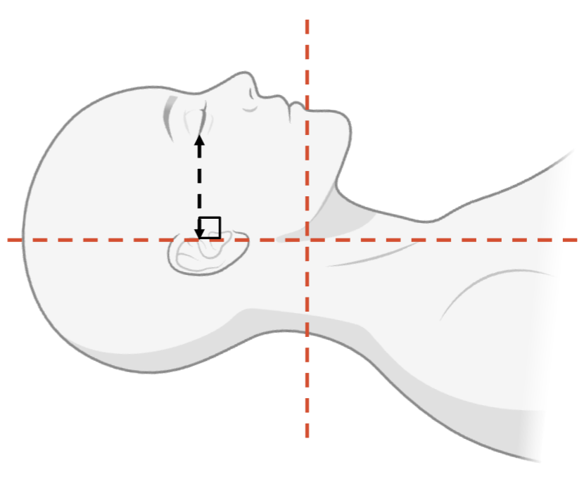
What is the correct patient positioning for CT brain protocol?
Head first
Supine
Head immobilised in cradle
EAM is perpendicular to the OML
What is the correct patient scan range (collimation) for CT brain protocol?
C2/C3 vertebra to above head
What is the correct patient breathing instruction for CT brain protocol?
Suspended breathing
What is the window width/level for a soft tissue (brain) reformat?
WW: 80
WL: 40
What is the window width/level for a bone reformat?
WW: 2800
WL: 600
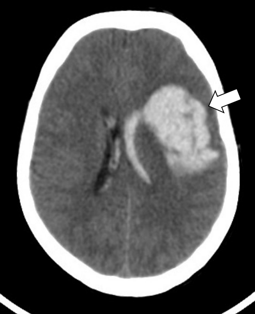
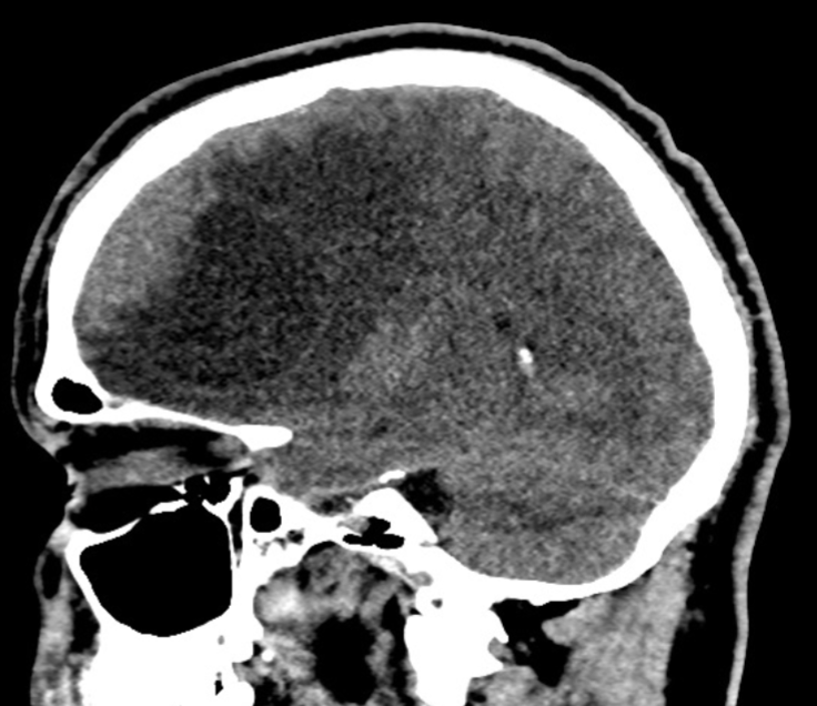
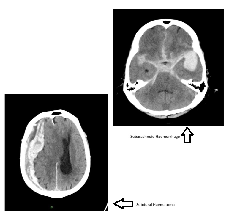
Name the pathology
Intracerebral haemorrhage (bleed)
Name the pathology
Name the pathology
MCA infarct (necrosis)
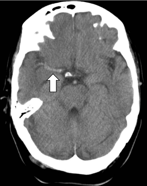
What dies this indicate?
Hyperdense MCA sign
Patient is actively having a stroke
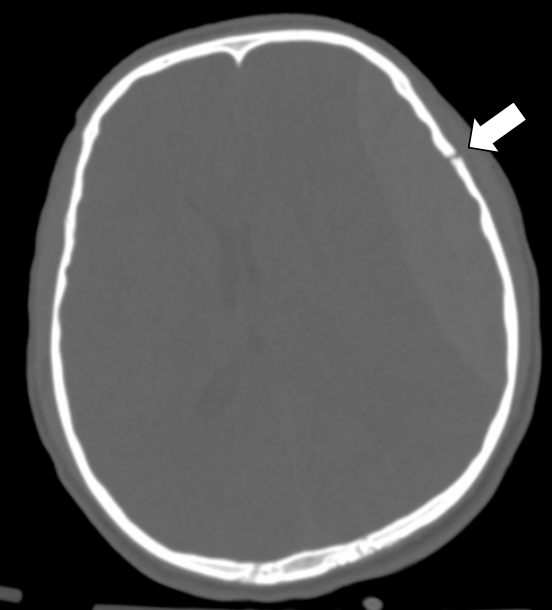
Name the pathology
Skull #
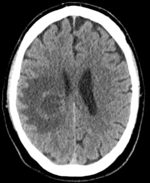
Name the pathology
Mass
Name the pathology
Hypodense mass
Contrast scanning protocol of brain
50mL ICM IV, 1mL/sec
5min delay before scan
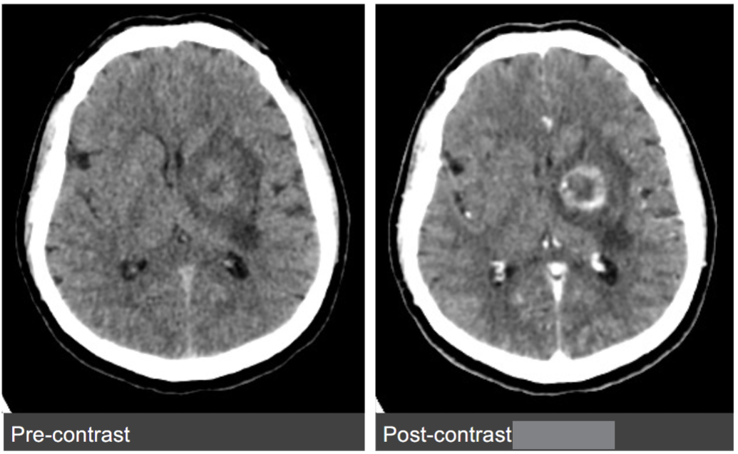
Name the pathology
Glioma
What is a stoke?
Interruption of blood supply to the brain, leading to rapid cell death (infraction - tissue death)
What is SMuRFs?
Standard modifiable cardiovascular risk factors
Hypertension
Diabetes
Hyperlipidaemia
Smoking
What is FAST?
Symptoms of a stroke
Face: facial asymmetry, mouth drooping
Arms: discoordination of motor function
Speech: slurring
Time: golden hour (hour to save patient, call for help immediately)
Not all symptoms need to be present to indicate stroke
Describe an ischaemic stroke
Clot
Brain not getting enough oxygen
Caused by blockage of arteries which supply/are in the COW
Blockage caused by clot (embolisation - clot in bloodstream travels)
Commonly caused by atherosclerotic plaque rupture in carotid arteries
80% of strokes are ischaemic
Describe atherosclerosis
Chronic disease
Plaque growth in the arterial vessel wall
Caused by vascular inflammation, cholesterol accumulation and stress
Leading cause of ischaemic strokes
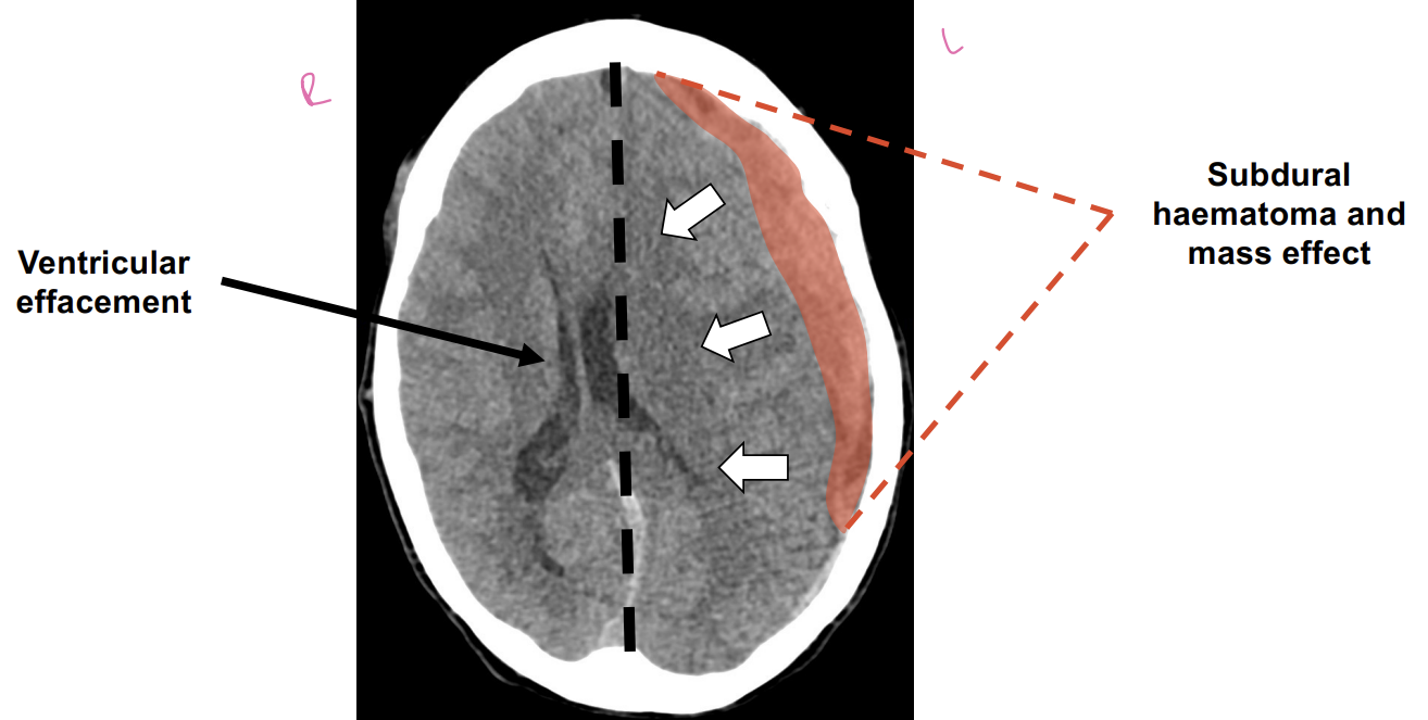
Describe an haemorrhagic stroke
Bleed
Can be caused by AVM rupture, hypertension, blood thinners, head trauma
Previous ischaemic stroke can turn into a haemorrhagic stroke
Caused by damage to damage to micro vessel during infarction
Describe the 2 types if haemorrhagic strokes
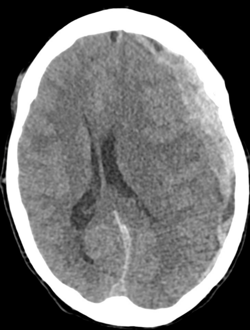
Subarachnoid haemorrhage: blood between the arachnoid membrane and pita mater
Subdural haematoma: blood between dura and arachnoid layers
Describe endovascular clot retrieval (ECR)
6 - 24 hours after symptom onset
Catheter passed from thigh to brain to retrieve the clot and reverse stroke
Describe thrombolysis
Clot busting and blood thinning drugs to rapidly dissolve clot
Only in ischaemic stroke not haemorrhagic
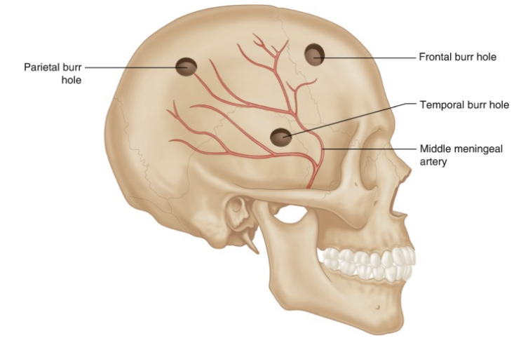
Describe a craniostomy
Holes are drilled into the skull to drain haematoma and reduce intracranial pressure
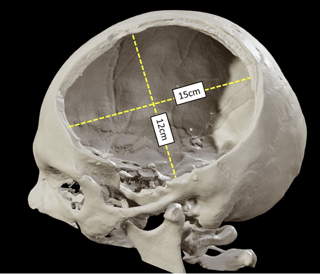
Describe decompressive craniectomy
Reduce intracranial pressure after malignant MCA stroke
Bone flap is removed until swelling is reduced
Describe CT perfusion
IV ICM at a fast rate (18 gauge)
Shuttle scanning (oscillating in and out of machine)
Heatmap generated to show mean transit time, time to peak and cerebral blood flow
Used to confirm stroke location (ECR and prognosis)
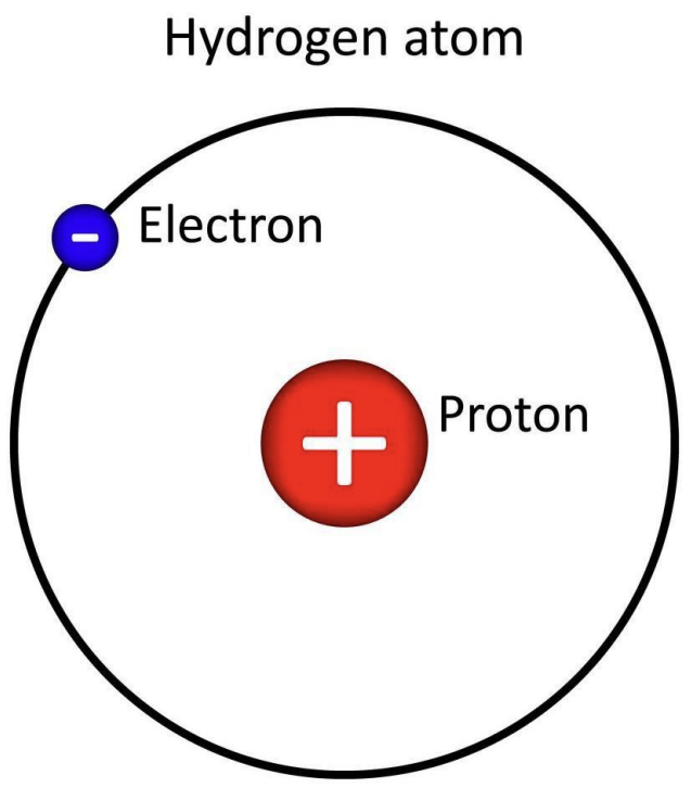
What is the common nucleus used in MRI medical imaging?
Hydrogen atom
What are the main electromagnetic components of an MRI?
Main magnet coils
3 gradient coils
Integral radiofrequency (RF) transmitter coil
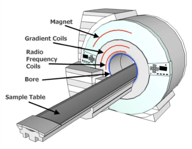
What is the function of the main magnet coils?
They create a strong constant magnetic field (B0)
What is the function of the three gradient coils?
Create changeable magnetic fields for spatial encodings
Acts as a GPS to help identify the location of the protons within the magnetic field
What is the function of the RP transmitter coil?
Creates oscillating RF field
Sends signals to protons in the body from the machine to collect returning signals
What is the strength of MRI machines used in clinical imaging?
0.2T - 3.0T
What is the isocentre?
The point within the scanner where the magnetic field is the strongest
ROI should be centred here
What are the 3 gradient coils and their axis?
Gx (X-axis)
Gy (Y-axis)
Gz (Z-axis)
Which gradient coils are on the transverse plane?
Gx (X-axis) and Gy (Y-axis)
Which gradient coils is on the longitudinal plane?
Gz (Z-axis)
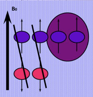
Describe how hydrogen in the body reacts to B0
B0 is applied across the body → majority of hydrogen atoms align with the main magnetic field (spin up direction) → some will align anti-parallel to B0
What is Zeeman Splitting?
Assesses the number of protons aligning parallel/anti-parallel to the main magnetic field
What is net magnetisation vector (NMV) in MRI?
Sum of the number of excess hydrogen protons aligned parallel to B0
Paired parallel and unpaired hydrogen protons cancel each other out, since majority of protons align parallel with B0, the sum of the left over unpaired parallel protons form NMV
These unpaired protons produce a longitudinal signal that is the basis of the MRI signal
NVM points in the same direction as the main magnetic field, Gz (longitudinal)
How is the NMV affected by the RF pulse in MRI?
The RF pulse tips the NMV from the longitudinal plane (Gz) to the transverse plane (Gxy)
What happens to the NMV immediately after the RF pulse?
The longitudinal magnetization (Mz) becomes zero
Transverse magnetization (Mxy) reaches its maximum and generates signal
Rotating transverse vector creates oscillating magnetic field → detected by RF receiver coil
What are the 2 processes of the 2 components of net magnetisation?
Longitudinal (T1 relaxation, recovery)
Transverse (T2 relaxation, decay)
What does T2 relaxation mean in MRI?
Happens after the RF signal
T2 relaxation is the process by which the transverse components of magnetization (Mxy) decay
Loss of phase coherence among proton spins (loss of synchronisation, losing rhythm)
Faster than T1
What does T1 recovery mean in MRI?
Happens after the RF signal
Protons release absorbed energy to their surrounding environment to realign with the main magnetic field (B0)
Along the longitudinal plane