A&P Ch. 13 Peripheral Nervous System
1/82
There's no tags or description
Looks like no tags are added yet.
Name | Mastery | Learn | Test | Matching | Spaced | Call with Kai |
|---|
No analytics yet
Send a link to your students to track their progress
83 Terms
Overview of Peripheral Nervous System
Sensory Receptors,
Afferent Nerves (Peripheral nerves & ganglia),
Efferent Nerves (Peripheral nerves & ganglia),
Motor Endings
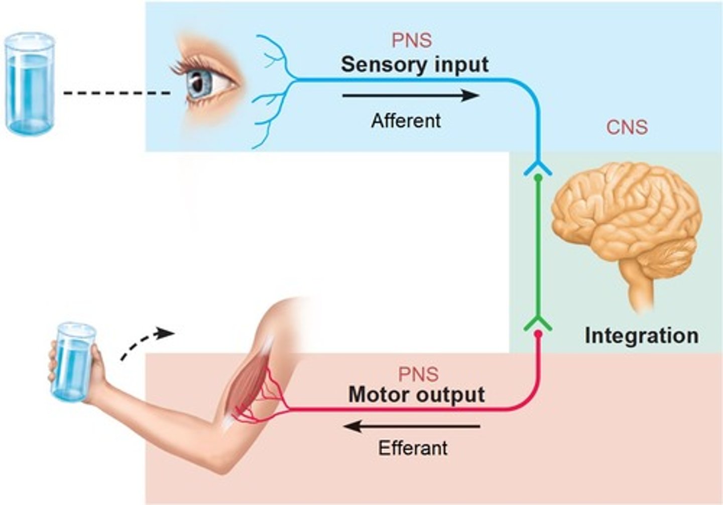
Sensory Receptors
Respond to changes in their environment called stimuli
-Stimulus initiates grades potential (local depolarization) that then in turn triggers nerve impulses along afferent PNS fibers to the CNS

Classifications of Sensory Receptors by Stimulus type
Mechanoreceptors,
Thermoreceptors,
Photoreceptors,
Chemoreceptors,
Nociceptors
Mechanoreceptors
mechanical force such as touch, pressure, etc.
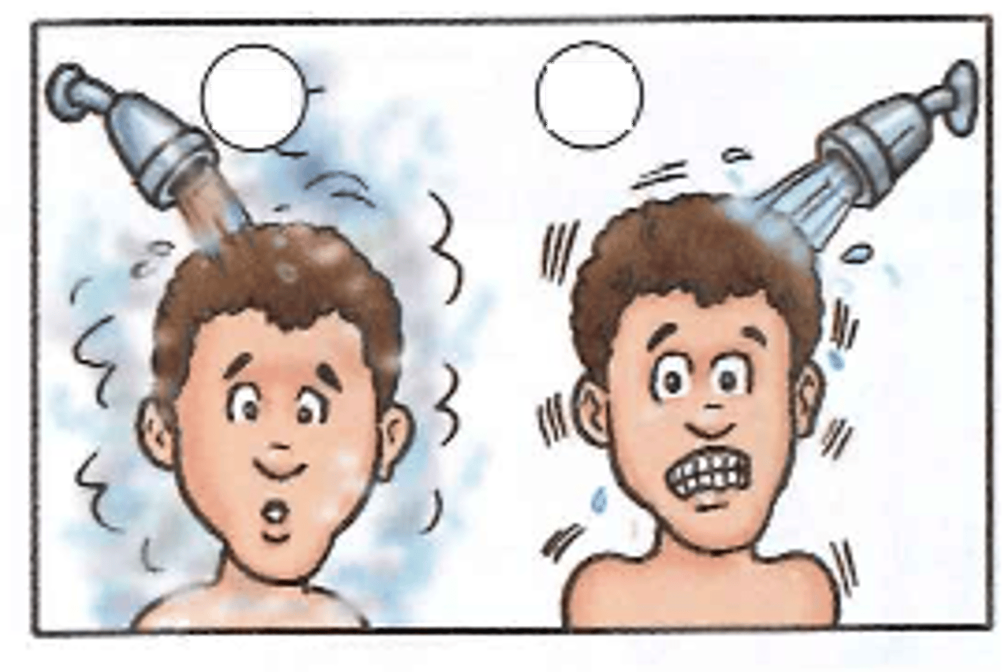
Thermoreceptors
temperature changes
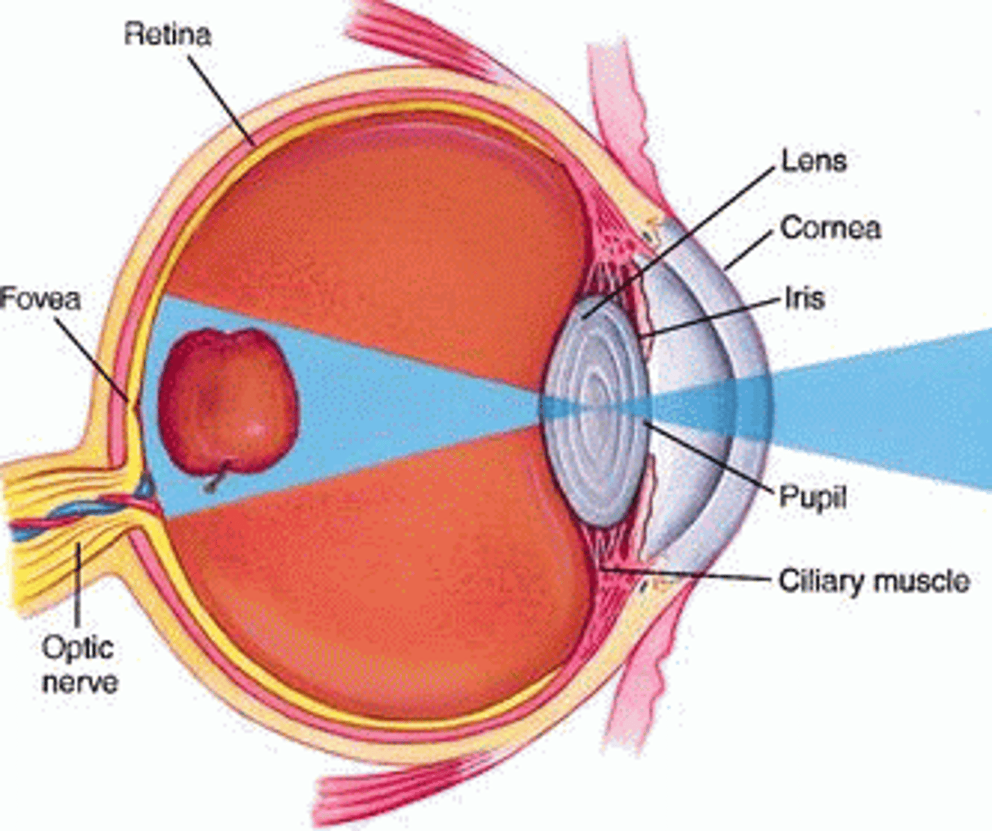
Photoreceptors
light energy
Chemoreceptors
chemicals in solution
Nociceptors
potentially damaging stimuli that result in pain
Classifications of Sensory Receptors by location
Exteroceptors,
Interceptors,
Proprioceptors

Exteroceptors
1) Located at or near body surface and respond to stimuli arising outside body
2) Include receptors for touch, pressure, pain, vibrations, skin temperature, limb motion, and special senses (vision, hearing, equilibrium, taste, smell)

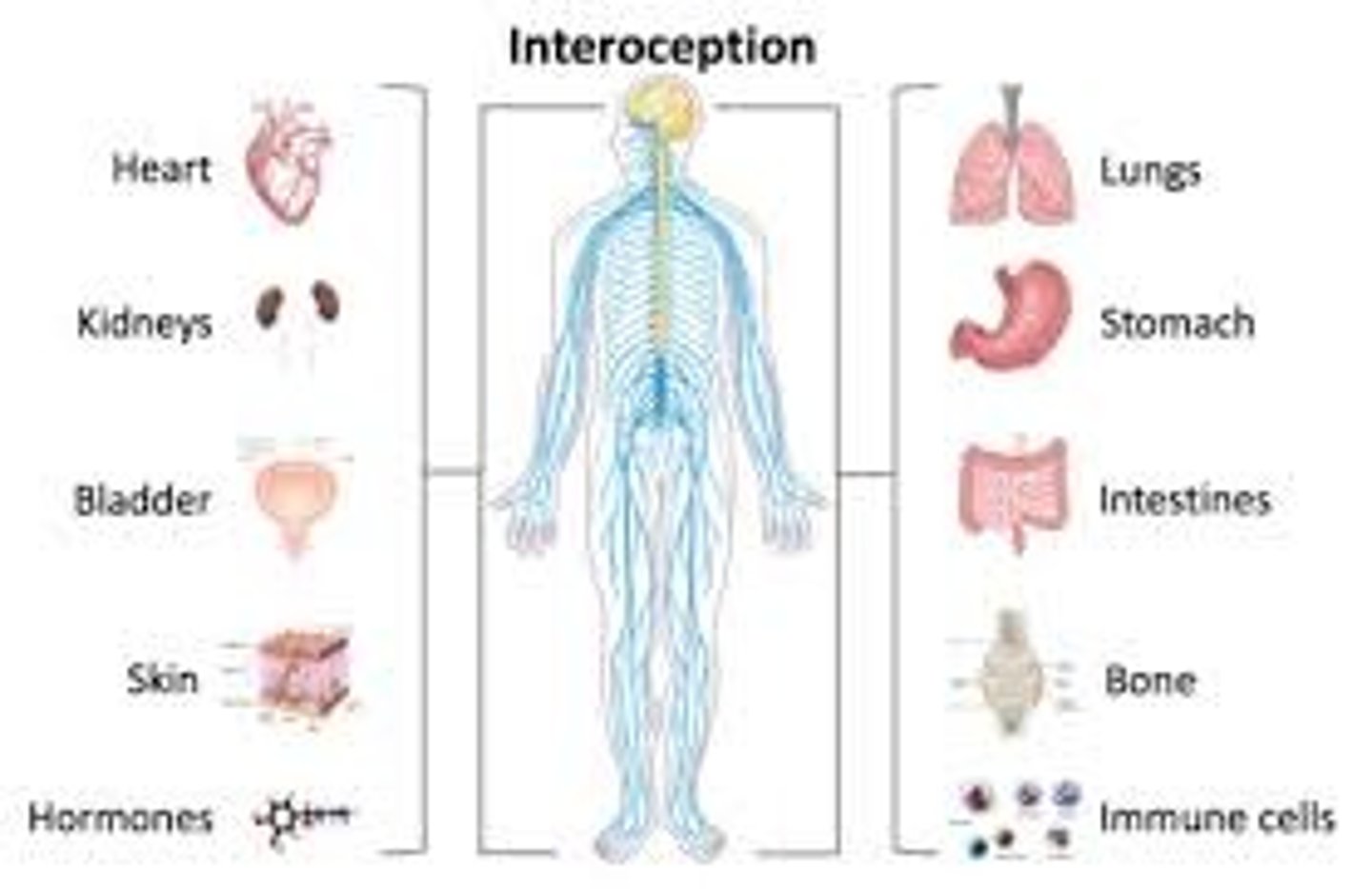
Interoceptors (visceroceptors)
1) Internal; respond to stimuli arising within body such as internal viscera and blood vessels
2) Include receptors for stretch, pain, temperature, chemical changes (nausea, hunger, etc.)
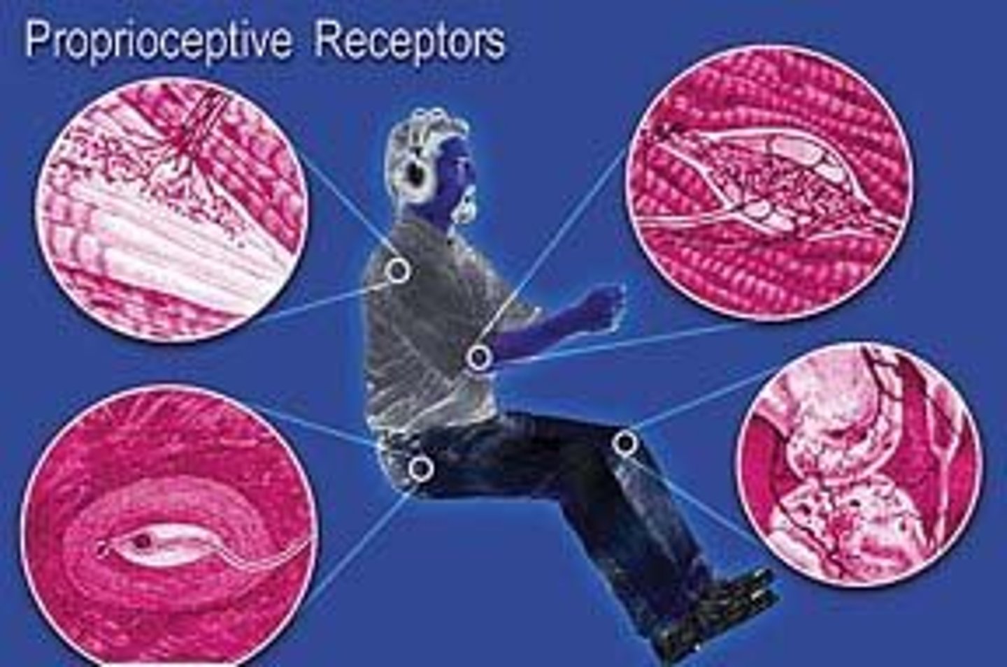
Proprioceptors
1)Respond to internal stimuli live above, but location is restricted to skeletal muscle, tendons, joints, and ligaments
2)Constantly advise brain on body movement
Classifications of Sensory Receptors by structural complexity
Simple Receptors,
Complex Receptors
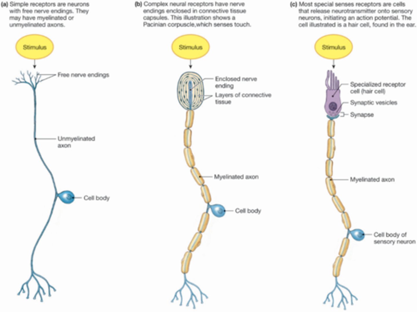
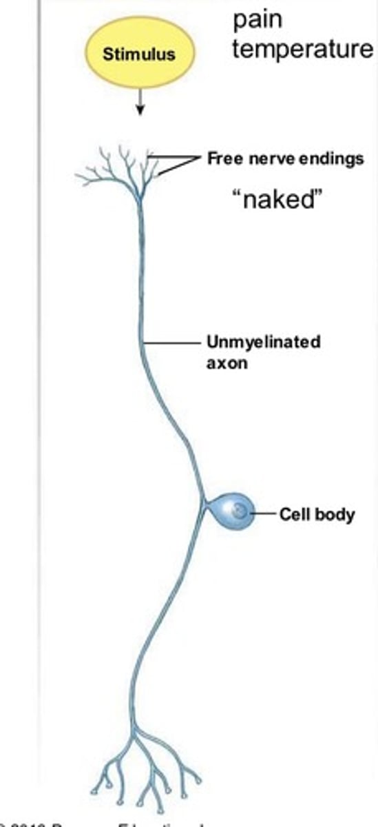
Simple Receptors
1) Modified dendritic endings of sensory neurons
2) Located throughout body and monitor general sensory info (touch, stretch, temp, pressure, chemicals, etc.)
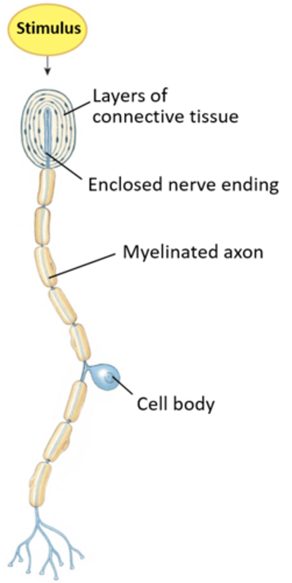
Complex Receptors
1)Clusters of multiple cell types; usually considered complex sensory organs
2)Associated with special senses: vision (eyes), hearing (cochlea), equilibrium (vestibule), smell, and taste
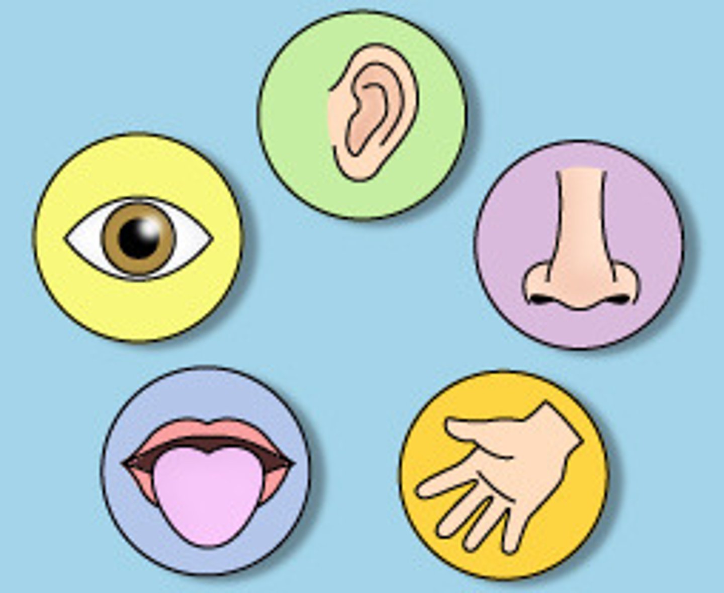
Special Senses
Equilibrium,
Hearing,
Smell,
Taste,
Vision
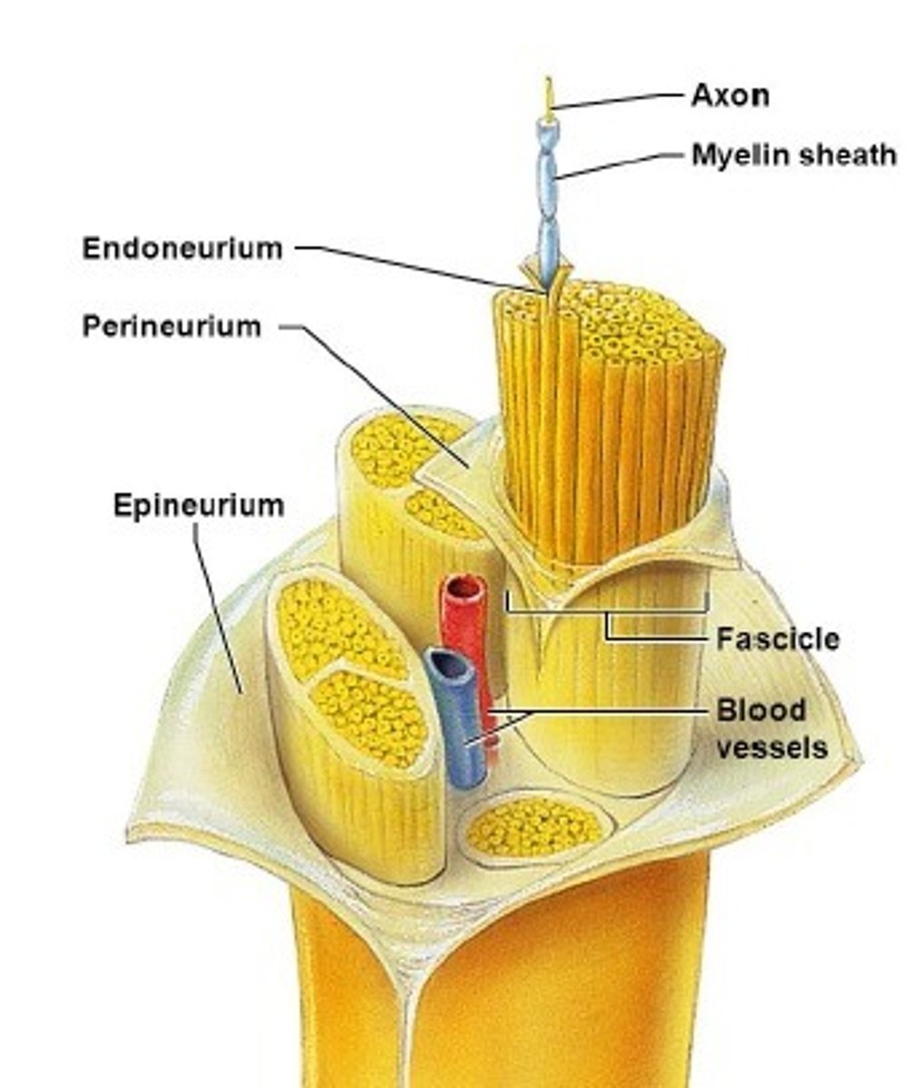
Structure of a nerve
Connective Tissue Wrappings (endoneurium, perineurium, & epineurium),
Blood Vessels,
Lymphatic Vessels

Connective Tissue Wrappings of nerve
Endoneurium, Perineurium, and Epineurium

Endoneurium
Bundles individual fibers,
Located around Schwann cells and Myelin Sheath

Perineurium
Bundles fibers into fascicles

Epineurium
Bundles fascicles into a nerve
Classification of Nerves
1) Mixed Nerves
2) Sensory (afferent) Nerves
3) Motor (efferent) Nerves
4) Regions they innervate: somatic afferent, somatic efferent, visceral afferent, & visceral efferent
Cranial Nerves
12 Pairs of nerves that mostly serve the head the head and neck,
Numbered in order, anterior to posterior, and paired with L and R
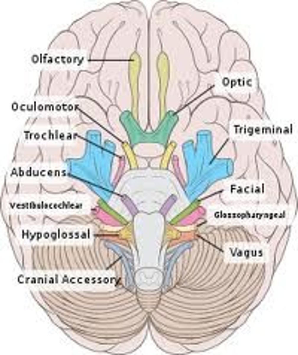
Olfactory (I)
Sensory: Smell
Optic (II)
Sensory: Vision (light)

Oculomotor (III)
Motor: Eye movements
Trochlear (IV)
Motor: Eye movements
Trigeminal (V)
Sensory: Face (skin & mucosa of nose and mouth);
Motor: Chewing
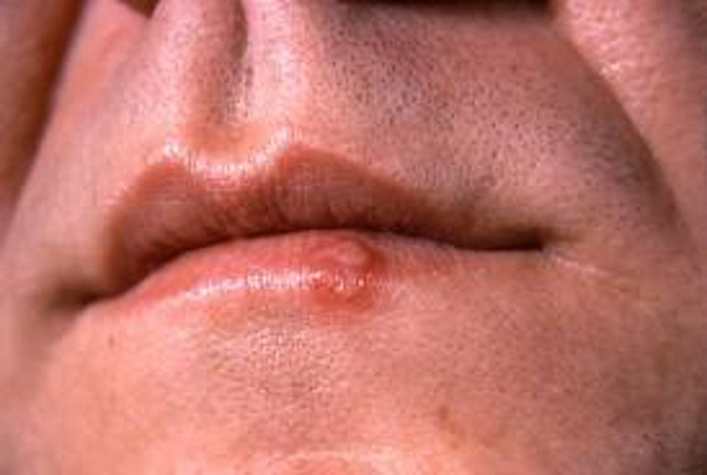
Clinical: HSV-1
HSV-1 (herpes simplex virus)
Causes cold sores and fever blisters,
Can also be found in facial and vestibular cranial nerves
(HSV-2 is found in spinal nerves serving genital area (causes genital herpes)
Abducens (VI)
Motor: Eye Movements
Facial (VII)
Sensory: Taste
Motor: Face (tears, expression, and salivary glands)
Clinical: Bell's Palsy
Vestibulocochlear (VIII)
Sensory: Hearing, Balance

Glossopharyngeal (IX)
Sensory: Taste
Motor: Pharynx (swallowing) and salivary glands

Vagus (X)
mixed sensory & motor:
From medulla to pharynx, larynx, and abdominal and thoracic viscera,
ANS controlled nerve

Accessory (XI)
Motor: Neck and Upper back

Hypoglossal (XII)
Motor: Tongue
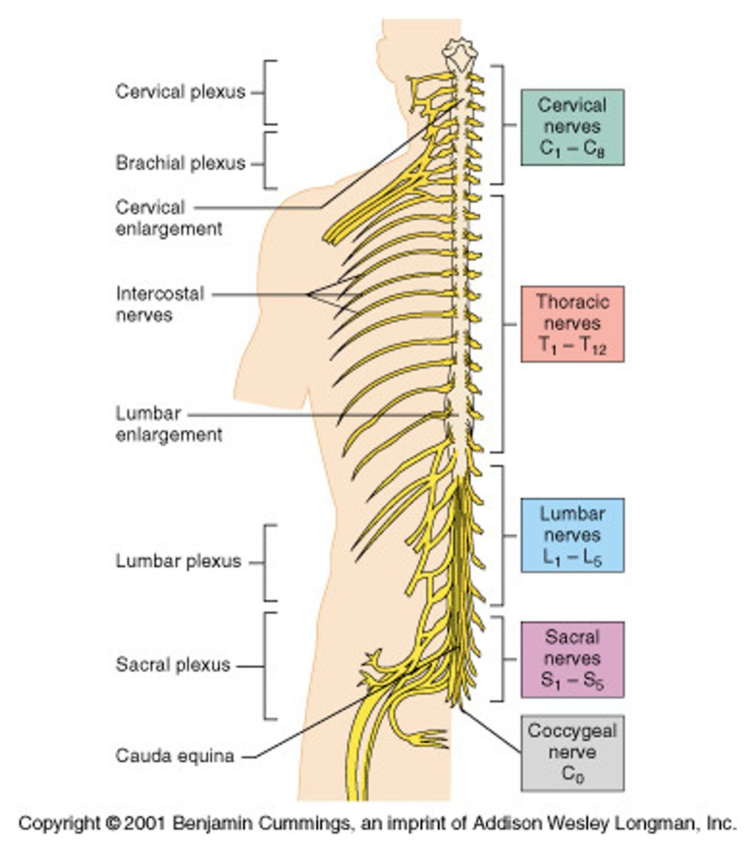
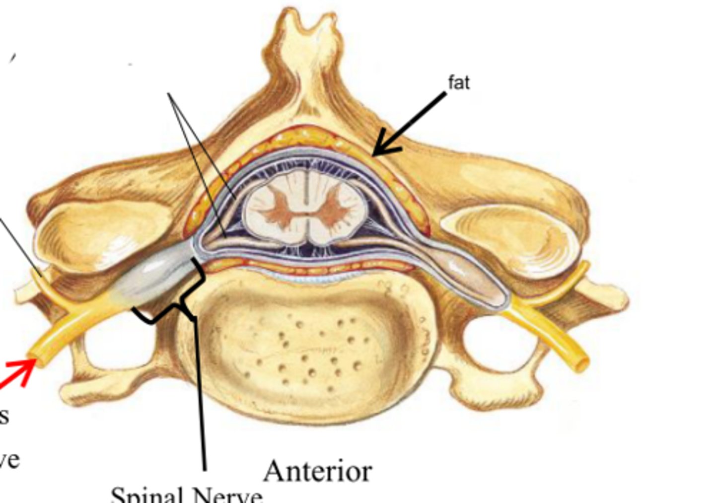
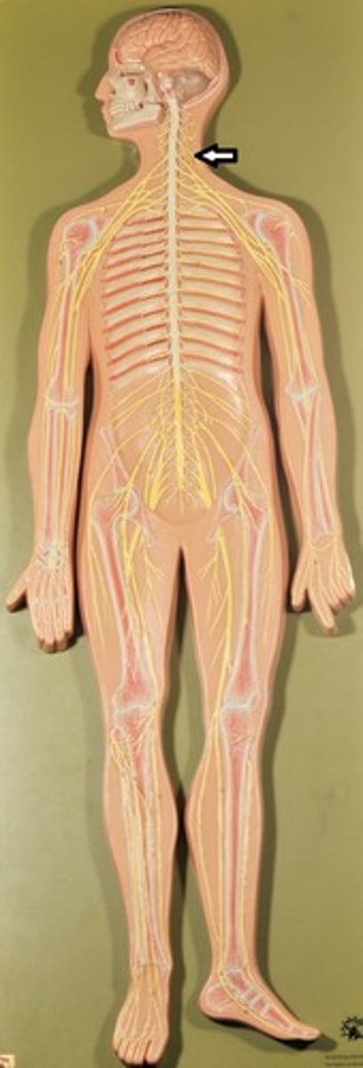
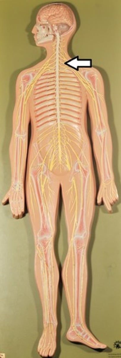
Spinal Nerves
1) 31 Pairs exiting through the intervertebral foramen.
2) Supply all parts of the body except head and some areas of neck.
3) Formed by dorsal and ventral roots of spinal cord (therefore, all are mixed)
4) Named for the region from which they arise
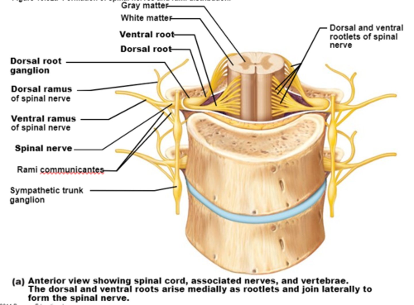
Anatomy of spinal nerves
Divide soon after leaving spinal cord into: Dorsal rami, Ventral rami, & Plexus
Singular form for "rami"
Ramus
Dorsal Rami
Serve the skin and muscles of the posterior trunk
Ventral Rami
Extended anterior to recombine with other ventrak rami superior and inferior to it (except in thoracic region) to form a complex network called a plexus
Ventral rami note:
Fibers from each ventral ramus travel to body periphery via several routes. As a result, each muscle in a limb receives its nerve supply from more than one spinal nerve. An advantage of this fiber regrouping is that damage to one spinal segment or root cannot completely paralyze any limb muscle.
Plexus
Formed by multiple ventral rami that will then give rise to individual spinal nerves
Dorsal Ramus
Innervates posterior trunk (the back),
Each dorsal ramus remains posterior to innervate narrow strip of muscle and skin in line with its emergence point from spinal column
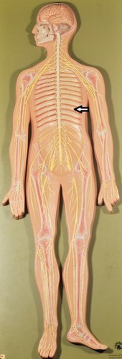
Intercostal Nerves
1)Ventral rami arranged in simple segmental pattern corresponding to dorsal rami (only location that doesn't recombine into a plexus) and extend anterior to form intercostal nerves.
2) Intercostal nerves and their branches then extend anterior to supply intercostal muscles, muscle and skin of anterolateral thorax, and most of abdominal wall

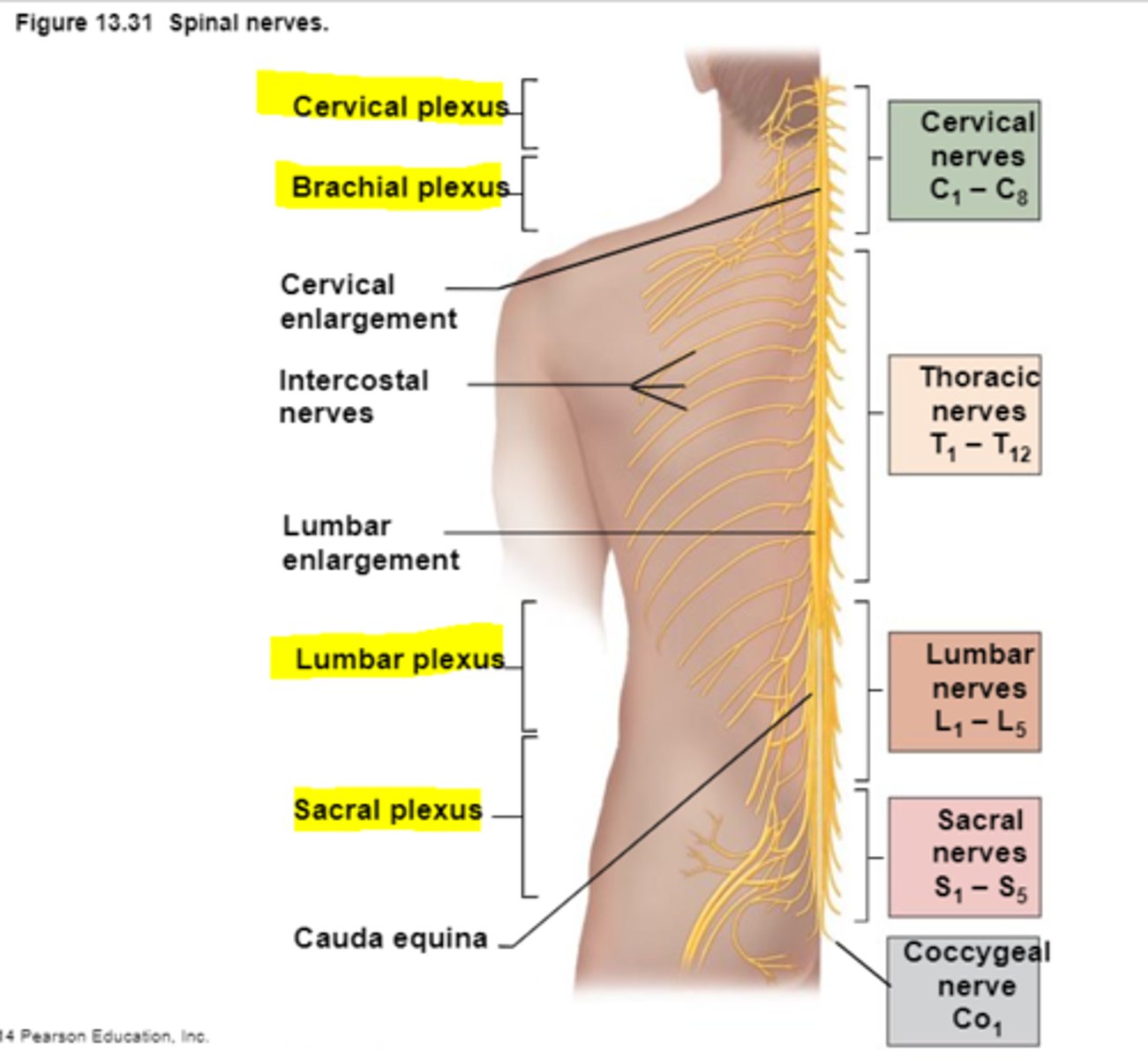
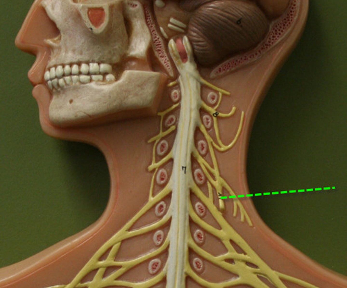
Cervical Plexus
Originated between C3-C5
1) Mostly cutaneous nerve innervation of neck and shoulder with exception of phrenic nerve
Phrenic Nerve
1)Supply both sensory and motor innervation to diaphragm
Clinical: Irritation to this nerve causes spasms of diaphragm known as hiccups,
If both phrenic nerves are severed, results in diaphragm paralysis and respiratory arrest occurs (patients kept alive by respirator)
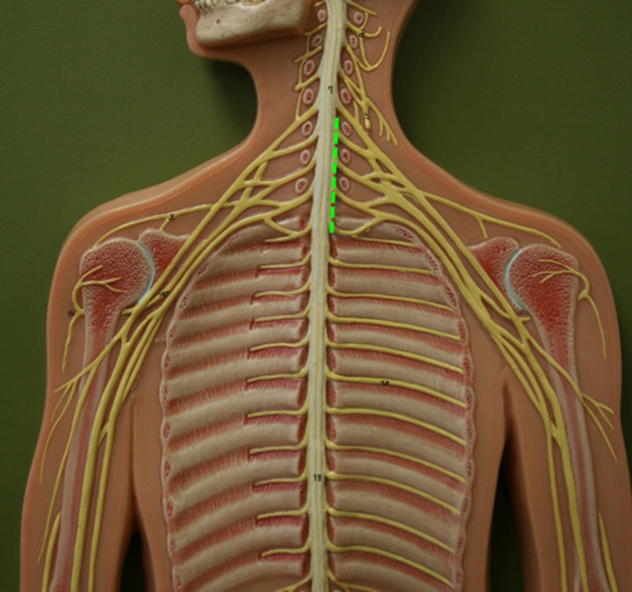
Brachial Plexus
1)Gives rise to virtually all nerves that innervate upper limb (arm).
Clinical: "Brachial block" blocks pain and movement in upper extremity while injuries can weaken or paralyze entire upper limb
2) Axillary, radial, median, and ulnar nerve.
Clinical: median nerve is compressed in carpal tunnel syndrome
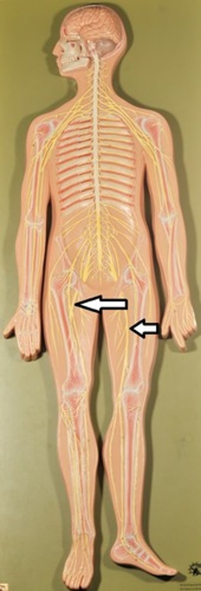
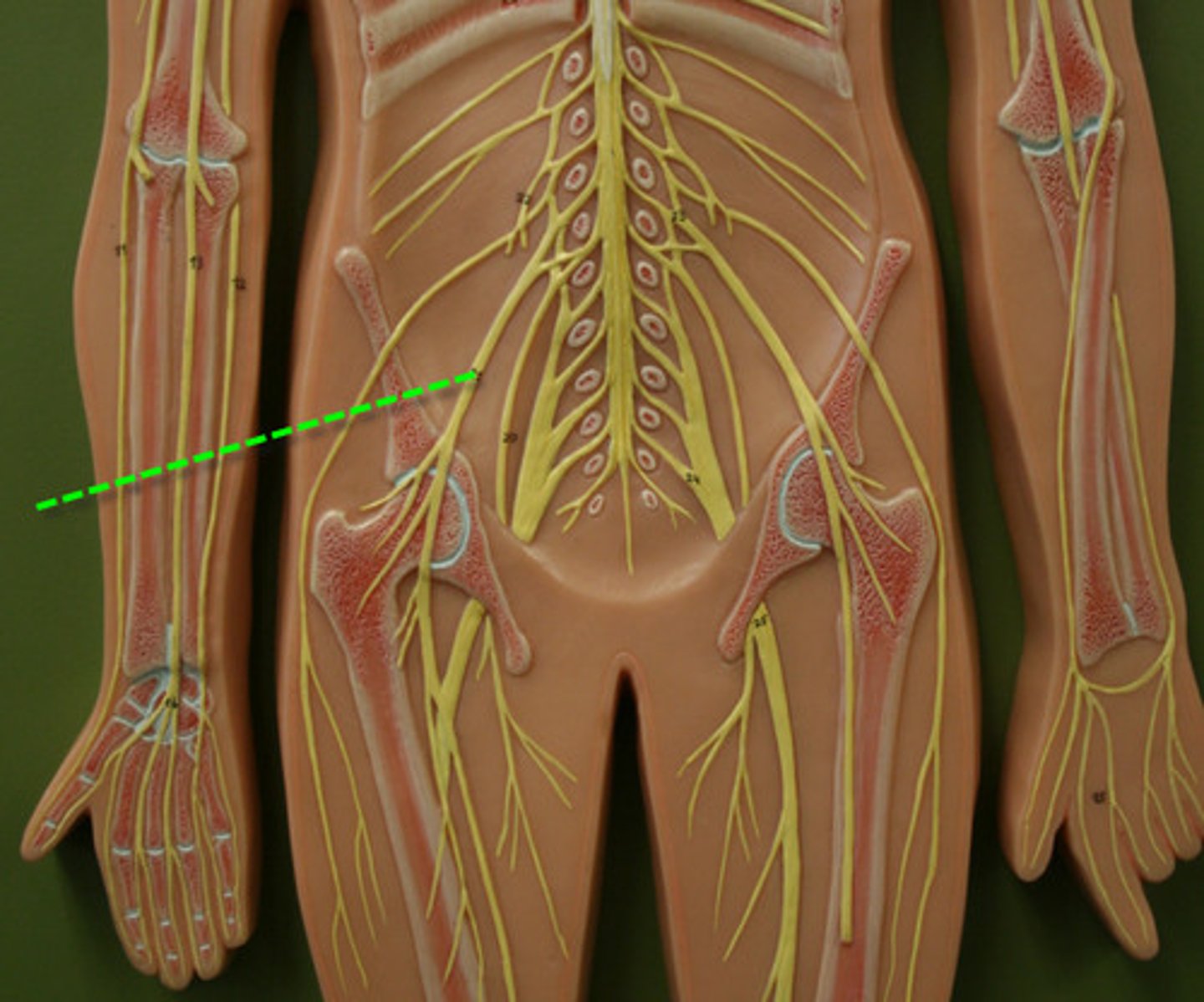
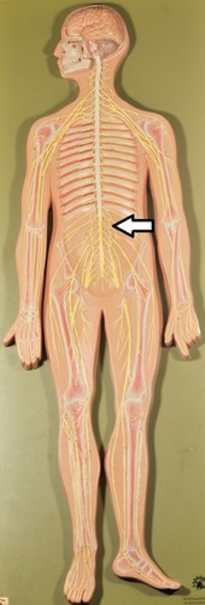
Lumbar Plexus
Origin: L1-L4
1) Proximal branches innervate parts of abdominal wall muscles but major branches descend to innervate anterior and medial thigh.
2) Femoral nerve
Clinical: Lumbar compression (as by a herniated disc) of spinal roots results in gait problems as well as pain or numbness of anterior thigh

Femoral Nerve
Innervates skin and muscles (quadriceps) of anterior thigh and medial surface of leg from knee to foot
Sacral Plexus
Origin L4-S4
1) Branches serve buttocks, lower limbs, pelvic structures and perineum
2) Sciatic nerve
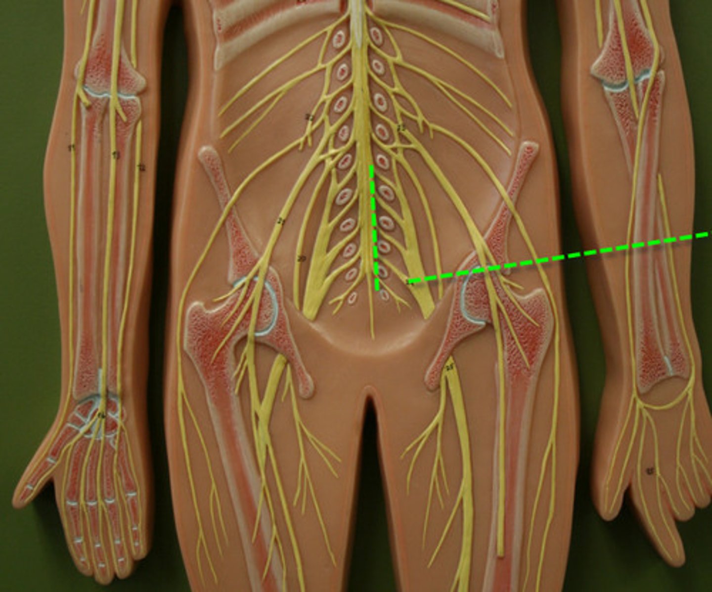
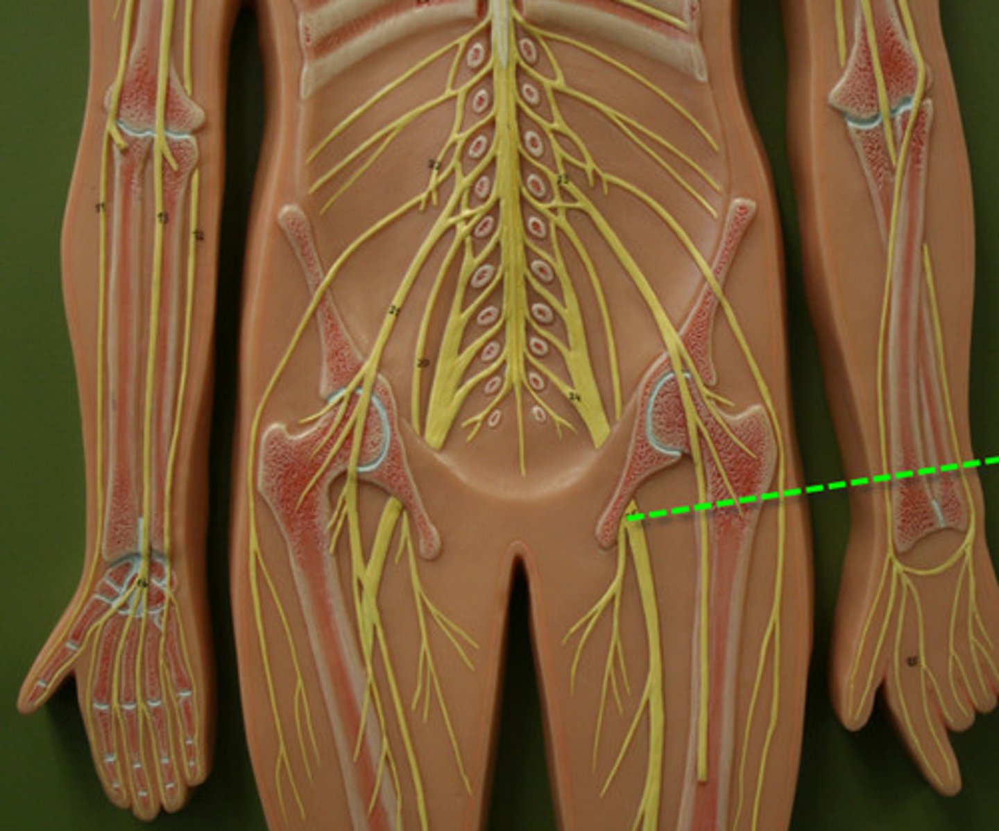
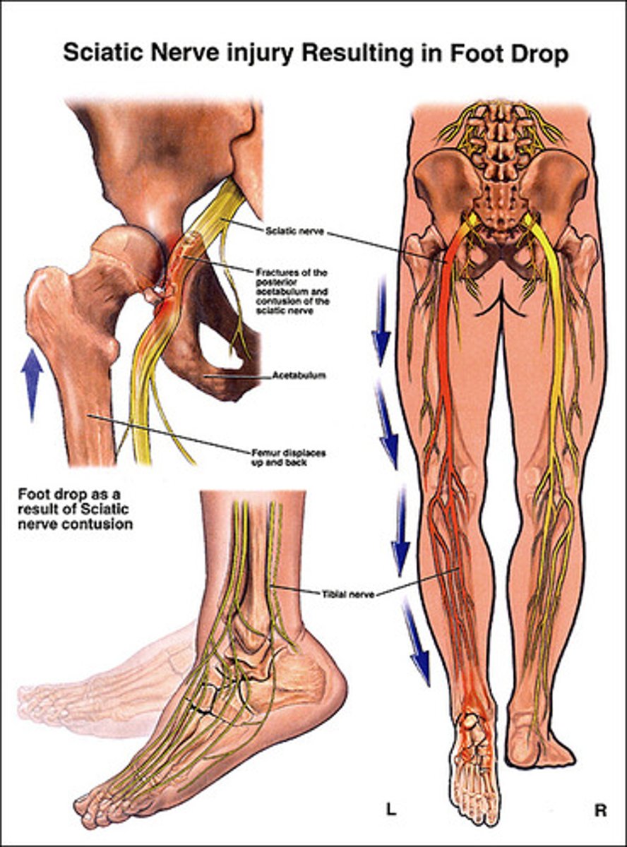
Sciatic Nerve
1) Thickest and longest nerve in body
2) Innervates posterior thigh to supply entire lower limb except anteromedial thigh
3) Has branches that innervate muscles and skin of perineum, help stimulate erection, and maintain voluntary control of urination
Sacral plexus clinical:
1) Lower lumbar-sacral compression of spinal nerves or damage from fall, disc herniation, or badly placed injection into buttock can produce pain, numbness, and impair lower limbs
2) Sciatica: stabbing pain radiating over course of sciatic nerve
3) Transection (serving) leaves leg nearly useless (hamstrings paralyzed and all foot and ankle movement is lost)
Dermatomes
1) Cutaneous branches of a single spinal nerve that innervate all areas of the skin.
2) Fairly uniform in width in body trunk and in direct line with their spinal nerves; different in lower limbs
-Lumbar nerves supply anterior leg surfaces
-Sacral nerves supply posterior leg surfaces
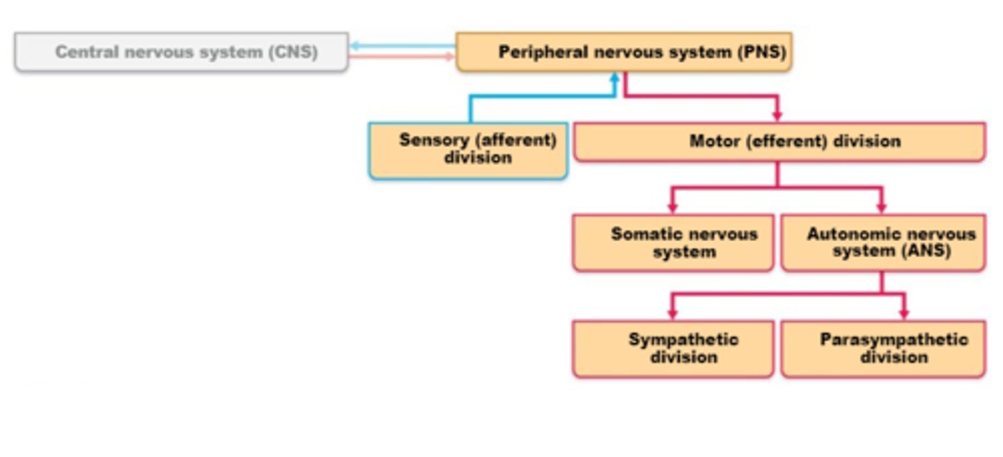
Motor Endings and the motor division
1) Efferent nerves in the PNS activate effectors by releasing neurotransmitters
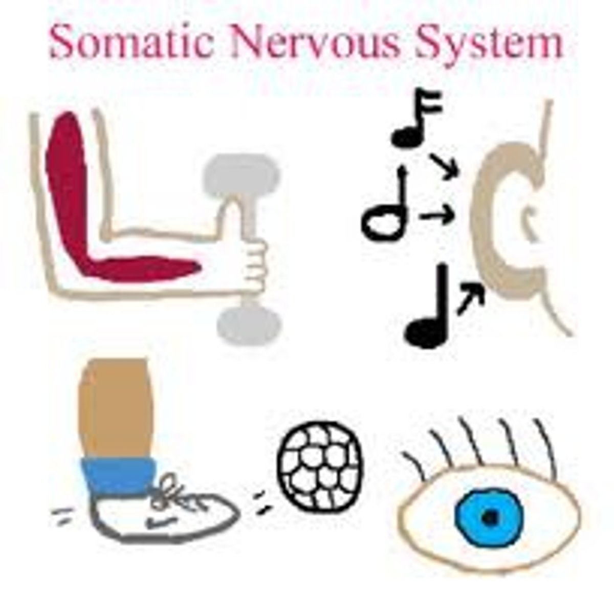
2) Somatic nerves to voluntary skeletal muscles (effectors)
3) Autonomic nervous system (involuntary) to internal organs (i.e. cardiac muscle, smooth muscle, and glands) has 2 pathways.
Efferent nerves in the PNS
1) Ach in the Somatic Nervous system
2) Typically Ach or NE in the ANS
Somatic nerves to voluntary skeletal muscles (effectors)
Motor endings at the neuromuscular junction concentrate neurotransmitters
- Very fast transmission of impulse across the synapse
Autonomic nervous system to internal organs pathways
1) Parasympathetic nerves
2) Sympathetic nerves
3) Motor endings are more diffuse on smooth muscle (slower transmission of impulse)

Parasympathetic Nerves
1)Routine daily activities with the ability to relax and slow down organ function
2)Local response (one foot on the brake)

Sympathetic Nerves
1) Vigorous exercise, excitement, emergency response.
2)Body wide mobilization (one foot on the gas)
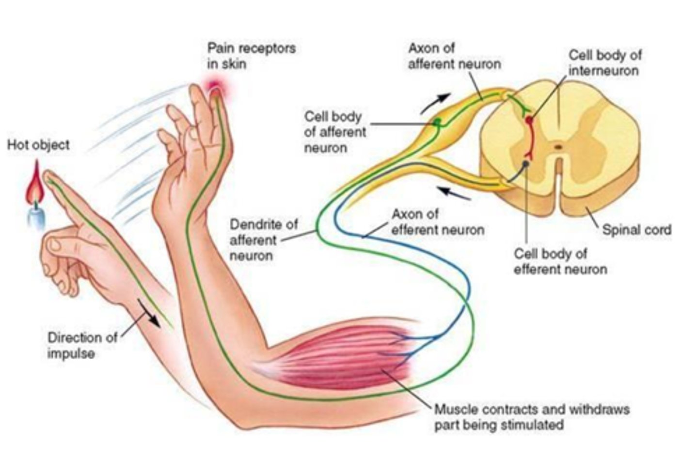
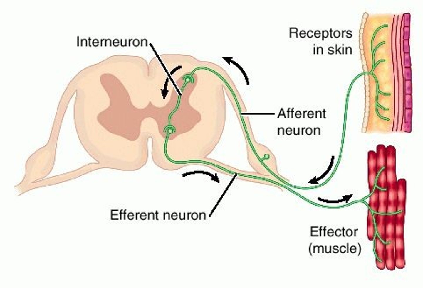
Reflex Arc
1) Basic functional unit of the nervous system (the neuron).
2) Capable of receiving a stimulus, integrating the information and sending out motor commands based on that stimulus
3)Seek to establish homeostatic balance and/or protection and are an automatic (involuntary) reaction to stimuli
4) Can be inborn (intrinsic) or learned (acquired from practice or repetition)
5) By testing reflex arcs, you gain knowledge of all 5 components

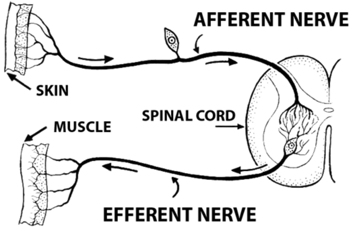
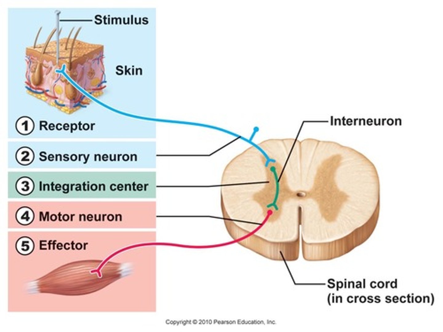
Components of Reflex Arc
Receptor,
Sensory (afferent) Neuron,
Integration Center (usually with an interneuron),
Motor (efferent) Neuron,
Effector
Functional classifications of Reflex Arcs
1) Somatic (activate skeletal muscle),
2) Autonomic (activate visceral effectors... smooth or cardiac muscle or glands)

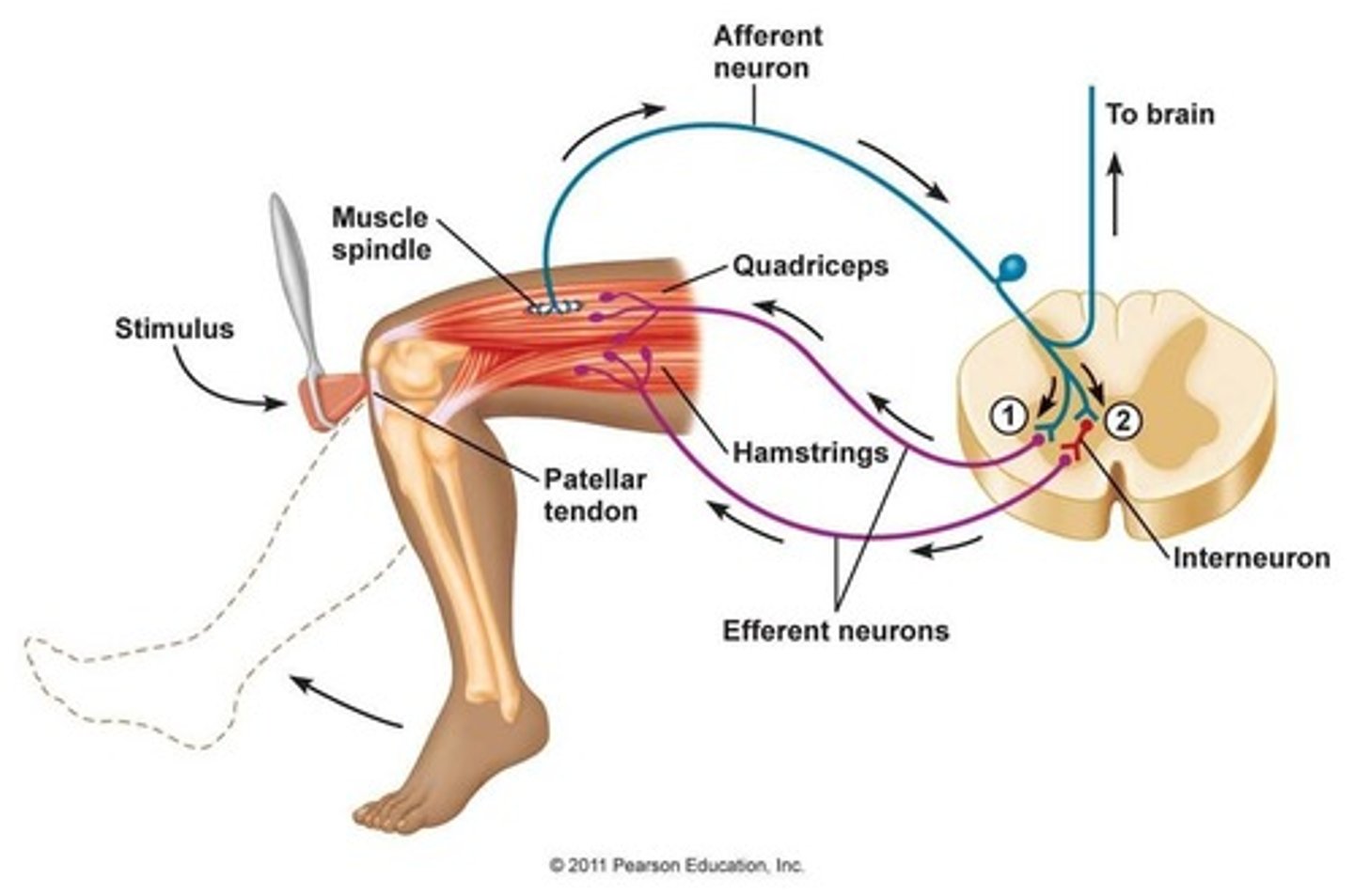
The Stretch Reflex
Muscle spindle receptors send action potentials in response to fibers stretching
ex: Knee jerk reflex-- As your knees begin to buckle, stretch receptor is activated and reflex causes quadriceps to contract without you having to think about it. Produces extension at the knee.
Reflex examples:
1) The stretch reflex
2) Withdrawal reflex
3) Cross-extensor reflex
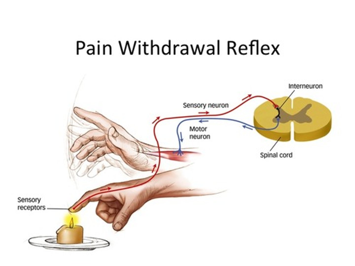
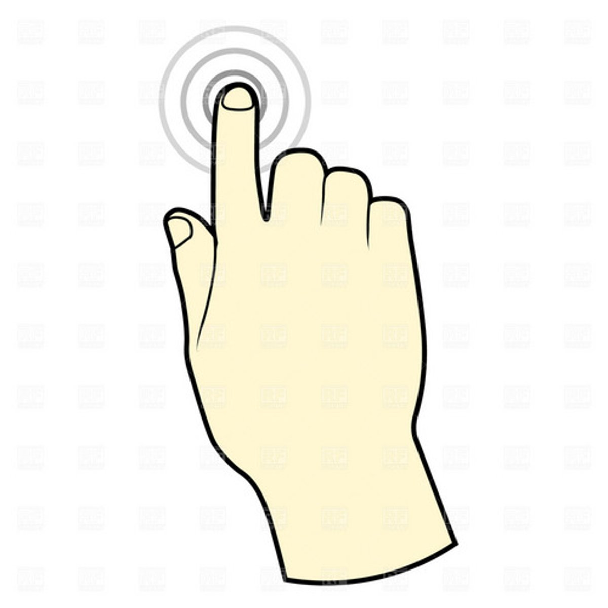
The Withdrawal Reflex
Causes automatic withdrawal of threatened body part from painful or unpleasant stimulus
ex) arm withdrawal reflex

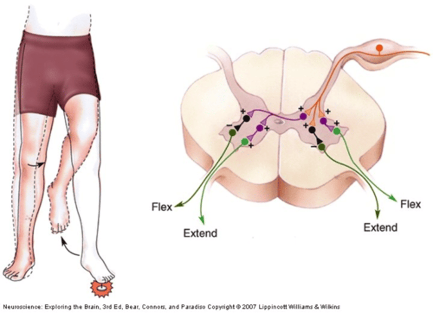
Cross-Extensor Reflex
1) One body part withdraws (flexion) while the opposite side of the body extends
2) Particular important in maintaining balance
ex: stepping on broken glass (rapid lifting of cut foot while activation of extensor muscles on opposite leg to support weight suddenly shifted to it)
Reflexes interact with other nerve pathways
In the withdrawal reflex, signals from pain receptors continue on to the brain to be interpreted as pain even though the reflex has already removed the body art

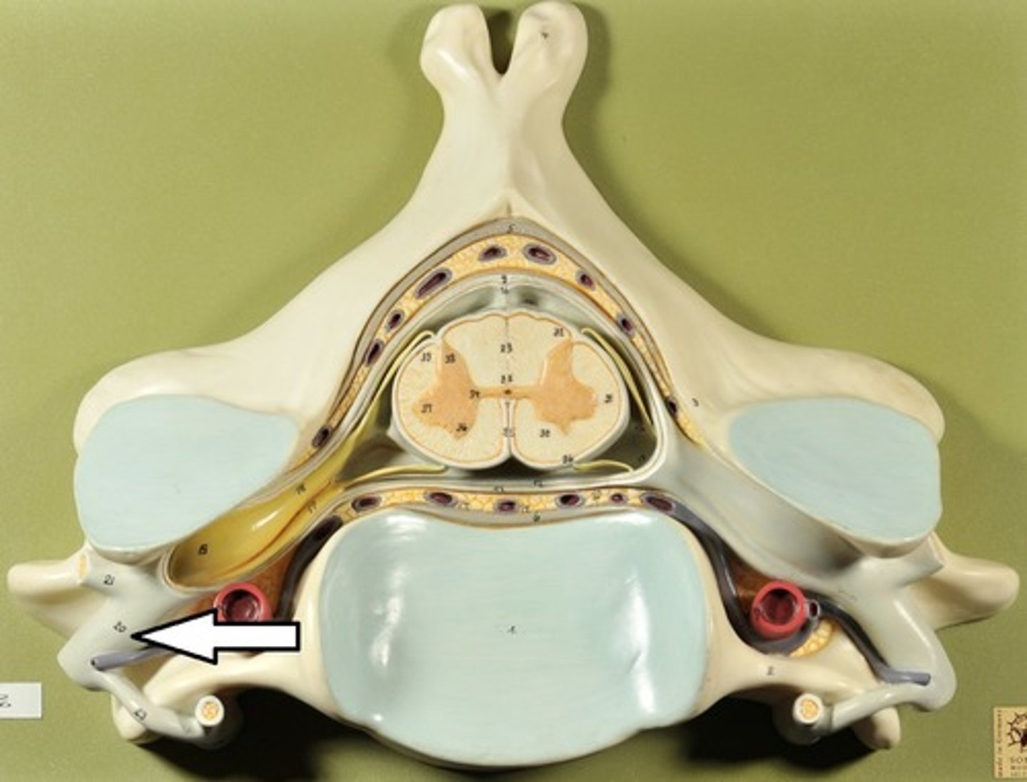
Ventral Root
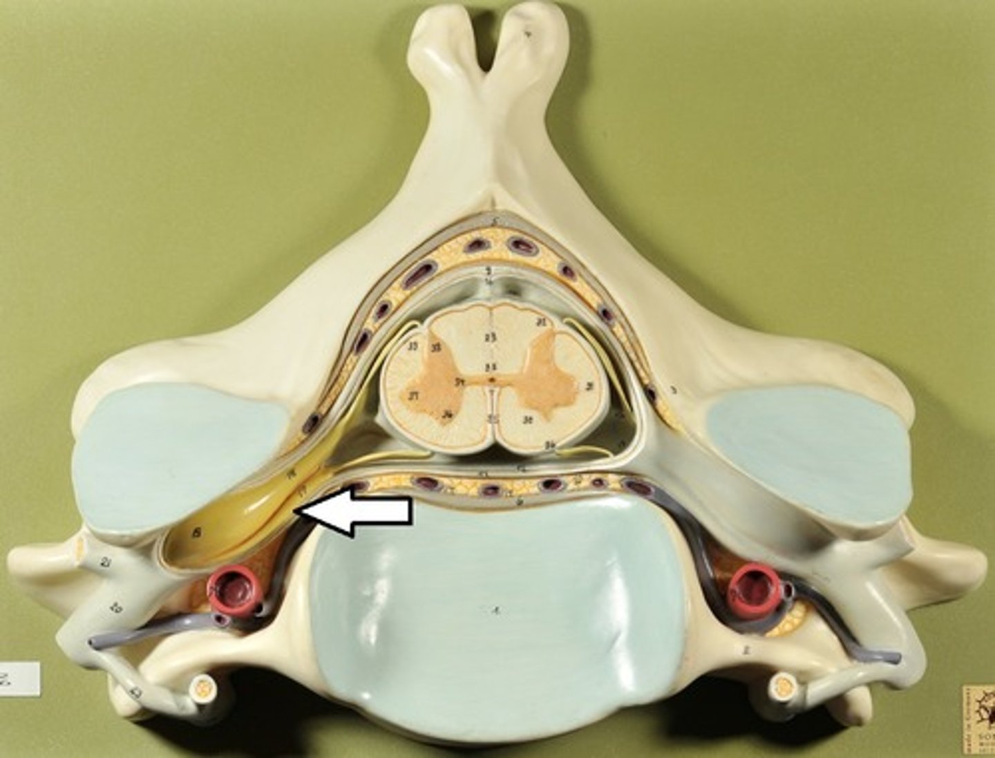
Dorsal Root
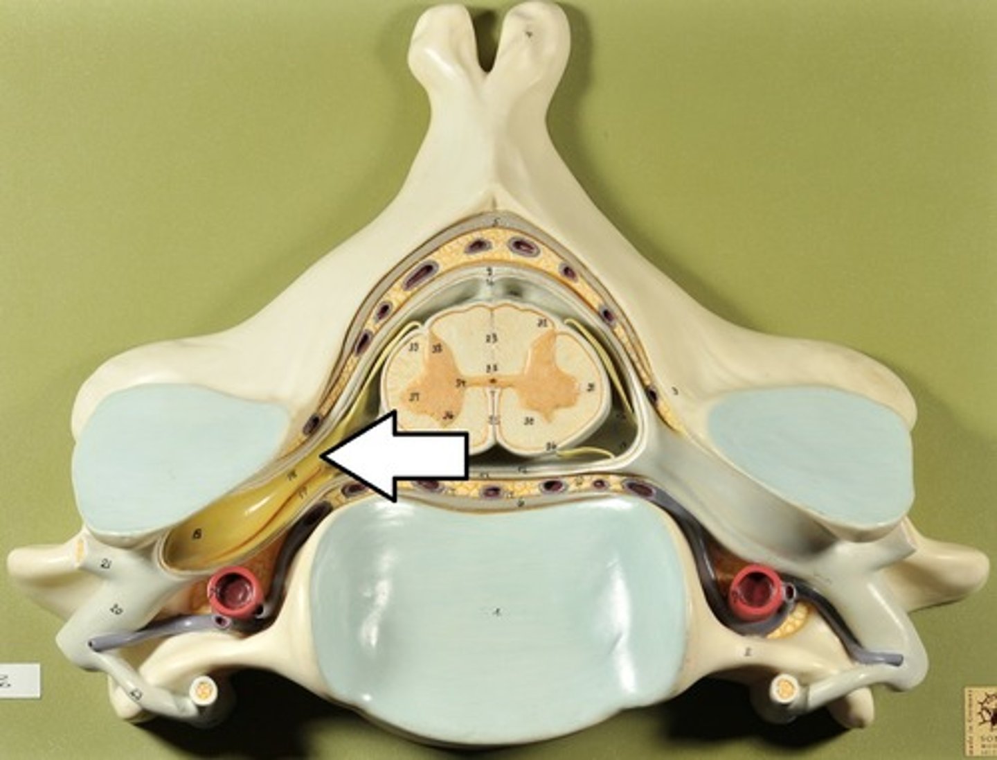
Spinal Nerve
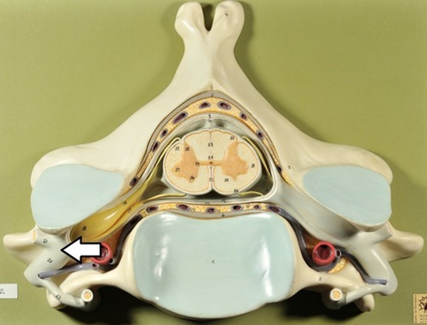
Dorsal Ramus
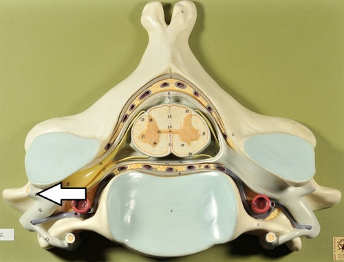
Ventral Ramus
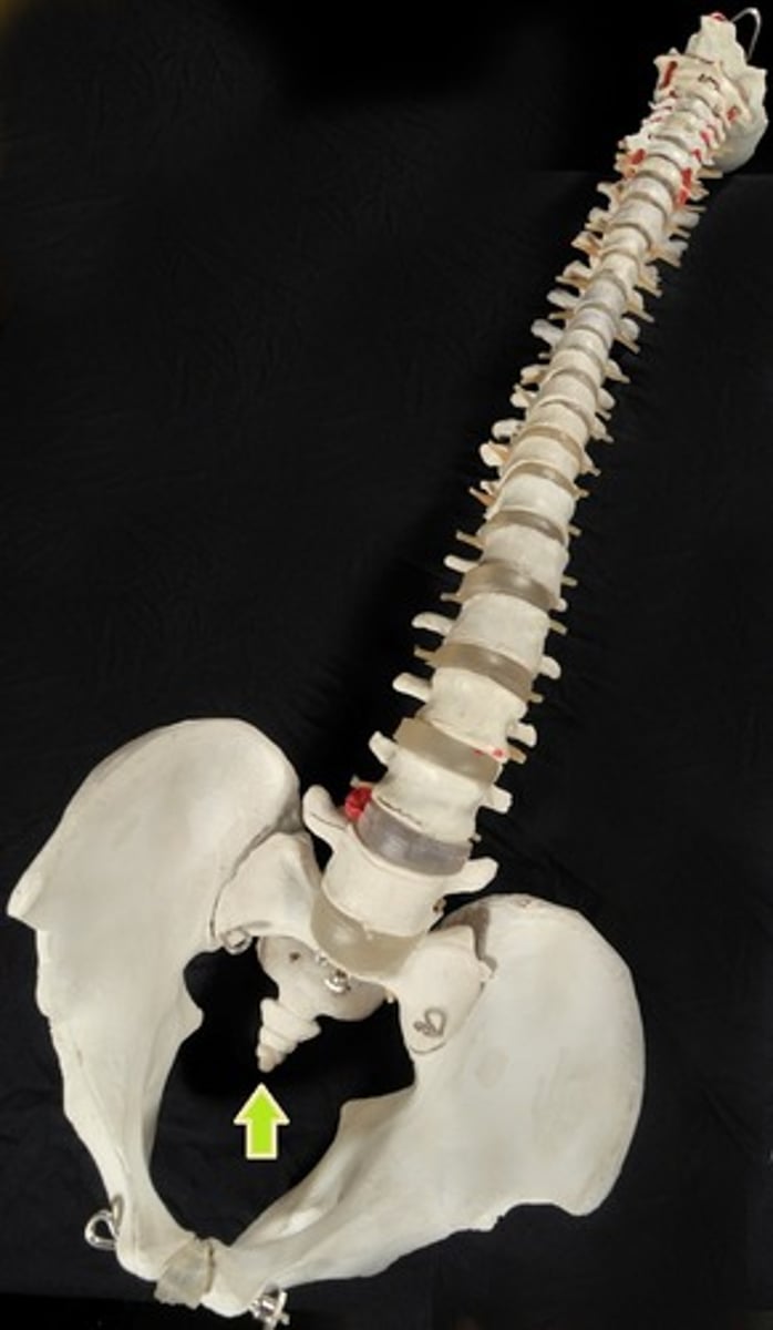
Coccygeal Nerve
Cervical Plexus

Phrenic Nerve
Brachial Plexus
Median Nerve

Intercostal Nerves
Lumbar Plexus
Femoral Nerve

Sacral Plexus
Sciatic Nerve