Neuroscience - Spinal Cord
1/177
Earn XP
Description and Tags
Name | Mastery | Learn | Test | Matching | Spaced | Call with Kai |
|---|
No analytics yet
Send a link to your students to track their progress
178 Terms
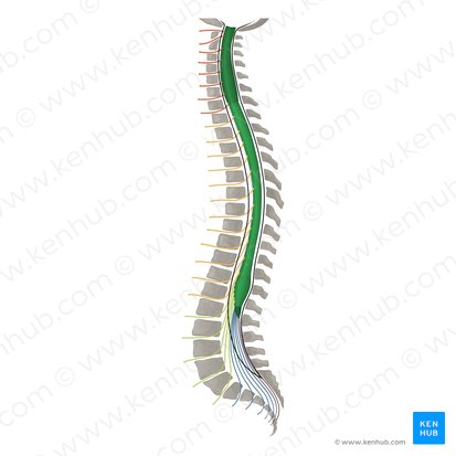
Where does the spinal cord extend?
from foramen magnum to L1/L2
What is the shape of the central area of the spinal cord and what is it made up of?
butterfly-shaped central area of gray matter
How is the gray and white matter of the spinal cord organized?
-gray matter on inside
-white matter on outside
(opposite of cerebrum)
What is the central canal of the spinal cord a remnant of in early development?
neural tube
How many pairs of spinal nerves are there?
31 (8 cervical, 12 thoracic, 5 lumbar, 5 sacral, 1 coccygeal)
What are the regions of enlargements of the spinal cord and why does this occur?
cervical and lumbar regions due to plexuses
What is the end of the spinal cord known as?
cauda equina ("horse's tail")
What is the location of a spinal tap?
lumbar cistern
Where does the spinal cord receive primary sensory info from?
via somatosensory receptors in the skin, skeletal muscles, tendons, via viscera
via viscerosensory receptors in the thoracic, abdominal, and pelvic viscera
What do somatic motor and visceral motor neurons innervate?
skeletal muscle, smooth muscles, cardiac muscle, glandular epithelium
What is the most important conduit between the body and the brain?
spinal cord
What fibers enter the spinal cord?
ascending somatosensory fibers
What fibers exit the spinal cord?
descending corticospinal fibers from the cerebral cortex to spinal neurons
What is neurulation?
process in which the neural plate, a flat structure of ectodermal tissue in the vertebrae embryo, folds to form the neural tube
In development of the spinal cord, what forms the alar plate?
cells on the dorsal side form the alar plate which subsequently becomes the dorsal horn (posterior)
In development of the spinal cord, what forms the basal plate?
cells at the ventral end form the basal plate which subsequently becomes the ventral horn
What is the ventral median fissure of the spinal cord?
a prominent longitudinal groove present on the anterior (ventral surface) of the spinal cord
Where does the ventral median fissure extend and what is its purpose?
-rungs along the midline, extending from the cervical to sacral region
-plays an important role in separating the spinal cord into two symmetrical halves
What is the dorsal median sulcus?
shallow groove found in the midline of the posterior aspect of the spinal cord
What is the dorsal median sulcus an external boundary of?
posterior median septum
What does the dorsal median sulcus separate?
two posterior funiculi
What is the dorsal funiculus/column?
region of sensory white matter located between the posterior median sulcus at the midline and the entry points of the posterior rootlets into the spinal cord (ex. posterolateral sulcus)
What does the dorsal funiculus/column form an integral component of?
forms an integral component of the dorsal column-medial leminiscal pathway
What kind of info does the dorsal column-medial leminiscal pathway facilitate the transmission of?
the transmission of vibration, fine touch, proprioception, and 2-point discrimination sensations from the periphery to the brain
What makes up the lateral funiculus/column?
several ascending and descending white matter tracts including the corticospinal and spinothalamic tract
What makes up the ventral funiculus/column?
white matter tracts including retriculospinal tract, vestibulospinal fibers, and anterior corticospinal tract
Where are the anterior and posterior grey commisures located?
within spinal area X
What is the anterior white commisure?
bundle of nerve fibers crossing the midline of the spinal cord
Where is the anterior white commisure located?
posterior to anterior median fissure and anterior to anterior gray commisure and central canal
What major neuronal pathways cross through the anterior white commisure?
-sensory spinothalamic tracts
-anterior corticospinal tract
What is the white matter of the spinal cord divided into?
3 funiculi
What are funiculi?
bundle of nerve fibers; white matter columns
What are the 3 funiculi that make up the white matter of the spinal cord?
-posterior (dorsal) funiculis/column
-lateral funiculus/column
-anterior (ventral) funiculus/column
What is located in the posterior funiculis/column?
-cuneate and gracile fasciculi (DCML)
-Lissaeur's tract (posterolateral tract)
What are fasiculi?
smaller grouping of axons
What is located in the lateral funiculus/column?
-lateral corticospinal tract
-anterolateral system (STT)
What is located in the anterior funiculus/column?
-reticulospinal tract
-vestibulospinal fibers
-anterior corticospinal tract
How is the gray matter of the spinal cord divided?
into Rexed laminae I to IX and Area X
How many Rexed laminae of gray matter in the spinal cord are there?
10
Where are Rexed laminae I - VI located?
posterior horn
Where is Rexed laminae VII located?
lateral horn/intermediate zone
Where are Rexed laminae VIII and IX located?
anterior horn
Where is Area X located?
around central canal
How are Rexed laminae characterized?
by the input they receive and the trajectory of axons arising there
What is located in the posterior horn of gray matter? (7)
-posterolateral tract
-Lamina I
-Lamina II
-Lamina III
-Lamina IV
-Lamina V
-Lamina VI
What is the posterolateral tract also known as?
Lissaeuer's Tract
What occurs in posterolateral tract (Lissauer's Tract)?
sensory nerves enter here, ascend or descend up to 4 segments and terminate in Rexed's laminae I through VI
What does Rexed Lamina I contain?
-posteromarginal nucleus
-noxious stimuli
What does Rexed Lamina II contain?
-substantia gelatinosa (of Rolando)
-noxious stimuli
What does Rexed Lamina III to IV contain?
-nucleus proprius
-position, light touch stimuli
What does Rexed Lamina V and VI contain?
-divided into medial and lateral portions
-noxious, visceral, proprioceptive stimuli
What is located in the lateral horn/intermediate zone of gray matter?
lamina VII
What does Rexed Lamina VII contain?
-dorsal nucleus of Clarke
-interomediolateral nucleus
What spinal levels is the dorsal nucleus of Clarke found in?
C8-L3
Where in the lateral horn/intermediate zone of gray matter can Rexed Lamina VII be found?
intermediate zone from central canal to lateral edge of spinal gray
What is the function of the dorsal nucleus of Clarke (spinocerebellar tract)?
relays unconscious proprioceptive info to the brain
What is the function of interomediolateral nucleus?
relays sensory info from viscera to the brain and autonomic signals from the brain to visceral organs
What spinal levels can the interomediolateral nucleus be found in?
T1-L2
What is located in the anterior horn of the gray matter of the spinal cord? (3)
-lamina VIII
-lamina IX
-lamina X
What is contained in Rexed Lamina VIII?
interneurons
What is contained in Rexed Lamina IX?
alpha, beta, and gamma motor neurons
Where is Rexed Lamina X located?
around the central canal (contains decusation)
What do alpha motor neurons innervate?
extrafusal muscle fibers
What is the largest motor neuron of the anterior horn cells?
alpha motor neurons
What stimulates alpha motor neurons?
Ia primary afferents and group II of muscle spindles or descending tracts
What type of motor neuron is associated with Renshaw cells (interneurons)?
alpha motor neurons
What is the function of Renshaw cells (interneurons)?
influence alpha motor neurons (inhibitory)
What do beta motor neurons innervate?
extrafusal and intrafusal muscle fibers
What is the diameter of beta motor neurons comparable to?
alpha motor neurons
What do gamma motor neurons innervate?
intrafusal muscle fibers
What regulates gamma motor neurons?
intersegmental pathways
Do gamma motor neurons receive inhibitory feed ack by Renshaw cells?
no
What does co-activation of gamma and alpha motor neurons ensure?
that muscle spindles remain sensitive to stretch during muscle contraction
What is the shape of the cervical level of the spinal cord?
oval
Which region of the spinal cord is proportionately larger than in other regions of the spinal cord?
cervical
What are cervical level characteristics of the spinal cord?
-increased area of white matter due to ascending and descending tracts
-gracile and cuneate fasciculi prominent
What is the shape of the thoracic level of the spinal cord?
round shape with relatively small posterior and anterior horns
Why does the thoracic level of the spinal cord have a progressive decrease in the amount of white matter?
due to gracile fasciculus only at lower levels
What makes the white matter of the thoracic level of the spinal cord appear larger?
small amount of gray matter
What are characteristics of the thoracic level of the spinal cord?
-dorsal nucleus of clarke
-lateral horn
Where does the dorsal nucleus of clarke (posterior thoracic nucleus) have projections to?
projections to the cerebellum via spinocerebellar tract
What is in the lateral horn at the thoracic level of the spinal cord?
-intermediolateral cell column
-preganglionic sympathetic neurons
What is the shape of the lumbar level of the spinal cord?
large round anterior horns
Does the lumbar level of the spinal cord have more white or grey matter?
more gray matter
In the lumbar level of the spinal cord, where is the dorsal nucleus of clarke obvious?
L1/L2 levels (upper lumbar regions)
What is the shape of the sacral level of the spinal cord?
round shape
Does the sacral level of the spinal cord have more white or gray matter?
gray matter
What does the intermediate gray matter at the S2-S4 (sacral level) contain?
preganglionic parasympathetic visceormotor neurons
What is obvious at the sacral level of the spinal cord?
substantia gelatinosa (lamina II)
What does the substantia gelatinosa (lamina II) convey?
thermal and nociceptive input
What are features of the dura mater of the spinal cord?
-dural sac
-filum terminale externum
What are features of arachnoid mater of the spinal cord?
-subarachnoid space filled with CSF
-lumbar cistern
What are features of pia mater of the spinal cord?
-denticulate ligaments
-filum terminale internum
What is the vasculature of the spinal cord?
-anterior and posterior spinal arteries
-radicular arteries
-spinal medullary arteries
What space are the radicular arteries a content of?
intervertebral foramen
What do the radicular arteries serve?
anterior and posterior roots
What do spinal medullary arteries bypass?
bypass the root to serve the cord
What do the terminal branches of the spinal medullary arteries join to form?
arterial vasocorona ("crown" of blood vessels around spinal cord)
How many possible types of fibers can the spinal nerves contain?
4
What are the 4 types of fibers of spinal nerves?
-general somatic afferent (GSA)
-general visceral afferent (GVA)
-general somatic efferent (GSE)
-general visceral efferent (GVE)