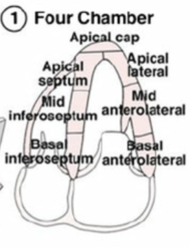
Wall Segments and Ultrasound Images
1/27
There's no tags or description
Looks like no tags are added yet.
Name | Mastery | Learn | Test | Matching | Spaced | Call with Kai |
|---|
No analytics yet
Send a link to your students to track their progress
28 Terms
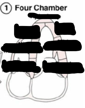
Identify this image.
A4C
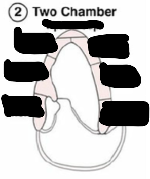
Identify this image.
A2C

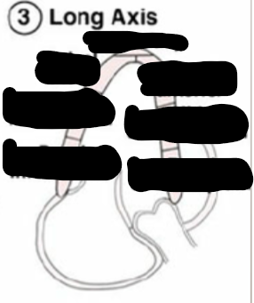
Identify this image.
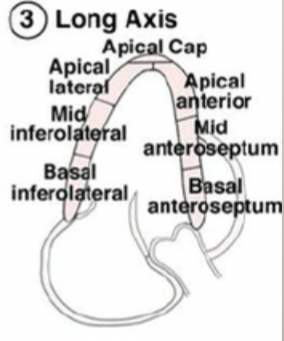
A3C or LONG AXIS
Identify this image.
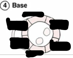
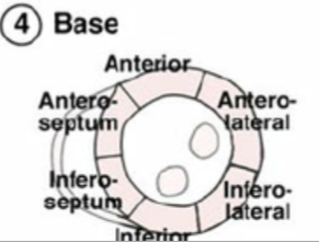
Base or top
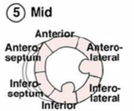
Identify this image.
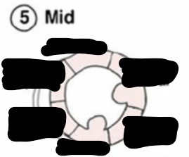
Mid
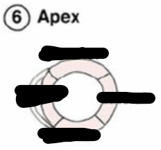
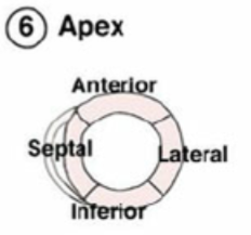
Identify this image.
Apex or bottom
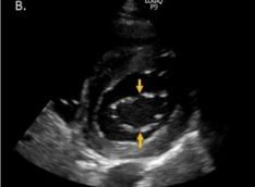
What is the “scout view”?
First image taken with an increased depth to visualize effusion
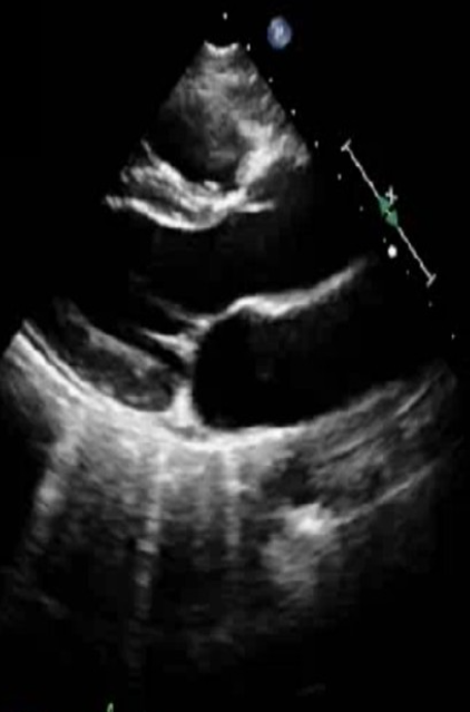
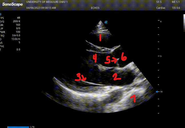
Identify this image.
PLAX
RV
LA
MV
LVOT
AV
Aortic root
Descending AO
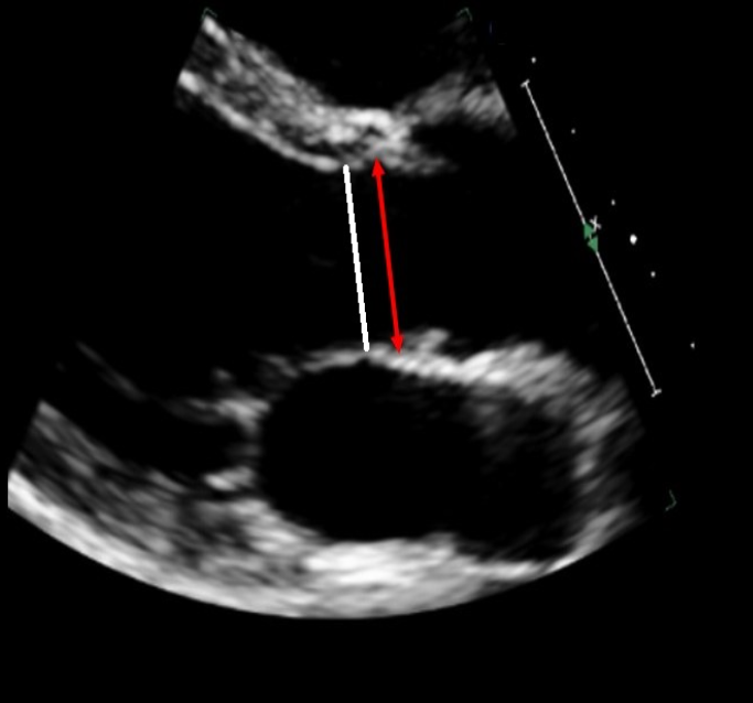
Identify this image.
PLAX AV measurements during MID SYSTOLE
White: LVOT
Red: Aortic annulus
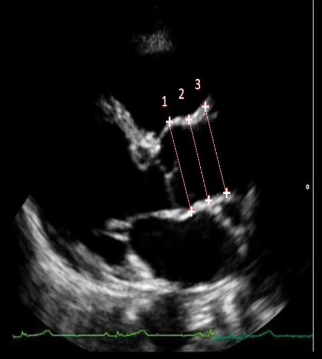
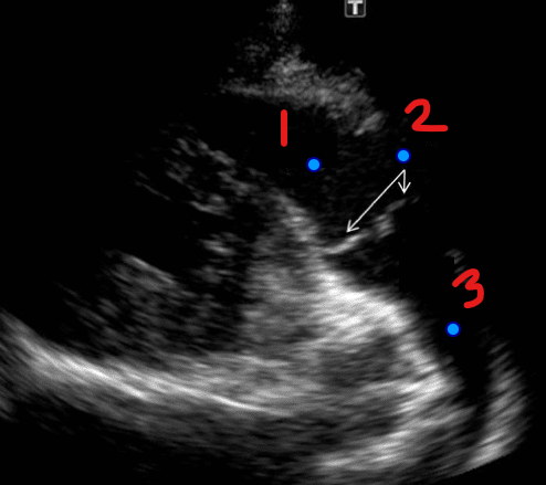
Identify this image.
PLAX AV measurements during END DIASTOLE
Sinus of Valsalva or aortic root
Sinotubular junction (STJ)
Ascending aorta
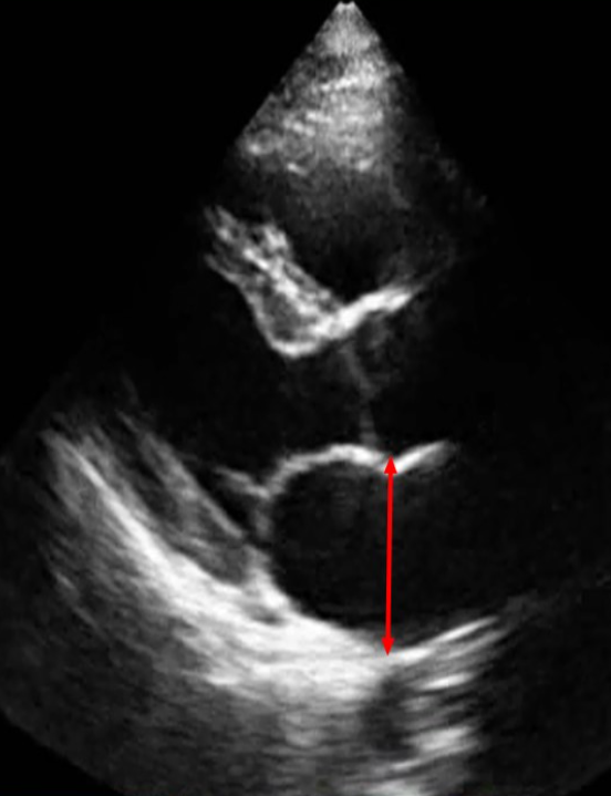
Identify this image.
PLAX LA diameter measured during END SYSTOLE
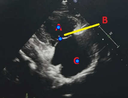
Identify this image.
PLAX RVIT
A. RV
B. TV
C. RA
Identify this image.
PLAX RVOT
RVOT
PV
Main pulmonary artery
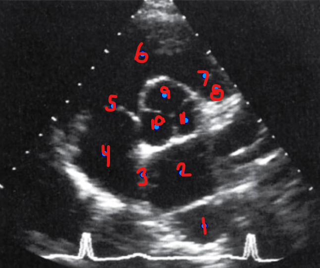
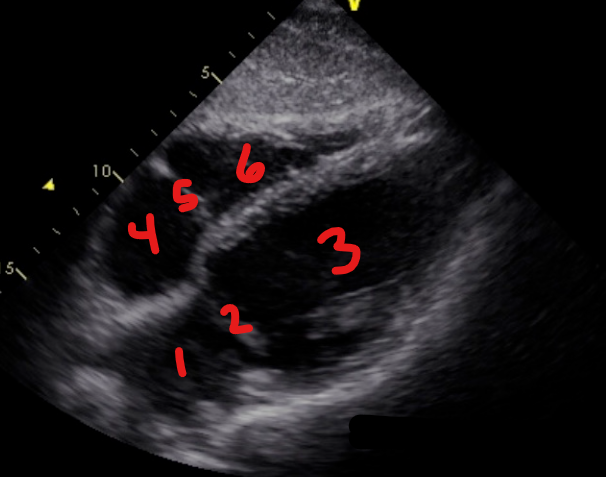
Identify this image.
PSAX AV Basal
Descending aorta
LA
IAS
RA
TV
RVOT
PV
Main pulmonary artery
Right coronary cusp (adjacent to RVOT)
Noncoronary cusp
Left coronary cusp (adjacent to LA)
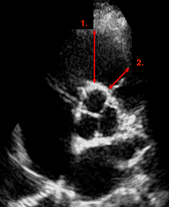
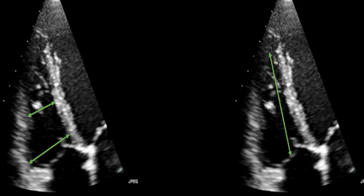
Identify this image.
Proximal RVOT measured at END DIASTOLE
Distal RVOT measured at END DIASTOLE
Identify this image.
PSAX MV level
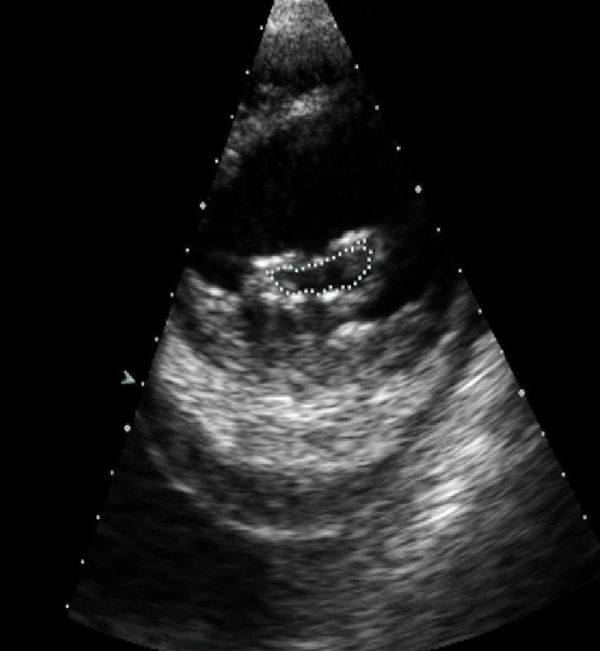
Identify this image.
Planimetry or most accurate way to measure MVA

Identify this image.
PSAX LV
Papillary muscle
LV
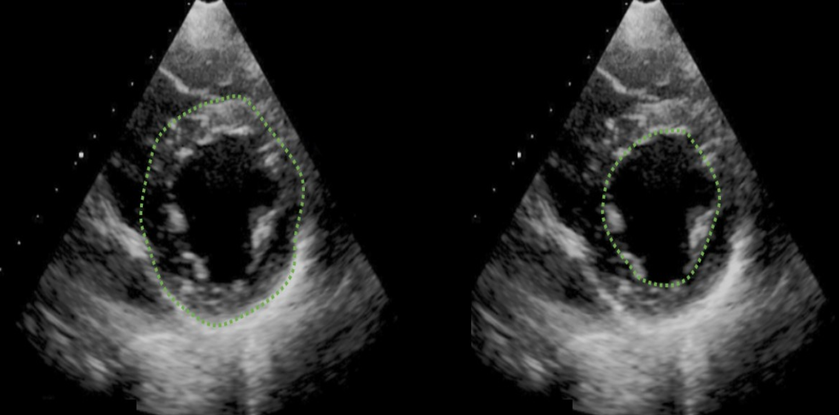
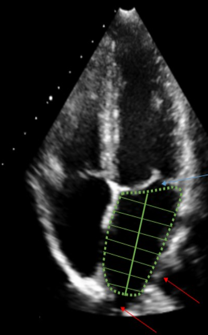
Identify this image.
LV mass measured at END DIASTOLE
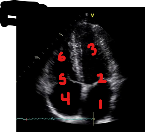
Identify this image.
A4C
LA
MV
LV
RA
TV
RV
Identify this image.
LA volume measured at END SYSTOLE
Identify this image.
RV dimensions measured at END DIASTOLE
Identify this image.
A2C
LA
MV
LV
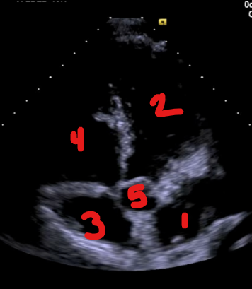
Identify this image.
A5C
LA
LV
RA
RV
Ao
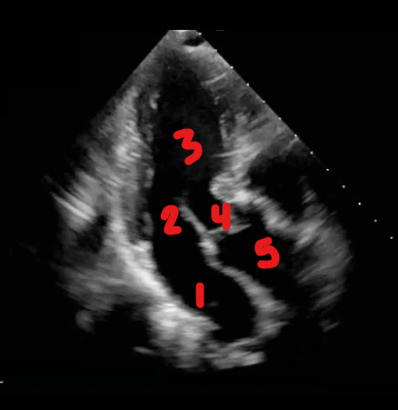
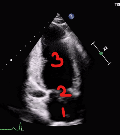
Identify this image.
A3C
LA
MV
LV
AV
Ao
Identify this image.
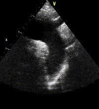
Suprasternal view of descending aorta
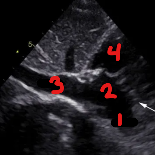
Identify this image.
IVC Subcostal
SVC
RA
IVC
RV
Identify this image.
Subcostal four chamber
LA
MV
LV
RA
TV
RV