Heart Anatomy
1/43
There's no tags or description
Looks like no tags are added yet.
Name | Mastery | Learn | Test | Matching | Spaced |
|---|
No study sessions yet.
44 Terms
Pericardium and types
double serous membrane
Visceral pericardium- next to the heart
Parietal pericardium- outside layer
What fills space between layers of pericardium?
Serous fluid
3 Heart Walls
Epicardium
Myocardium
Endocardium
Epicardium
Epicardium
Outside layer (visceral pericardium)
Connective tissue layer
Myocardium
Middle layer (cardiac muscle)
Endocardium
Inner layer (endocardium)
Continuous within linings of blood vessels leaving and entering the heart
How many Chambers are there?
4, right and left sides act as seperate pumps
Atria
recieving chambers

Right and left ventricle
discharging chambers

Interventricular septum
longitudinal separaion

How may valves and function
4, allows blood to flow in one direction
Atrioventricular valves
- between atria and ventricles
Bicuspid (left)
Tricuspid (right)

Semilunar valves
between ventricle and artery

Pulmonary and aortic semilunar valve
• valves open as blood pumped through
• Held in place by chordae tendineae
(Heart strings)
• close to prevent back flow

Heart Strings
chordae tendineae
Aorta
leaves left ventricle; oxygenated blood
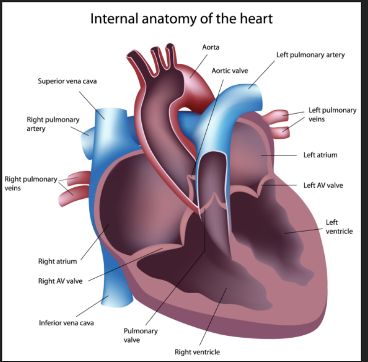
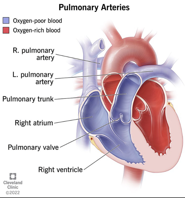
Pulmonary artery
leaves right ventricle detox blood to lungs
Vena Cava
enters right atrium

Pulmonary veins (4)
Enter left atrium oxygenated blood from lungs

Coronary circulation
nourshing the circulatory system- involves coronary arteries, cardiac veins, blood empties into the right atrium via coronary sinus
2 types of Crornary circulation
Pulmonary circulation - heart to lungs
Systemic Circuit- heart to body
Conduction System
intrinsic conduction system (nodal) system heart muscles, cells contract, without nerve impulses, in a regular, continuous way
• special tissue sets the pace
Sinoatrial node (pacemaker maker)
Atrioventricular node
Atrioventricular bundle
Bundle branches
Purkinje fibers
Cardiac Cycle
Atria contract simultaneously
• Atria relax, then ventricles contract
• Systole = contraction
• Diastole = relaxation
Systole
contraction
Diastole
relaxation
Ventricular systole
blood pressure builds before ventricle contracts pushing out blood
Early diastole Ventricular systole
atria finish re-filling, ventricular pressure is low
ECG EKG
P Wave - atrial depolarization (Contraction)
QRS - ventricular depolarization (Contraction)
T wave - ventricular repolarization (Relaxation)
U wave - repolarization of the purkinje fibers (Relaxation)
P Wave
P Wave - atrial depolarization (Contraction)
QRS
QRS - ventricular depolarization (Contraction)
T wave
T wave - ventricular repolarization (Relaxation)
U wave
U wave - repolarization of the purkinje fibers (Relaxation)
cardiac output (CO)
Amount of blood pumped by each side of the heart in one minute
stroke volume
volume of blood put out by each ventricle in one contraction
healthy heart pumps out about ___ of blood present ventricles ___
healthy heart pumps out about 60% of blood present ventricles (2oz)
Starling’s law of the heart
the more that the cardiac muscle is stretched, the stronger the contraction
What increase stretching
Venous return increases stretching
Exercise and muscular pump
tachycardia
tachycardia- 100+ pm
Bradycardia
Bradycardia- -60 pm
Fribrillation
Fribrillation- lack of blood supply to the heart muscle
shuddering
Useless pump
stroke volume constant
declines when blood volume suddenly drops or weakened heart rate
How can you modify Basic heart rate?
Cardiac output is maintained by faster heartbeat
• HR is also modified by chemicals, hormones and ions
Neural controls that control heart rate
• physical and emotional stress
stimulates SA and AV nodes
Heart rate increases
• congestive heart failure
”worn out” or weak
Prescription meds can enhance contractile force and stroke volume
• hormones and ions (electrolytes)
epinephrine and thyroxine increases HR
Ion calcium in blood
Depresses and slows the heart
Physical Factors of HR
• Age, gender, exercise, body temperature
• Fetus resting HR- 140-160
• Females 72-80