Electrocardiogram (EKGs)
1/35
There's no tags or description
Looks like no tags are added yet.
Name | Mastery | Learn | Test | Matching | Spaced |
|---|
No study sessions yet.
36 Terms
Toward, upward
If sum of electrical vectors on EKG is __ a lead, it is positive in direction and spike is __
P wave
SA node fire and depolarization of atria → Atrial systole
(EKG component)
PR Interval
Time between atrial depolarization and ventricular depolarization; reflects conduction through AV node
(EKG component)
Q
Depolarization of interventricular septum (QRS complex)
R
Depolarization of main mass of ventricles (QRS complex)
S
Depolarization towards base of heart (QRS complex)
ST segment
Reflects plateau of action potentials in ventricles → ventricular contraction
(EKG component)
T wave
Ventricular repolarization immediately before ventricular relaxation
(EKG component)
Contraction, initiates
The P-wave does NOT equal atrial __, rather it __ this process
Depolarization, contraction, Ca2+
Events in P-wave
Represents atrial __ and initiates atrial __
Electrical activity going thru atria with high __ (ion) concentration
AV, slowly, contracting
Events in PR Segment
Delay as signal moves through the __ node
AV node __ depolarizing → Not visible since amount of tissue is so small
Atria still __
Contraction, spike, atrial
Events in QRS Complex
Ventricle depolarization and ventricular __
More cardiac muscle (myocytes) means more charge = illustrated by a visible __
Also __ repolarization technically, not visible
Contraction, Ca2+
Events in S-T Interval
Ventricular __, no net electrical forces
Ventricles contracting with high intracellular __ levels
Repolarization, Ca2+
Events in T-wave
Ventricular __
ventricles still contracting with high intracellular __ levels
Bump, +, -, inversion
__ patterns in EKG represent differences in charge
Upward bump reflects _ dipole movement
Downward bump reflects _ dipole (or an __)
Same, angle, perpendicular
EKG Function
Each EKG lead is “looking” at the __ electrical activity from a different __/perspective
If net electrical current vector is __ to the lead, there is no deflection on ECG
RA, LA, LL
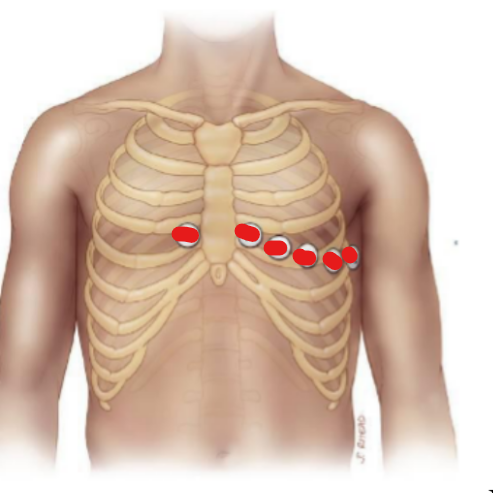
Limb leads (3)
RA LA, RA LL, LA LL
Classic leads formed by limb combinations - think left/lower more +
__ (-) to __ (+) arm-arm
__ (-) to __ (+) arm-leg
__ (-) to __ (+) arm-leg
aVL
Core lead to LA
left
aVF
Core lead to LL
floor
aVR
Core lead to RA
right
Right, lateral
V1 is (left/right-most)
V6 is (medial/lateral-most)
Positive QRS, vectors
Axis is the degree in the limb leads that has most net __ (+/-) __ complex current
helps localize net current movement (__) in heart and even localize pathology
Ischemia
When oxygen delivery does not meet tissue demand
Infarct
Occlusion of blood to region of myocardium → necrosis
300/(#big boxes between QRS)
Calculation of rate in EKG done by (math)
P wave
Arrythmias on EKGs are often discernable from an absent _ __ right before QRS complex
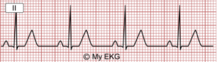
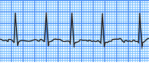
60-100, narrow, flat, normal
In NSR
Rate is between __-__ bpm
QRS is __, ST is __, T-wave is __
100, narrow, flat, shorter, normal
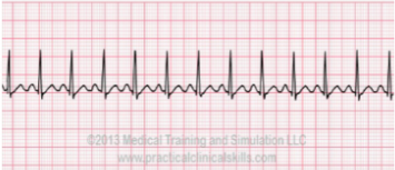
In Sinus Tachycardia
Rate is faster than __ bpm
QRS is __, ST is __ and slightly __, T-wave is __
60, narrow, flat, normal
In Sinus Bradycardia
Rate is slower than __ bpm
QRS is __, ST is __ , T-wave is __
Disorganized, ventricular, P, narrow, flat, normal
In Atrial Fibrillation
__ atrial activity, with coordinated __ contraction
No _-wave before QRS
QRS is __, ST is __, T-wave is __
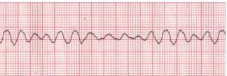
Disorganized, cardiac output, P, QRS, T
In Ventricular Fibrillation
__ ventricular activity, means no contraction and minimal __ __
No identifiable _ waves, __ spike, or _ waves
Ischemia, disconnect, P, QRS, widen, inversion
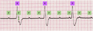
In Complete Heart Block
__ to vessels suppling AV node
electrical __ between SA/AV nodes and His-Purkinje-ventricles
_ waves and __ are unrelated, QRS spike __, and notable T wave __
P, QRS, ST, depressions
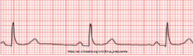
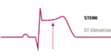
In STEMI
Normal _ wave, narrow __, except __ segment is elevated
May present with reciprocal __ to ST segment in other leads (complete 12 lead)
ischemic, troponins, depressions, inversion
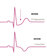
NSTEMI
Suspected cardiac __ injury from elevated __ (protein)
Instead of ST elevations, commonly seen are ST __ or T-wave __
Asystole, PEA
2 conditions of arrest (no pulse = no oxygen delivery)