Untitled
5.0(1)
5.0(1)
Card Sorting
1/156
There's no tags or description
Looks like no tags are added yet.
Study Analytics
Name | Mastery | Learn | Test | Matching | Spaced |
|---|
No study sessions yet.
157 Terms
1
New cards
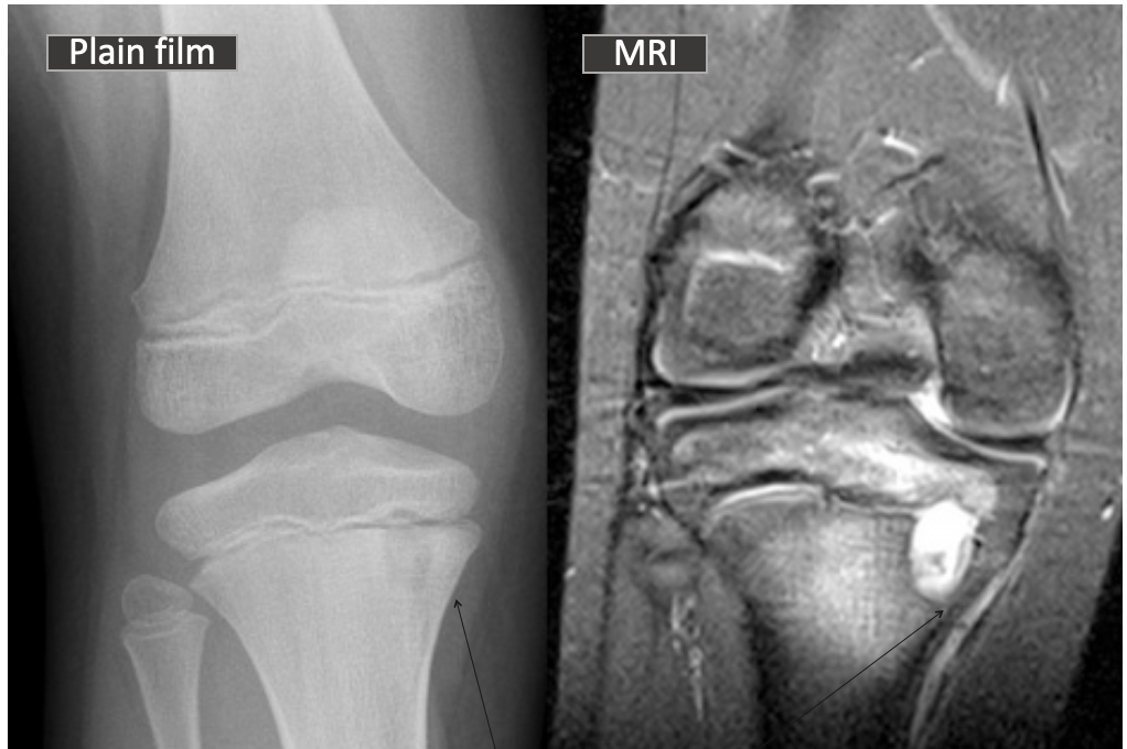
What is this?
Intraosseous Abscess
2
New cards
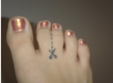
What is this?
Syndactyly
3
New cards
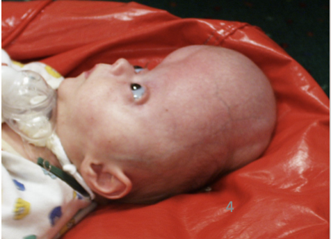
What is this?
Craniostynostosis
4
New cards
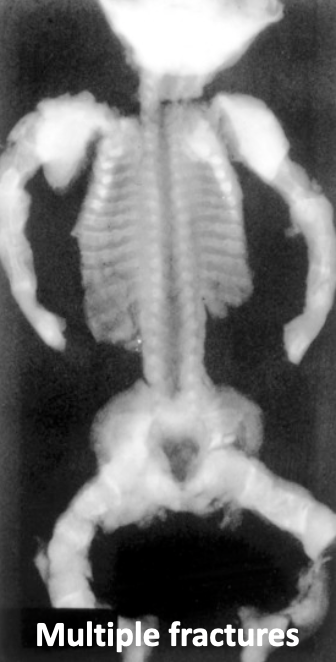
What is this showing?
Fractures-found in osteogenesis imperfecta
5
New cards
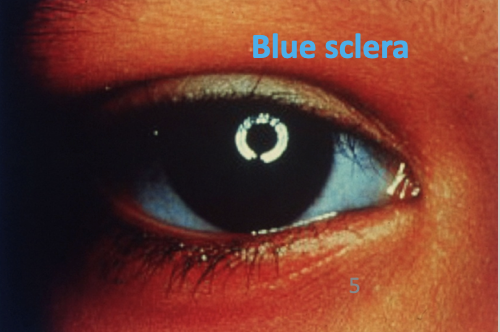
What is this? What disease?
Blue sclera-found in osteogenesis imperfecta
6
New cards
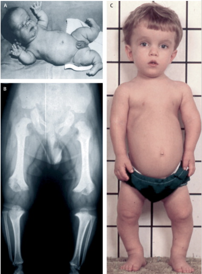
What disease?
Achondroplasia
7
New cards
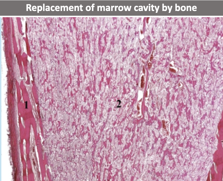
What does this histo slide show?
Osteopetrosis
8
New cards
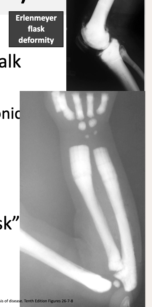
What disease?
Osteopetrosis
9
New cards
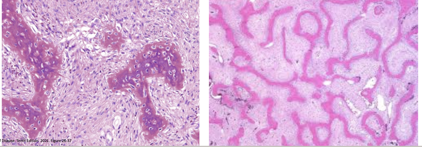
What does this histo slide show?
* curvilinear shapes of the trabeculae mimic alphabet characters
* Found in Fibrous Dysplasia
* Found in Fibrous Dysplasia
10
New cards
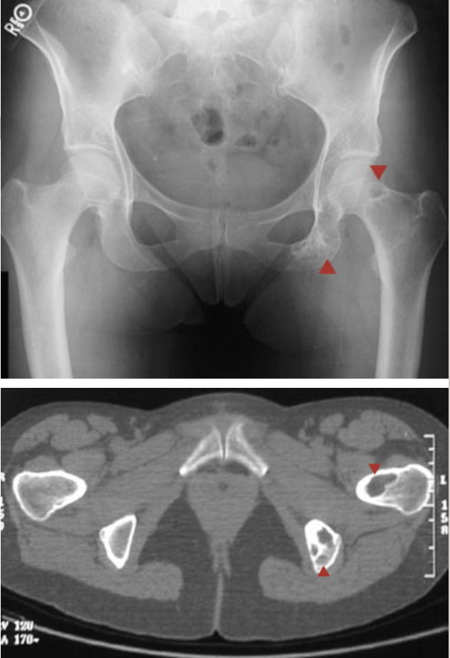
What does this X-ray show?
Fibrous Dysplasia
11
New cards
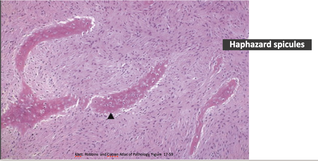
What does this histo slide show?
Haphazard Spicules found in Fibrous Dysplasia
12
New cards
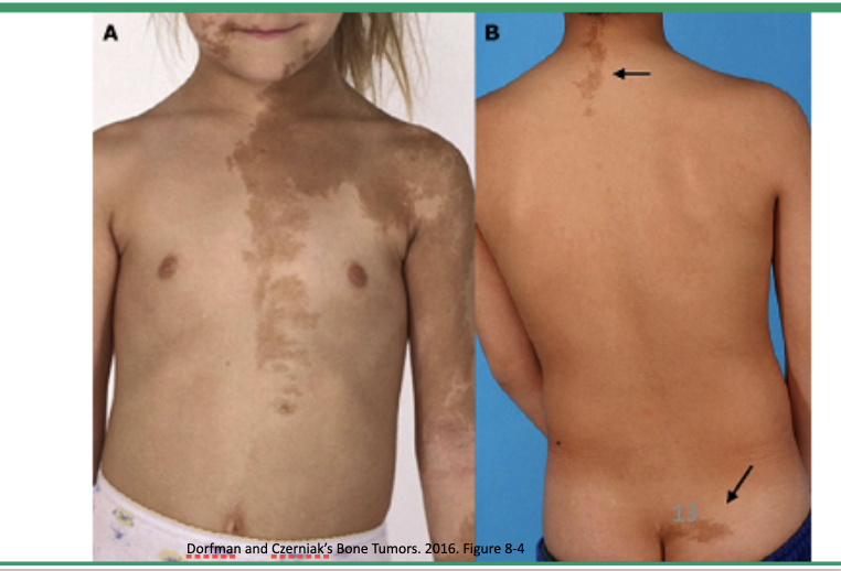
What are the brown spots? What disease?
Cafe au lait spots found in McCune-Albright Syndrome- associated with Polyostotic Fibrous Dysplasia
13
New cards
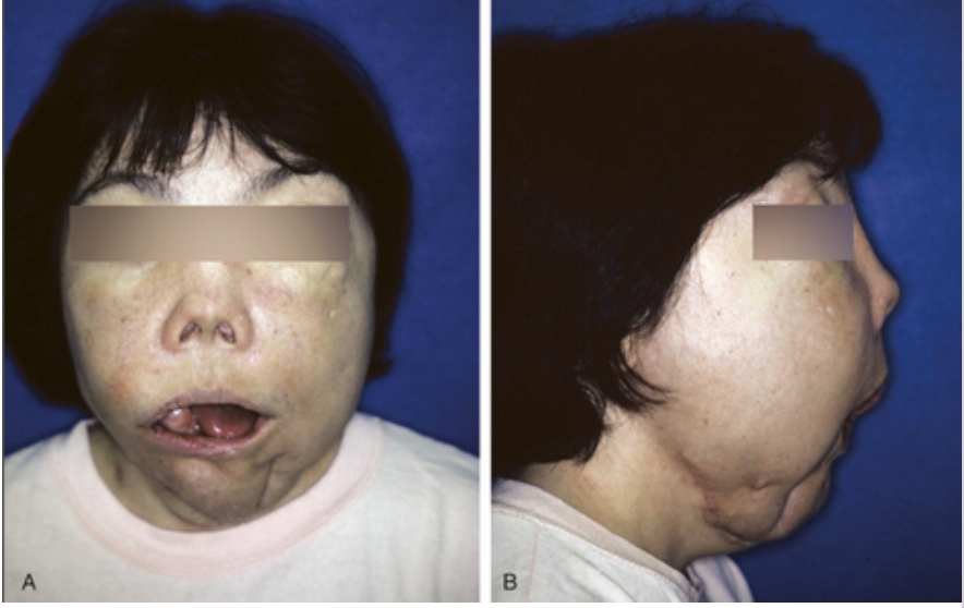
What disease?
Craniofacial involvement with polyostotic disease -Fibrous Dysplasia
14
New cards
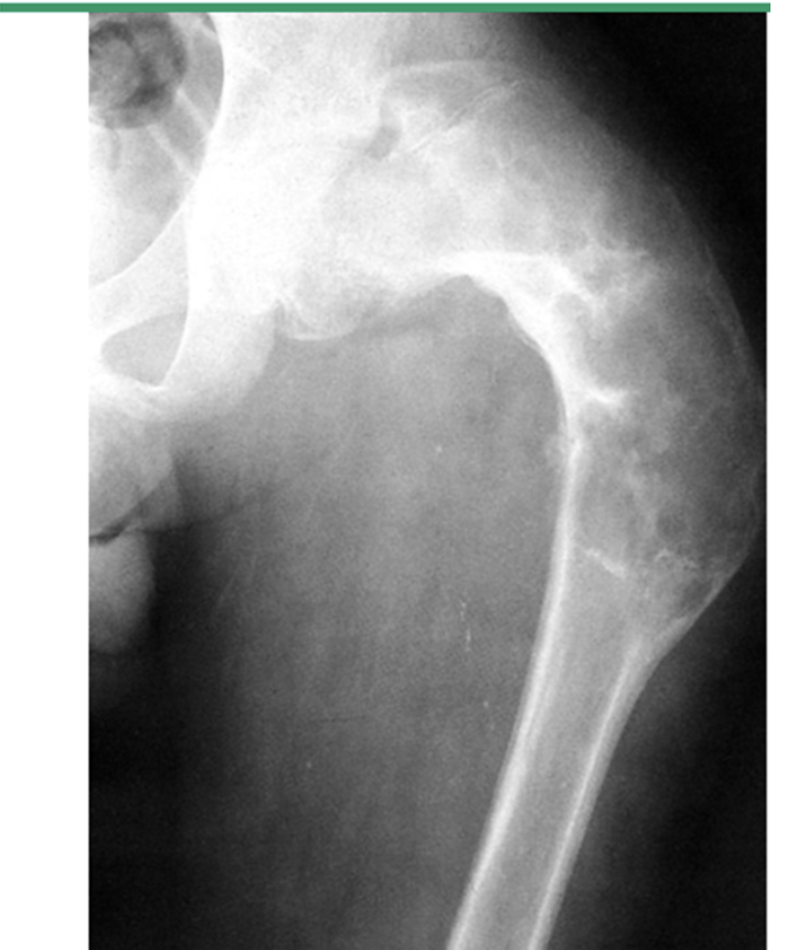
What does this xray show?
Fibrous Dysplasia of the Proximal femur with Shepherd’s Crook Deformity
15
New cards
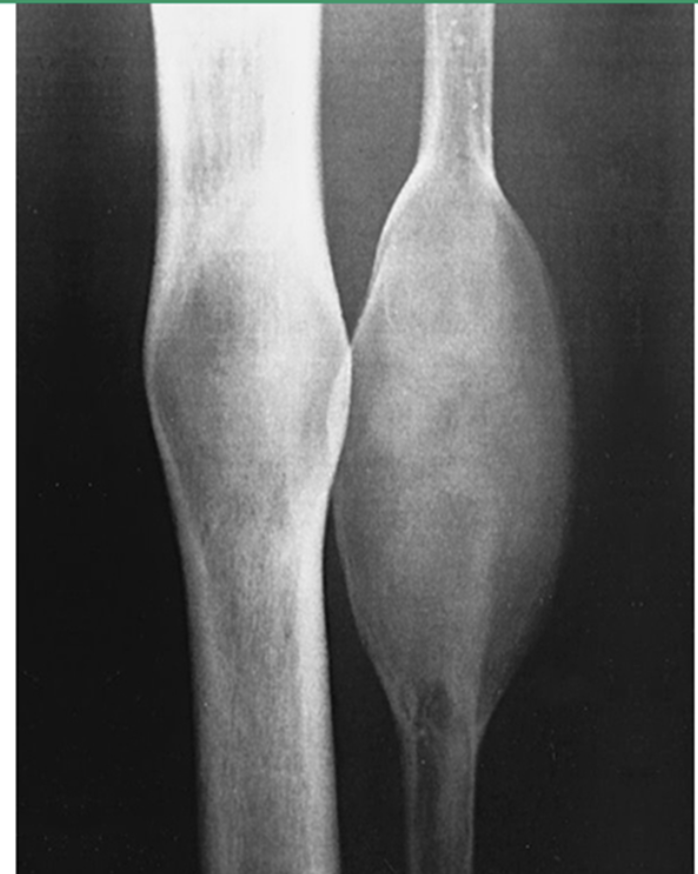
What does this xray show?
Polyostotic Fibrous Dysplasia
16
New cards
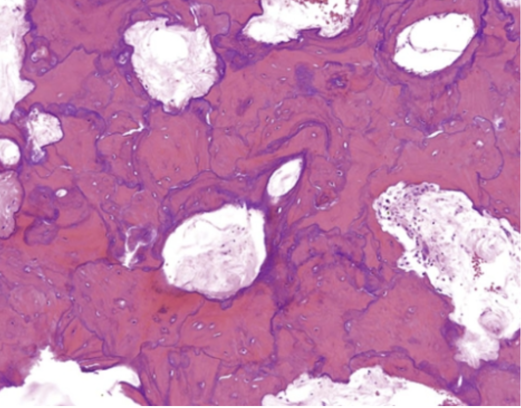
What does this histo slide show?
Osteitis Deformans - Jigsaw puzzle-like mosaic pattern appearance to lamellar bone
Sclerotic phase- prominent cement lines
Sclerotic phase- prominent cement lines
17
New cards
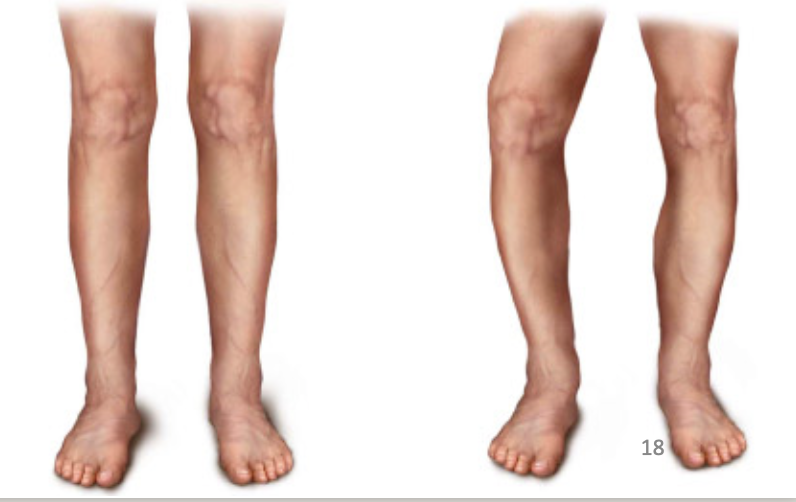
What disease is this found in?
Left: normal Right: Paget’s Disease
Osteitis Deformans
Osteitis Deformans
18
New cards

What disease?
Cause of Headaches in Osteitis Deformans
19
New cards

What is this xray showing? What disease?
Bowing of weakened Pagetoid Bone
Sclerotic, irregular thickening of both cortical and cancellous bone - found in Osteitis Deformans
Sclerotic, irregular thickening of both cortical and cancellous bone - found in Osteitis Deformans
20
New cards
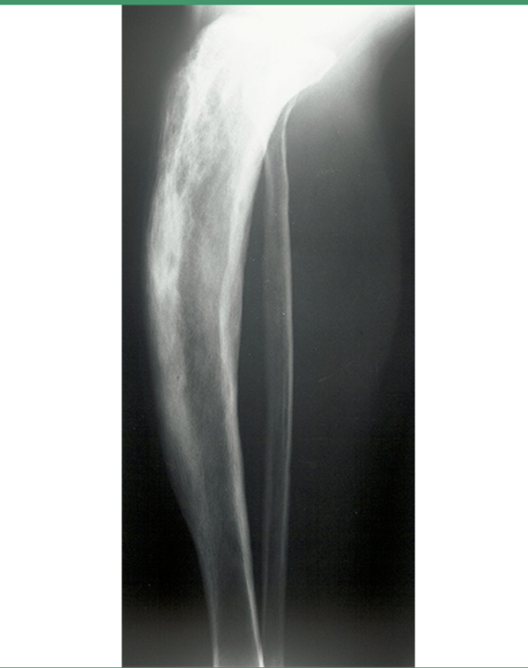
What is this xray showing?
Pagetic Tibia- Osteitis Deformans
21
New cards
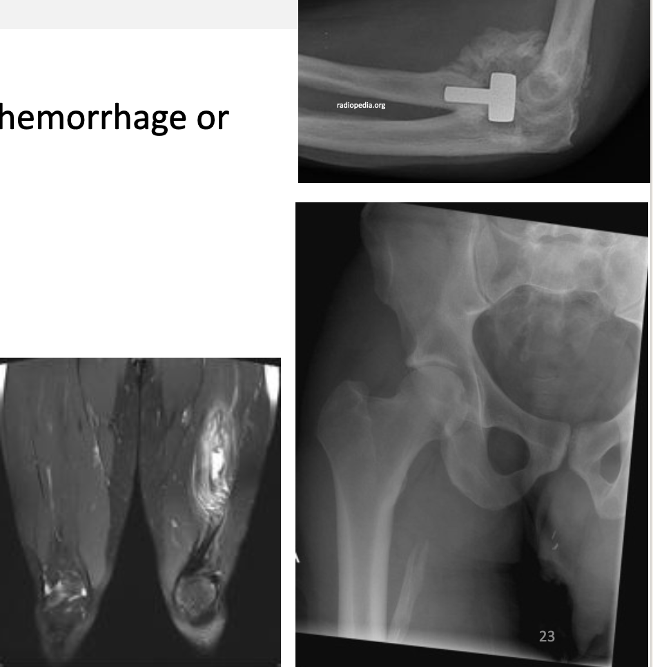
What is this showing?
Myostitis Ossificans-bone formation in muscle
22
New cards
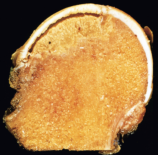
What is this?
Femoral Head with wedge shaped infarct- Osteonecrosis
23
New cards
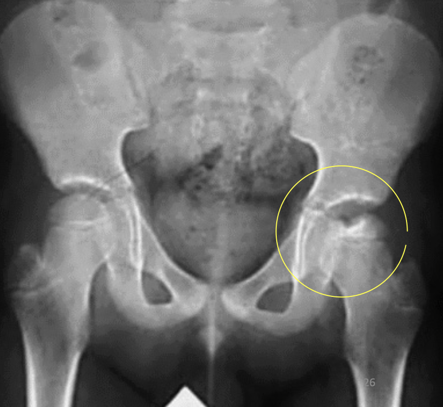
What is this xray showing?
Legg-Calve-Perthes Disease
Idiopathic Avascular Necrosis of the hip
Idiopathic Avascular Necrosis of the hip
24
New cards
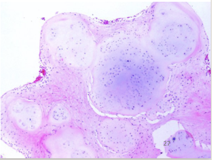
What does this histo slide show?
Synovial Chondromatosis
25
New cards
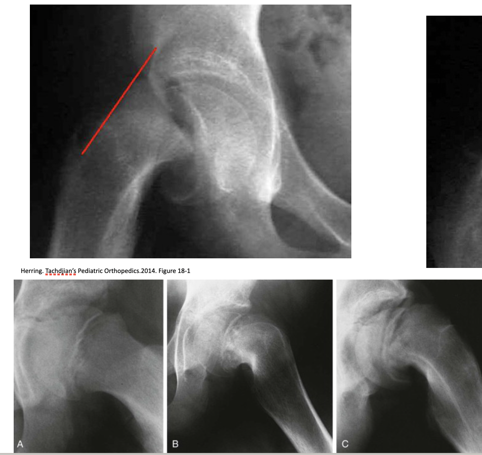
What is this showing?
Slipped Capital Femoral Epiphysis
26
New cards
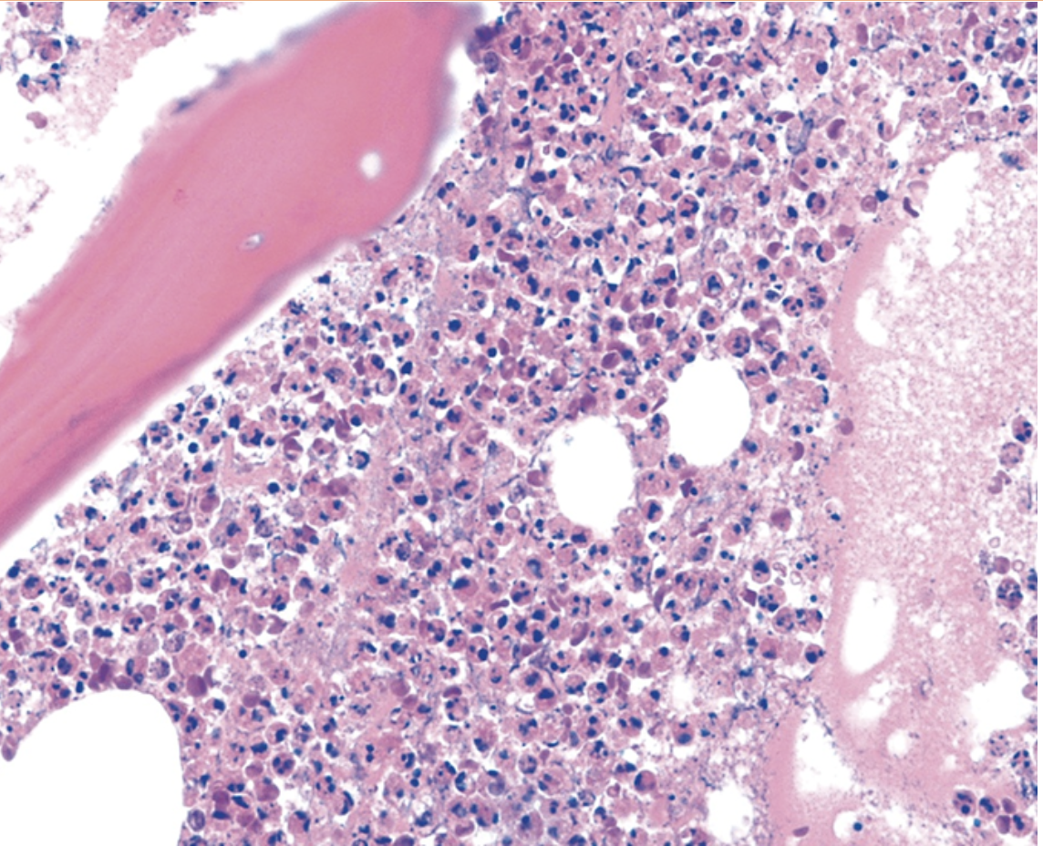
What does this histo slide show?
Acute Osteomyelitis
27
New cards
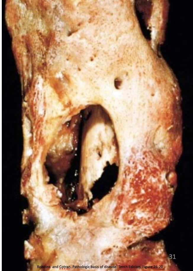
What is this?
Acute Osteomyelitis
28
New cards

What is this?
Brodie Abscess found in Acute Osteomyelitis
29
New cards
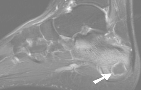
What is this?
Intraosseous Abscess (Brodie Abscess) found in Acute Osteomyelitis
30
New cards
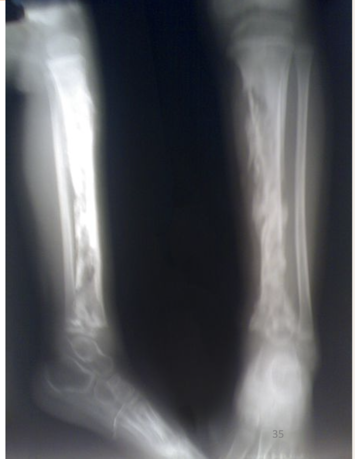
What is this xray showing?
Acute Osteomyelitis - Long bones showing radiolucencies
31
New cards
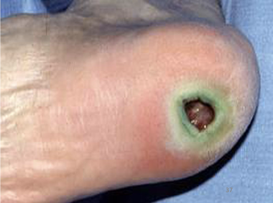
What is this? What is the green pigment called?
Pseudomonas Aeruginosa Osteomyelitis
Shows Green pigment - Pyocyanin
Shows Green pigment - Pyocyanin
32
New cards
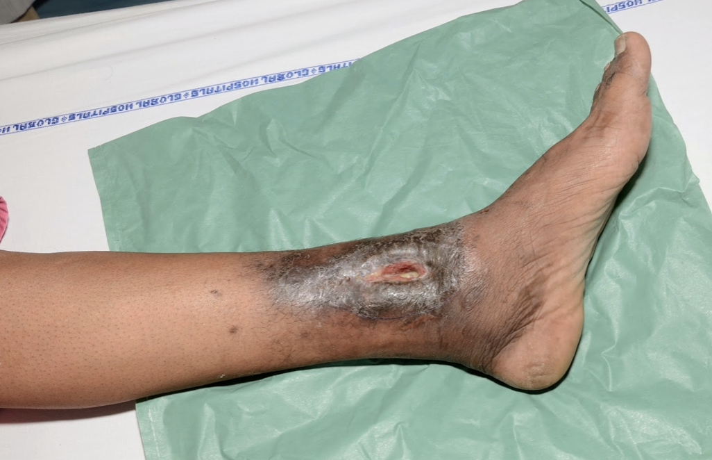
What is this showing? What disease is associated with this?
shows a draining sinus tract
Chronic Osteomyelitis
Chronic Osteomyelitis
33
New cards
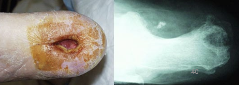
What is this xray showing and what disease is this associated with?
Osteomyelitis- Characteristic Xray: lytic focus of bone destruction surrounded by a zone of sclerosis
34
New cards
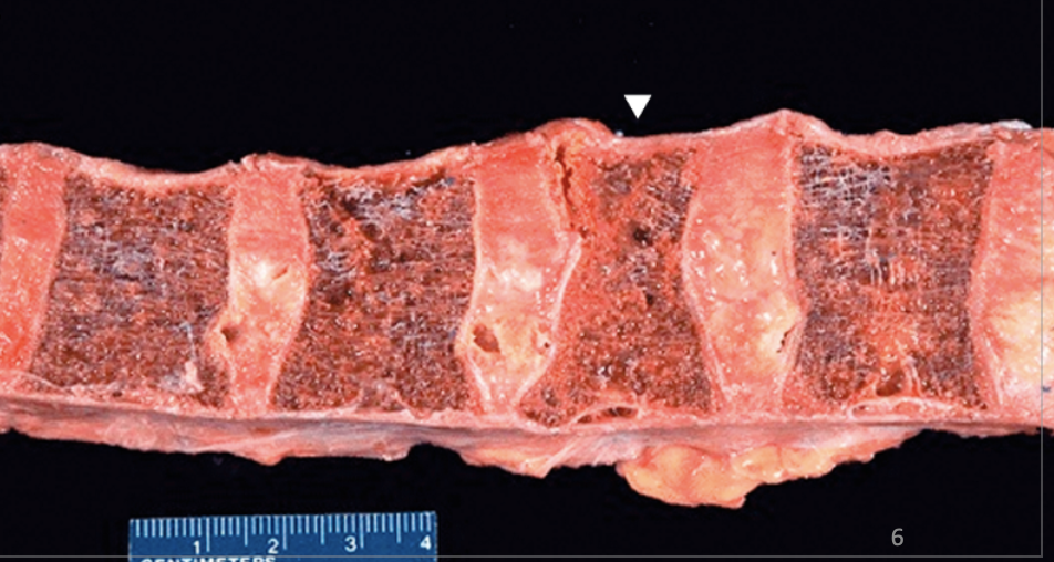
What is the arrow pointing at? What disease is this associated with?
Osteoporosis
Arrow = increased risk of fracture
Arrow = increased risk of fracture
35
New cards
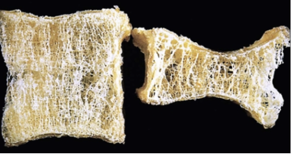
What is this showing and what disease is this associated with?
Osteoporotic Vertebral Body-Osteoporosis
36
New cards
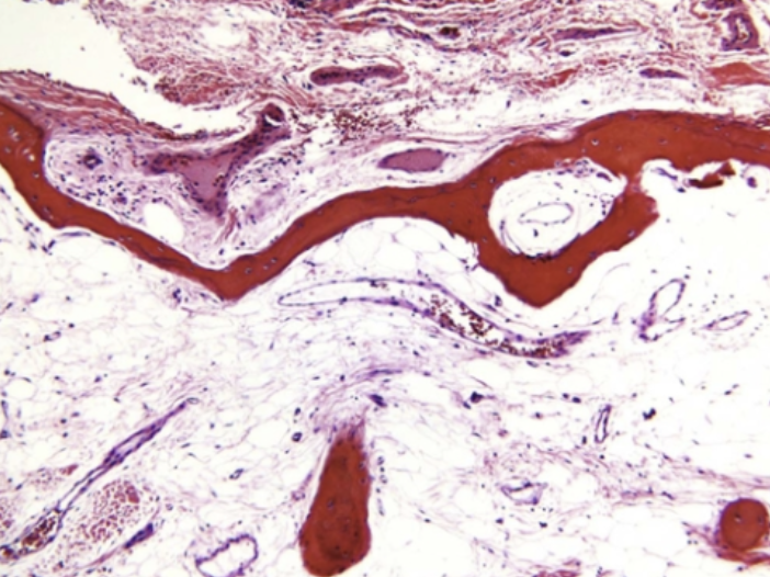
What does this histo slide show?
Osteoporotic Bone
37
New cards
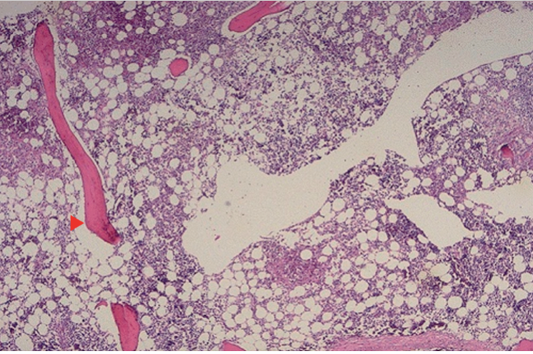
What does this histo slide show?
Fracture
38
New cards
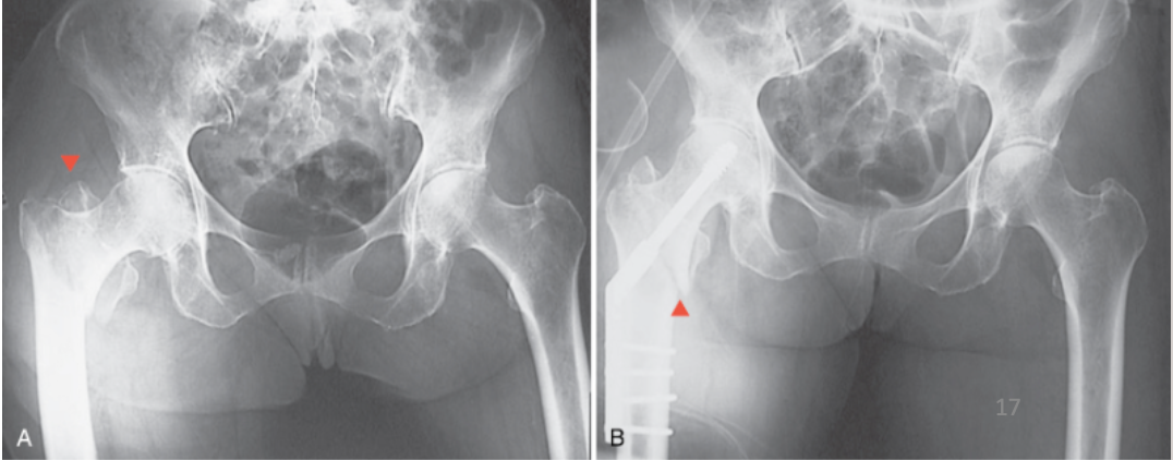
What is this showing?
Fracture
39
New cards
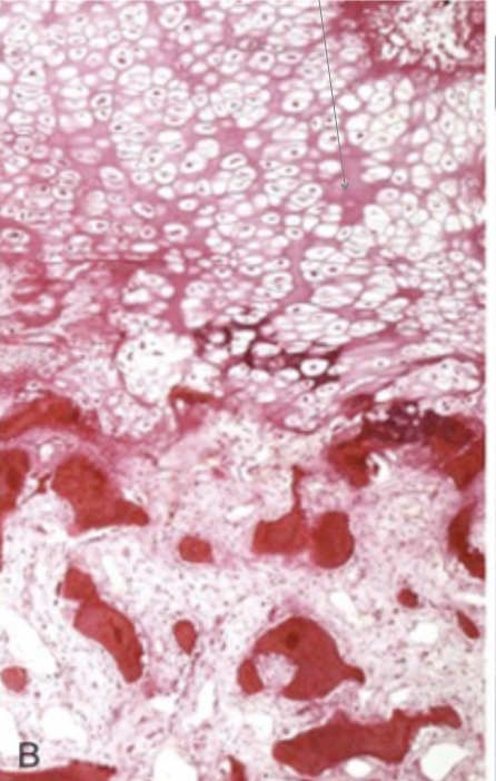
What does this histo slide show?
Rickets - Showing loss of normal cartilage palisades at costochondral junction
40
New cards
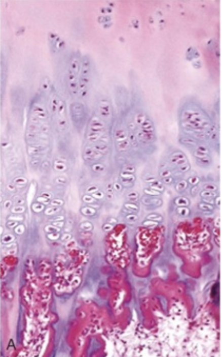
What does this histo slide show?
Normal bone matrix
41
New cards
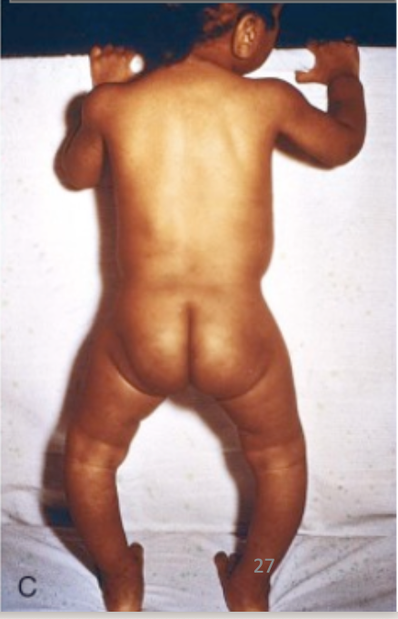
What is this picture showing? What disease is this associated with?
Leg bowing due to poor mineralization - Rickets
42
New cards
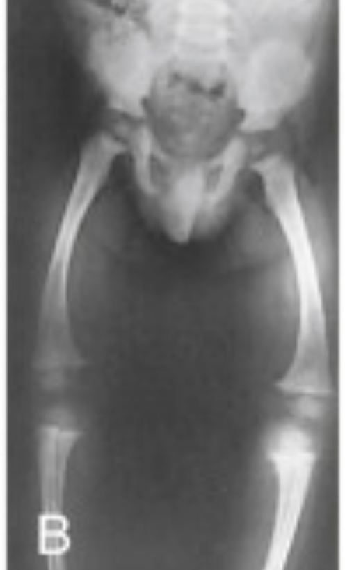
What is this X-ray showing? What disease is it associated with?
Rickets- Epiphyses are open, mottled and overgrown
43
New cards
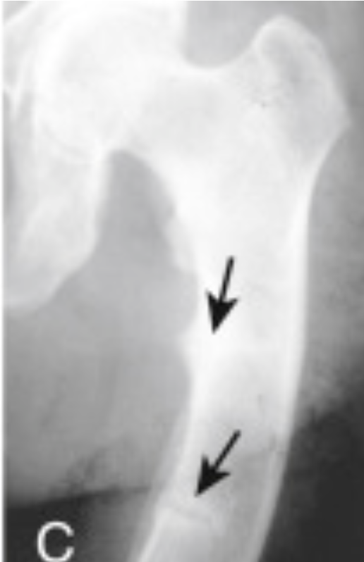
What is this X-ray showing? What disease is it associated with?
Adult with Osteomalacia and pseudofractures
44
New cards
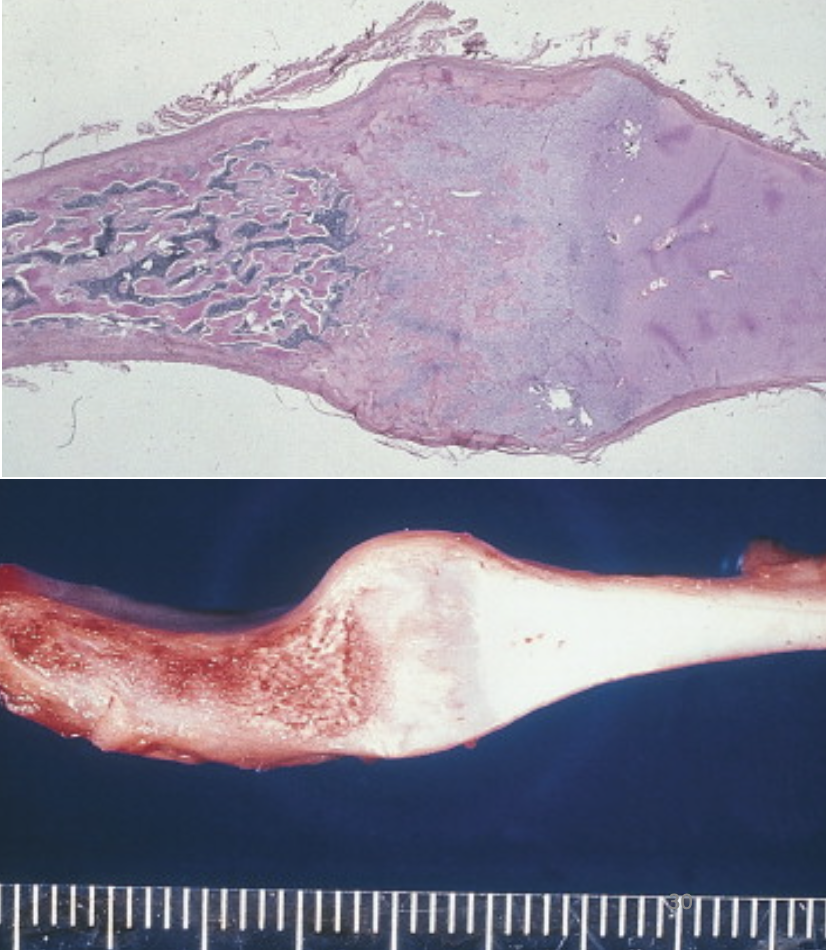
What does this histo slide show?
Rickets- enlarged mass of uncalcified cartilage at costchondral junction
45
New cards
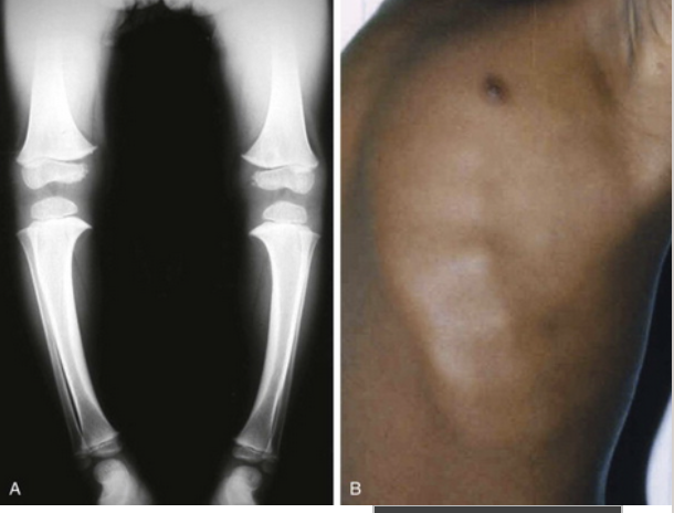
What is this X-ray showing? What disease is it associated with?
Rickets- Rachitic Rosary: beadlike prominences at junction of a rib and its cartilage
46
New cards
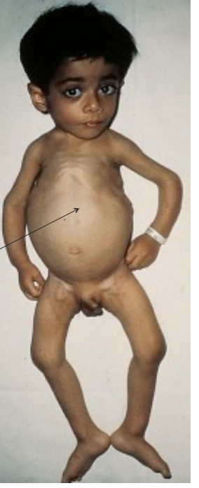
What disease does this child have?
Rickets- child has Rickets secondary to gluten sensitive enteropathy
Can also see Harris Sulcus groove (the arrow)
Frontal Bossing/squared head is prominent
Can also see Harris Sulcus groove (the arrow)
Frontal Bossing/squared head is prominent
47
New cards
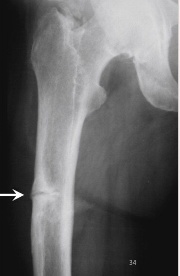
What is this X-ray showing? What disease is it associated with?
Arrow shows Milkman Fracture
Aka: Looser Zone
Aka: Looser Zone
48
New cards
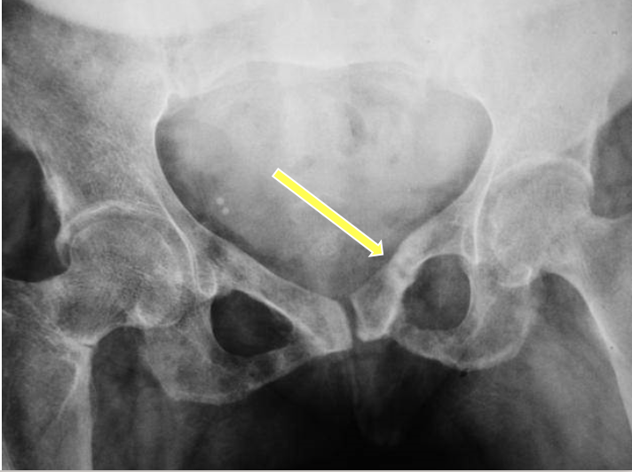
What disease is this associated with?
Osteomalacia
49
New cards
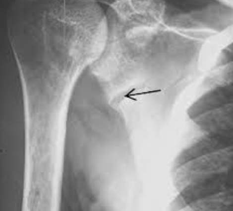
What disease is this xray showing?
Osteomalacia
50
New cards
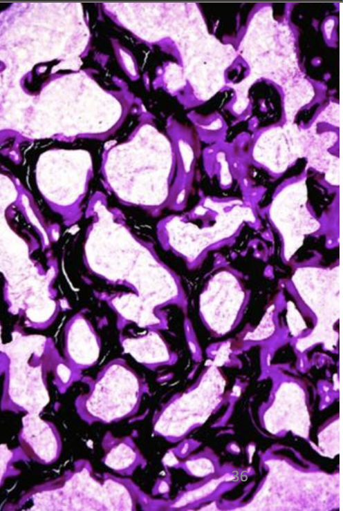
What does this histo slide show?
Von Kassa Stain (calcium) - Osteomalacia
surfaces of the bony trabeculae (*black*) are covered by a thicker than normal layer of osteoid (*pink/purple*)
Exaggeration of Osteoid Seams
surfaces of the bony trabeculae (*black*) are covered by a thicker than normal layer of osteoid (*pink/purple*)
Exaggeration of Osteoid Seams
51
New cards
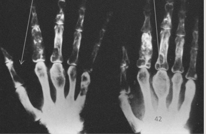
What is the arrow on left showing? on the right?
Arrow on left hand: Subperiosteal bone resorption
Arrow on right hand: Bulbous bulging from Brown Tumors
Arrow on right hand: Bulbous bulging from Brown Tumors
52
New cards
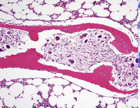
What does this histo slide show?
Hyperparathyroidism
53
New cards
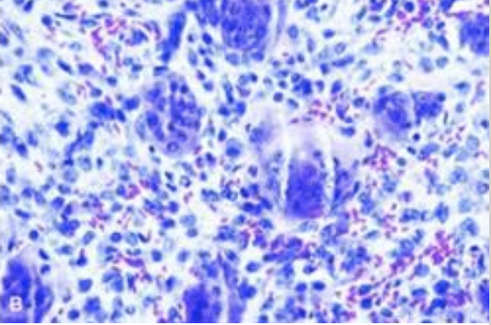
What does this histo slide show?
Hyperparathyroidism
54
New cards
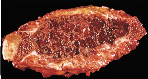
What is this gross path showing?
histologic changes of hyperparathyroidism are known as osteitis fibrosa (refers to accelerated bone remodeling)
This can progress to *cystica*
This can progress to *cystica*
55
New cards
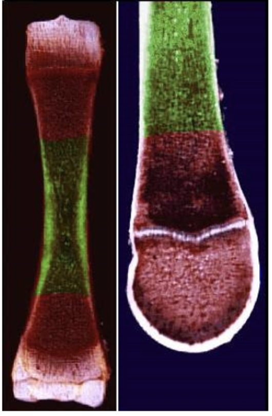
What area in bone is this called?
Diaphysis
56
New cards
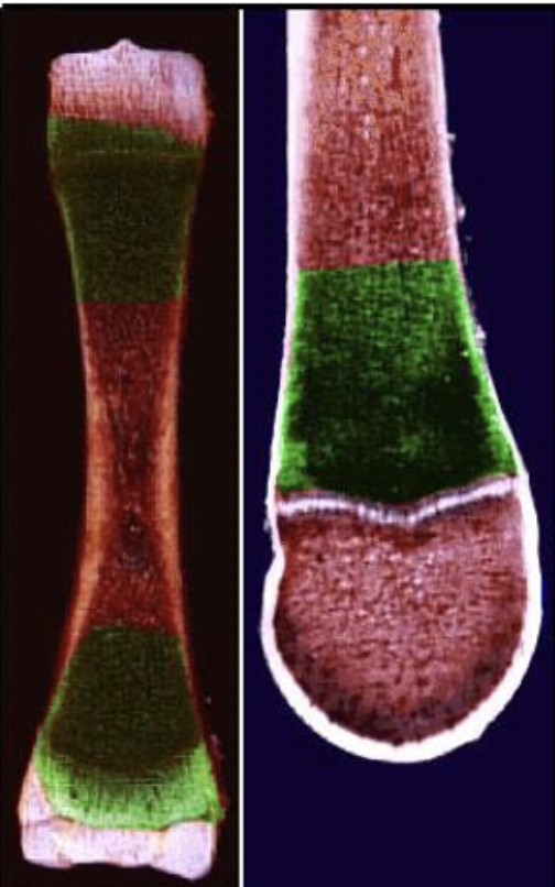
what area in the bone is this called?
Metaphysis
57
New cards
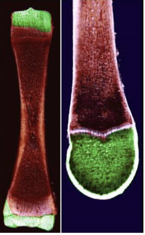
what area in the bone is this called?
Epiphysis
58
New cards
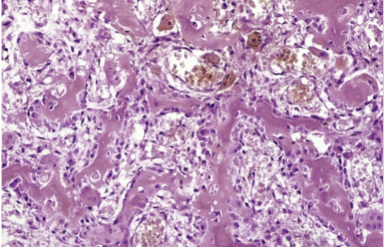
What does this histo slide show?
Osteoid Osteoma
59
New cards
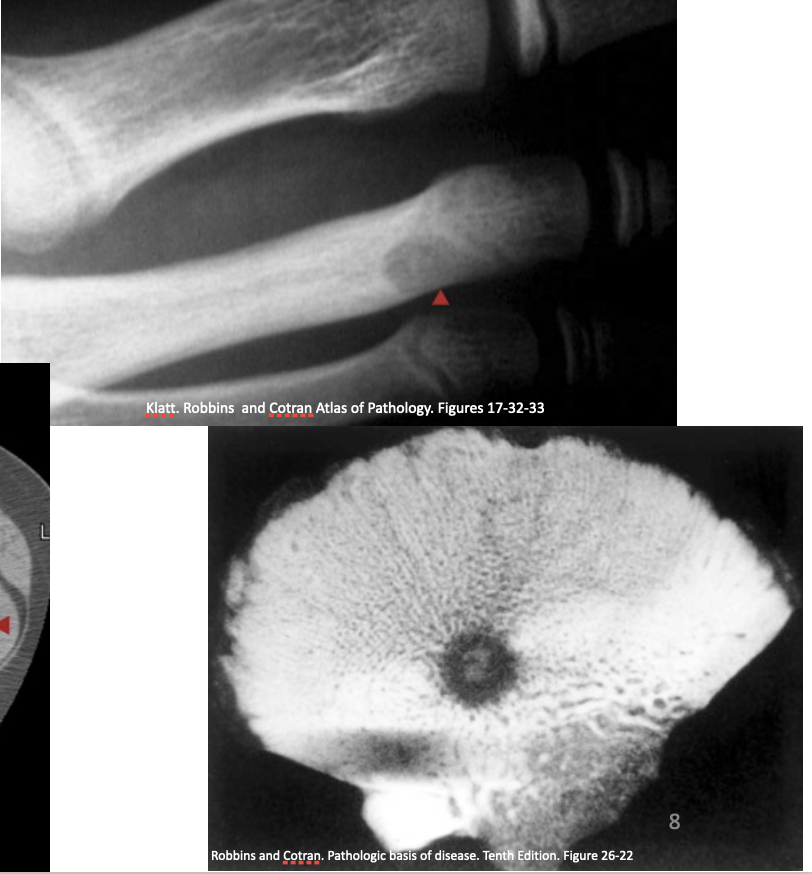
What is this showing?
Osteoid Osteoma- Well-circumscribed lesion characterized by radiolucency with mineralized nidus (usually
60
New cards
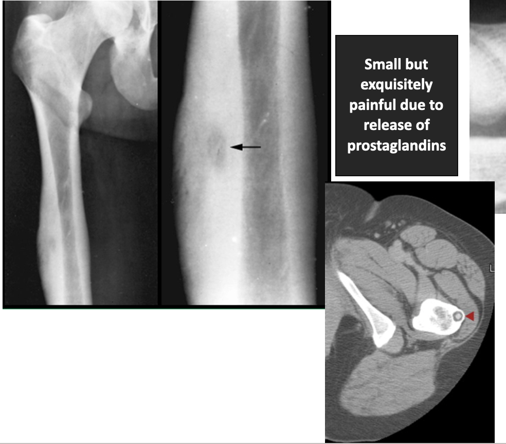
What is this showing?
Osteoid Osteoma
61
New cards
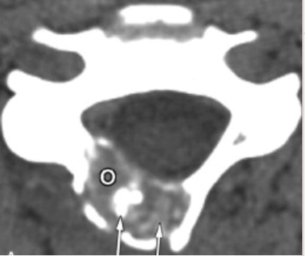
What is this showing?
Osteoblastoma
62
New cards
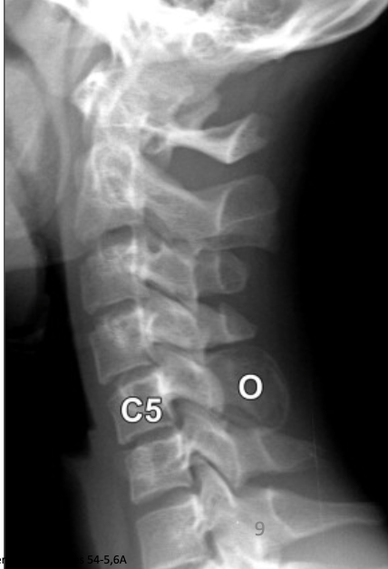
What is this showing?
Osteoblastoma - Lucent, expansile mass involving spinous process at C5
63
New cards
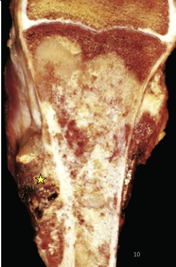
What is this showing?
Osteosarcoma
64
New cards
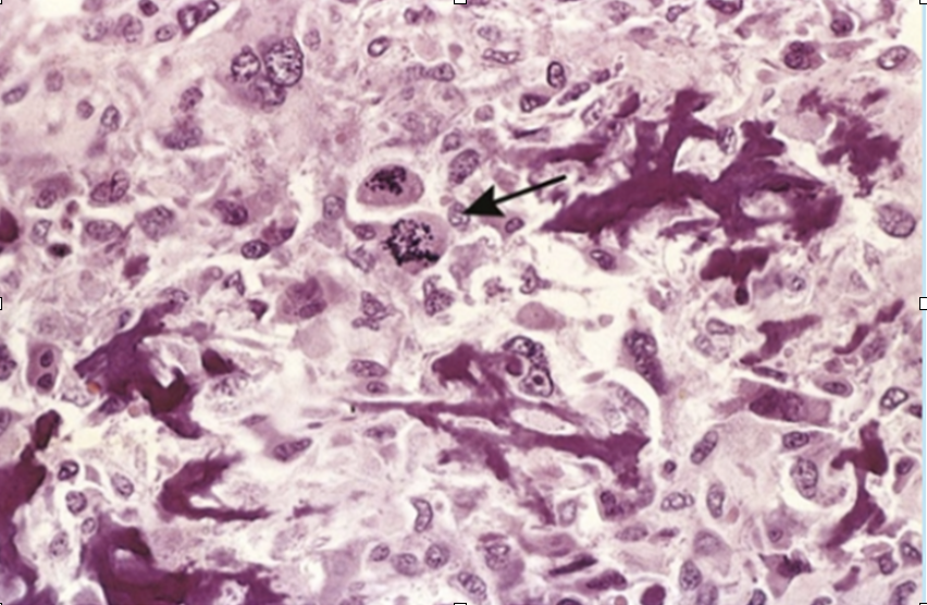
What is this showing?
Osteosarcoma
65
New cards
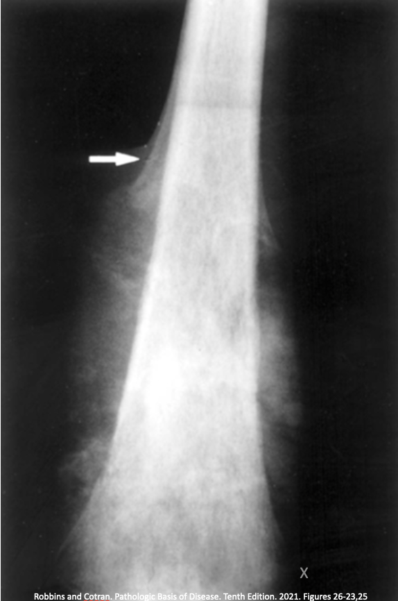
What is this showing?
Osteosarcoma-
* Arrow: Radiographs show “sunburst” pattern, disruption of the cortex and lifting of periosteum forming “Codman triangle”
* Arrow: Radiographs show “sunburst” pattern, disruption of the cortex and lifting of periosteum forming “Codman triangle”
66
New cards
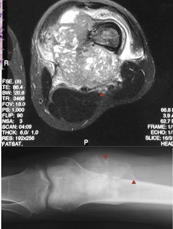
What is this showing?
Osteosarcoma
67
New cards
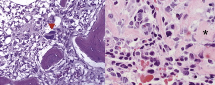
What is this showing?
Osteosarcoma
68
New cards
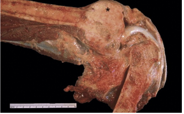
What is this showing?
Osteosarcoma
69
New cards
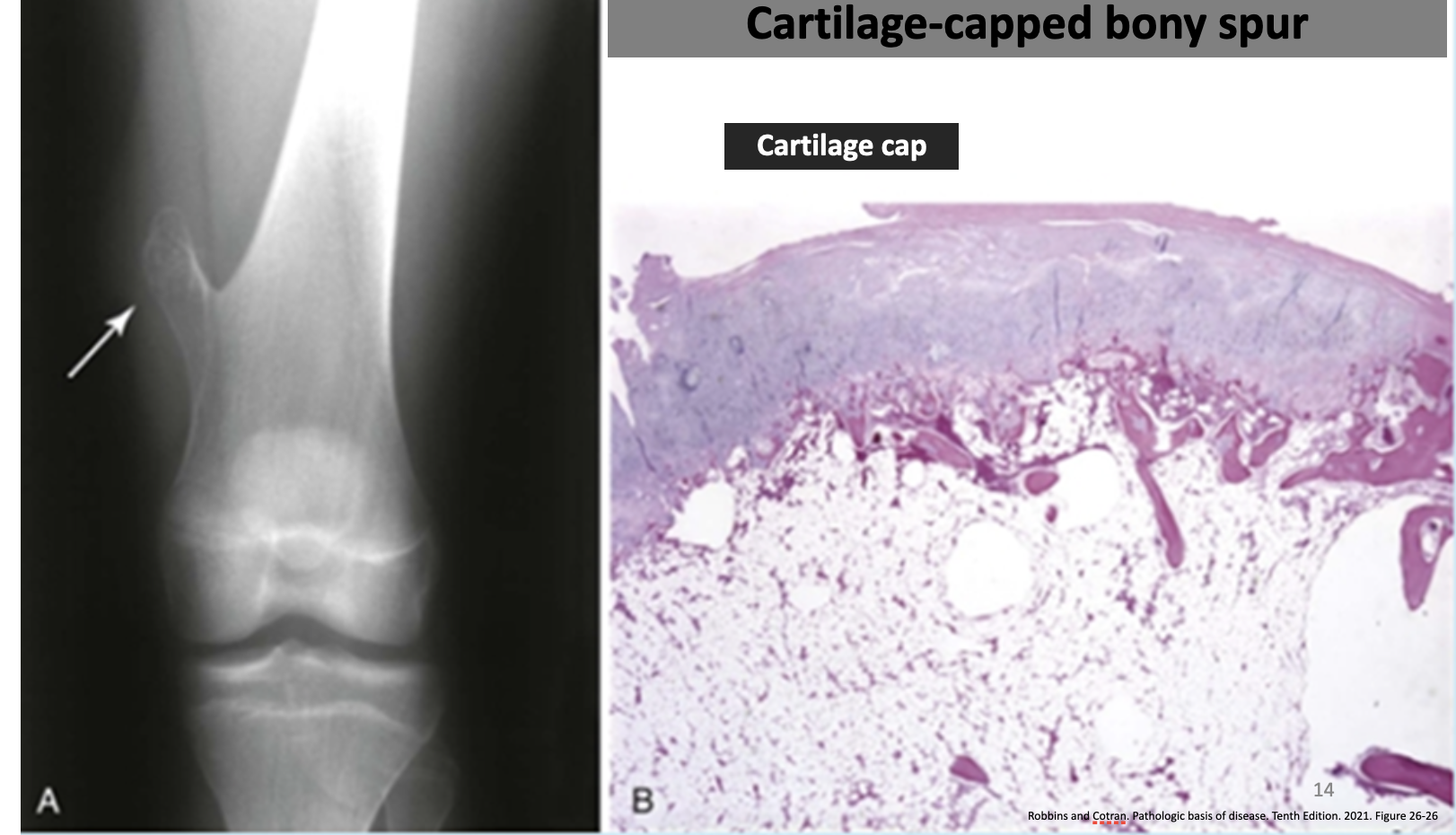
What is this showing?
Osteochondroma
70
New cards
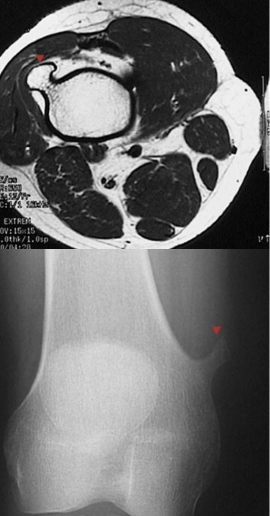
What is this showing?
Osteochondroma
71
New cards
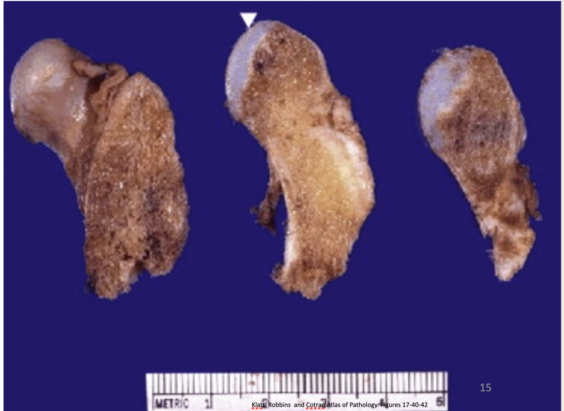
What is this showing?
Benign cartilage Cap- Osteochondroma
72
New cards
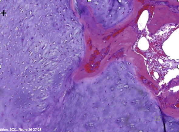
What is this showing?
Chondroma- Histologically comprised of well-circumscribed nodules of hyaline cartilage
73
New cards
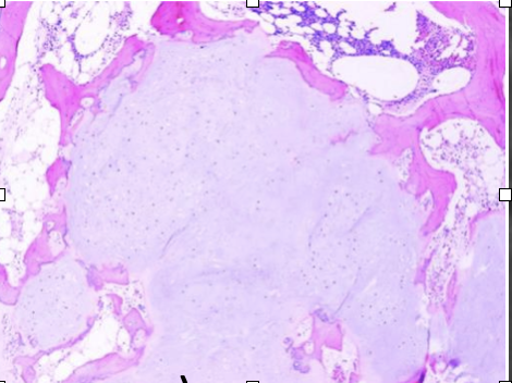
What is this showing?
Chondroma
74
New cards
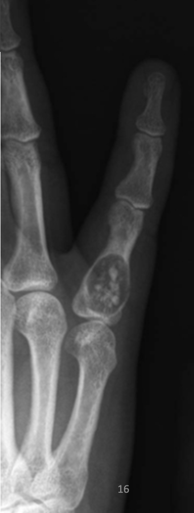
What is this showing?
Chondroma
75
New cards
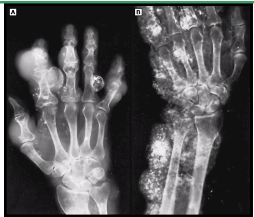
What is A showing? B?
Maffucci Syndrome
A: multiple soft tissue masses and calcified thrombi
B: Contrast material filling cavernous hemangiomas of the soft tissues
A: multiple soft tissue masses and calcified thrombi
B: Contrast material filling cavernous hemangiomas of the soft tissues
76
New cards
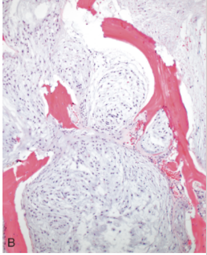
What disease is this showing?
Chondrosarcoma
77
New cards

What disease is this showing?
Histology of grades 1, 2, 3 chondrosarcoma
78
New cards
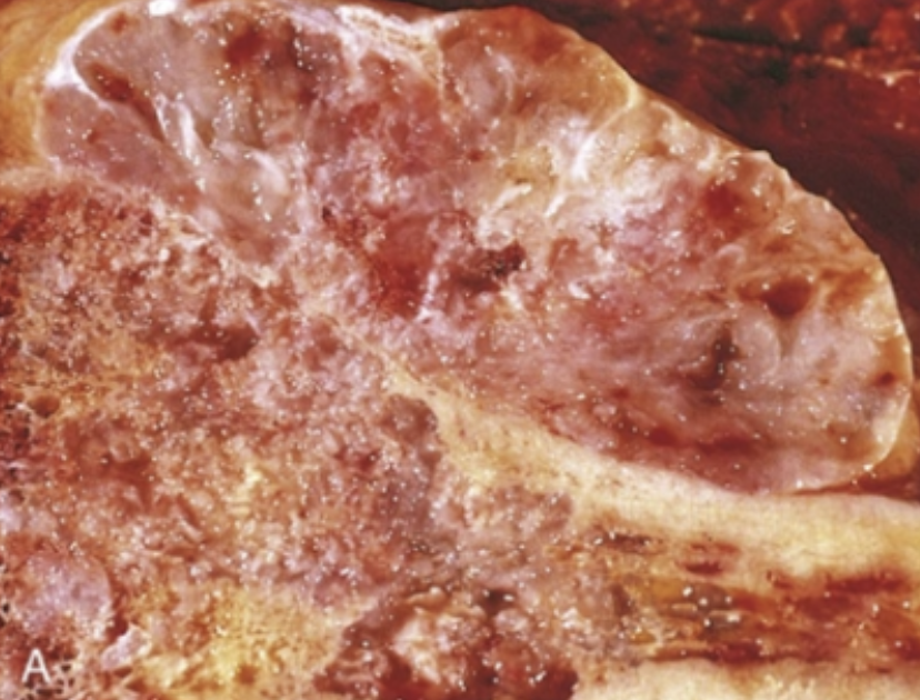
What is this? What disease is this showing?
Cartilage growing within medullary cavity and through the cortex
Chondrosarcoma
Chondrosarcoma
79
New cards
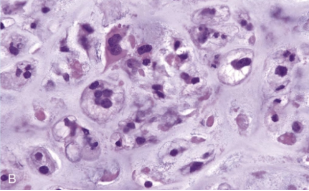
What is this? What disease is this showing?
Malignant cells producing cartilage
Chondrosarcoma
Chondrosarcoma
80
New cards
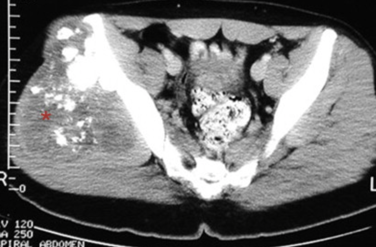
What disease is this showing?
Chondrosarcoma - “cotton wool" appearance characteristic plain film radiograph finding
“popcorn” appearance on CT
“popcorn” appearance on CT
81
New cards
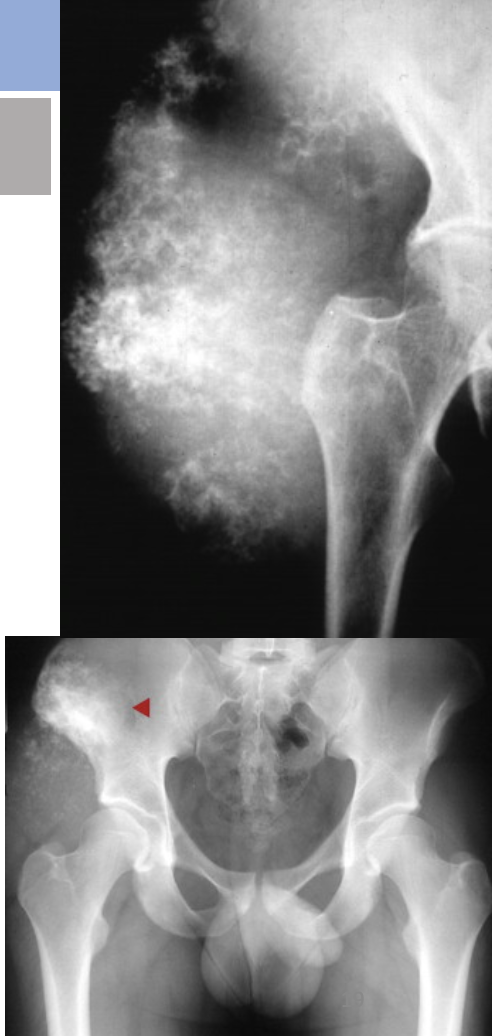
What disease is this showing?
“cotton wool" appearance characteristic plain film radiograph finding - Chondrosarcoma
82
New cards
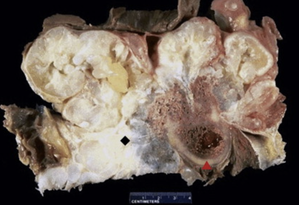
What disease is this showing?
Chondrosarcoma- Tumor arising from pelvic bone
83
New cards
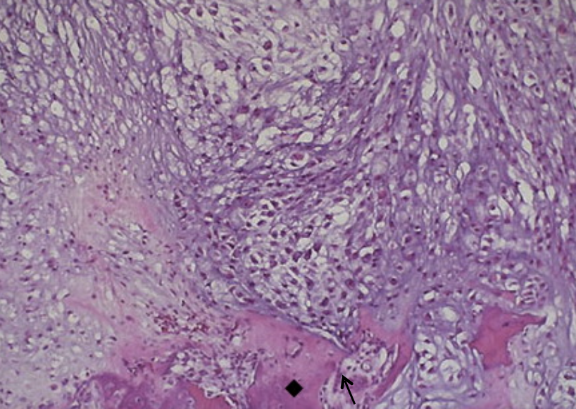
What disease is this showing?
Chondrosarcoma- Tumor destroying bone
84
New cards
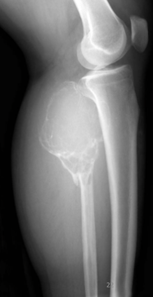
What disease is this showing?
Giant Cell Tumor of bone
Soap Bubble Appearance
Soap Bubble Appearance
85
New cards
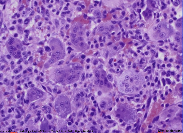
What disease is this showing?
Giant Cell Tumor of Bone Histology
86
New cards
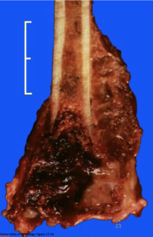
What disease is this showing?
Giant Cell Tumor of Bone- Gross Path Image
87
New cards
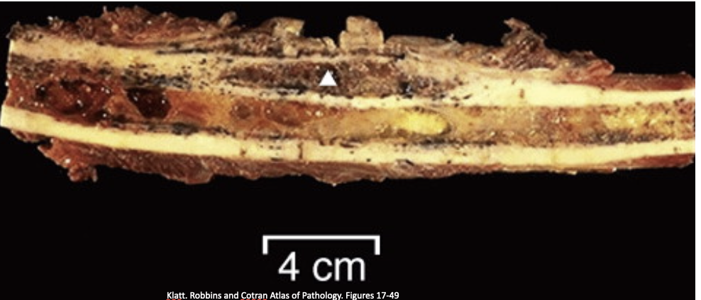
What disease is this showing?
Ewing Sarcoma
88
New cards
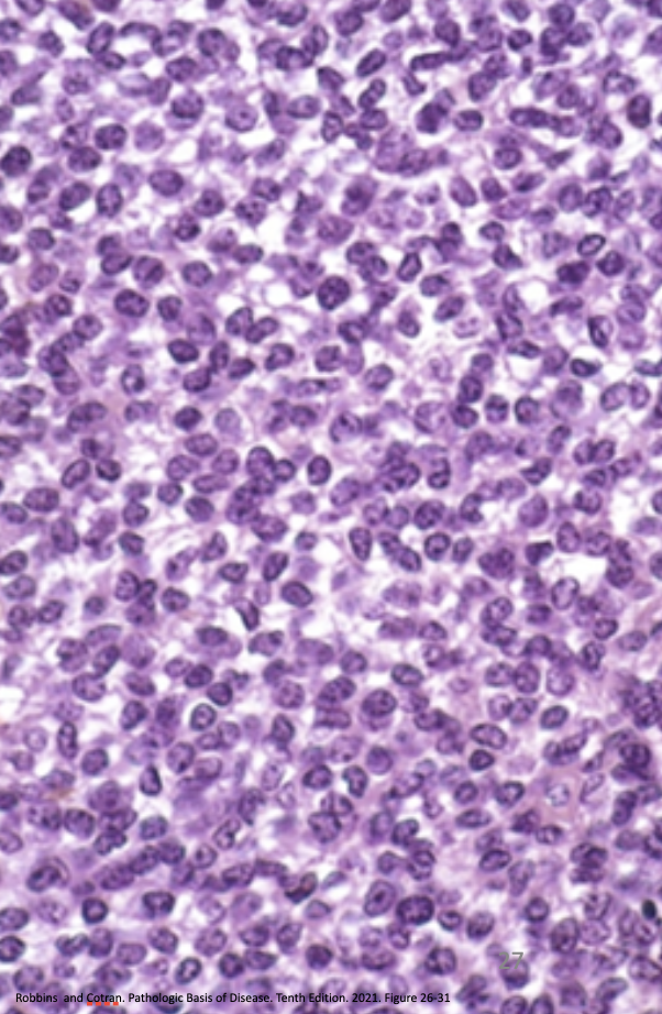
What disease is this showing?
Ewing Sarcoma Histology
89
New cards
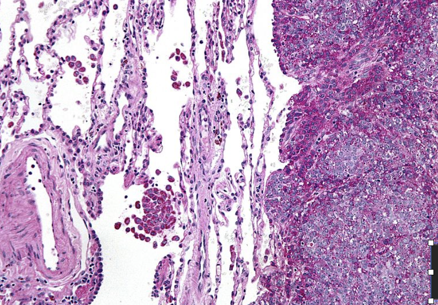
What disease is this showing?
Ewing Sarcoma Histology
90
New cards
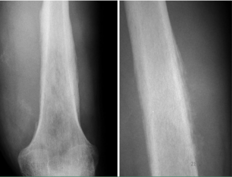
What disease is this showing?
Ewing Sarcoma
91
New cards
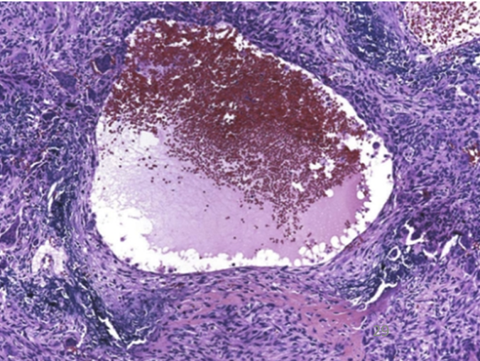
What disease is this showing?
Aneurysmal Bone Cyst
92
New cards
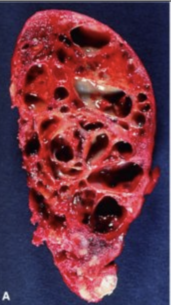
What disease is this showing?
Aneurysmal Bone Cyst
93
New cards
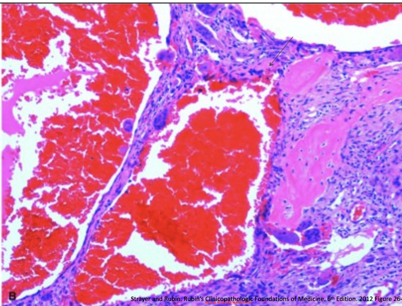
What disease is this showing?
Aneurysmal Bone Cyst
94
New cards
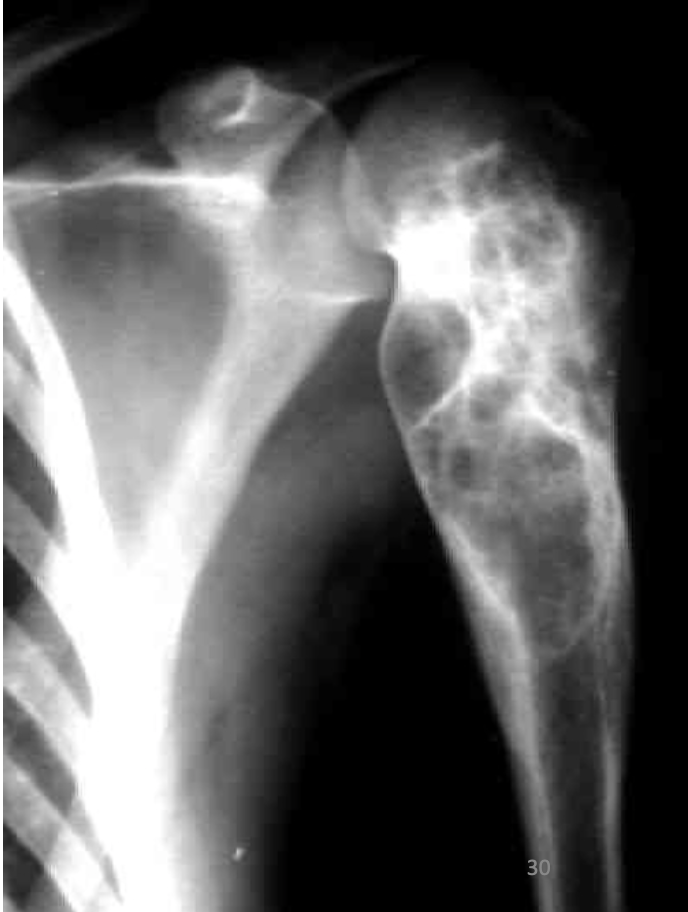
What disease is this showing?
Aneurysmal Bone Cyst
95
New cards
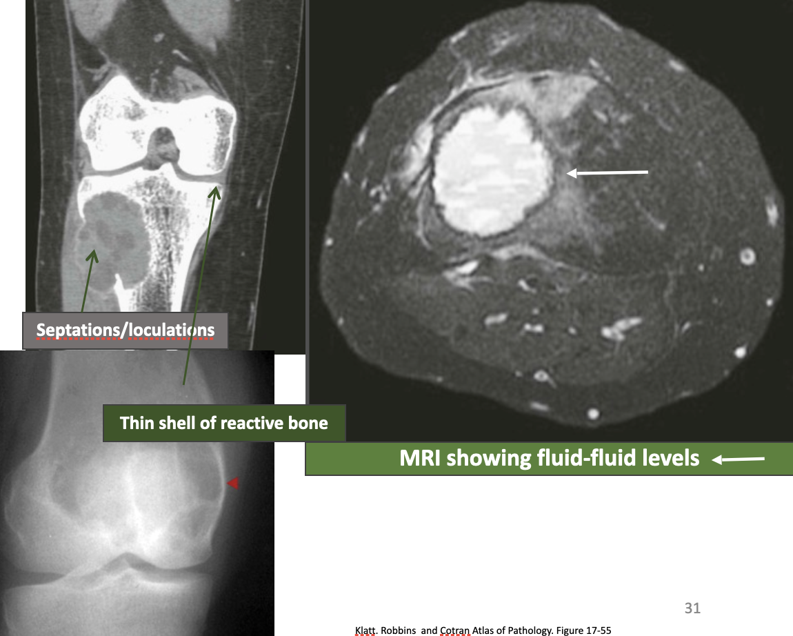
What disease is this showing?
Aneurysmal Bone Cyst
96
New cards
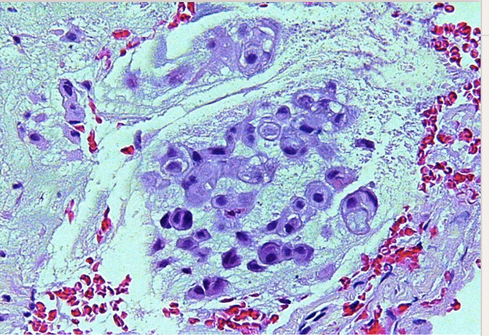
What disease is this showing?
Chordoma
97
New cards
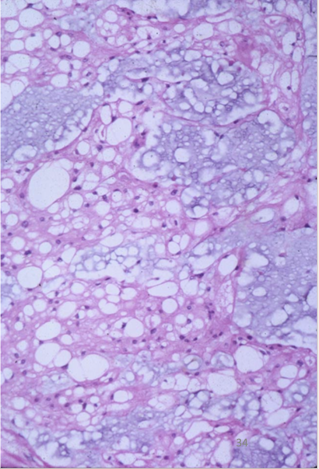
What disease is this showing?
Chordoma
98
New cards
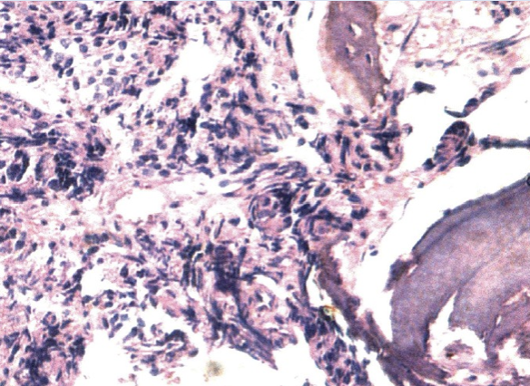
What disease is this showing?
Metastatic Tumor
99
New cards
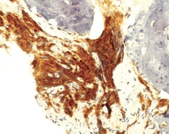
What disease is this showing?
Metastatic Prostate Carcinoma
100
New cards
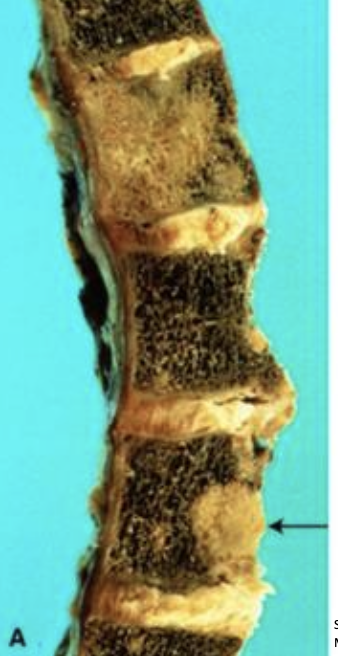
What disease is this showing?
Metastatic Breast Carcinoma to vertebral body