Tissue Quiz: Urinary and Reproductive Systems
1/50
There's no tags or description
Looks like no tags are added yet.
Name | Mastery | Learn | Test | Matching | Spaced | Call with Kai |
|---|
No analytics yet
Send a link to your students to track their progress
51 Terms
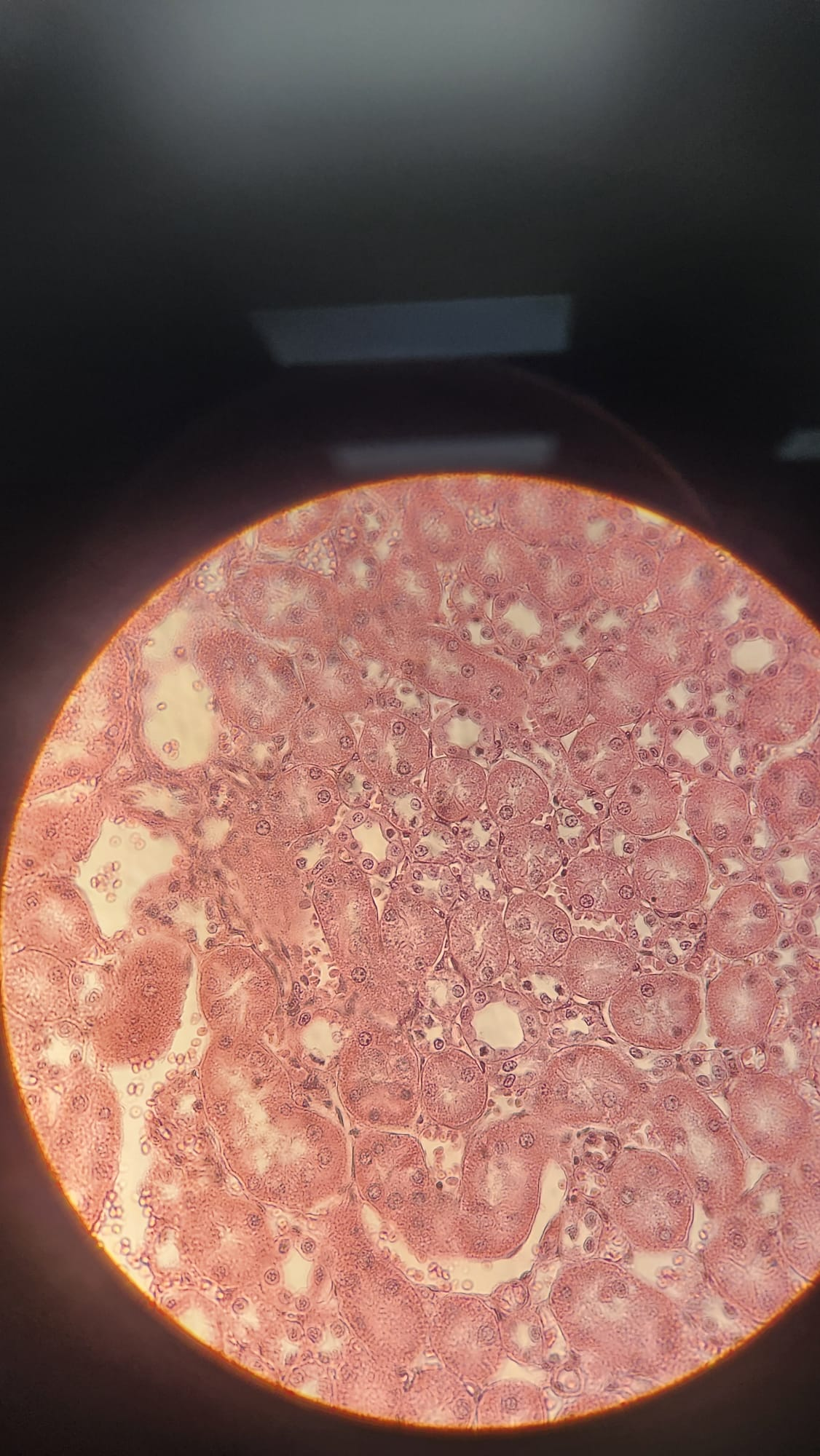
Which organ is this?
Kidney
Which organ is this?
Kidney
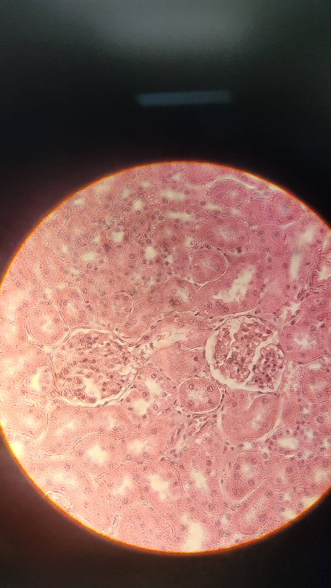
What are the 2 round structures seen here?
Renal corpuscles (glomerulus + Bowman’s capsule)
Which organ is this?
Ureter
Which organ is this?
Bladder
Which organ is this?
Bladder
What is this structure?
Epididymis
Which organ is this?
Epididymis
Which organ is this?
Epididymis
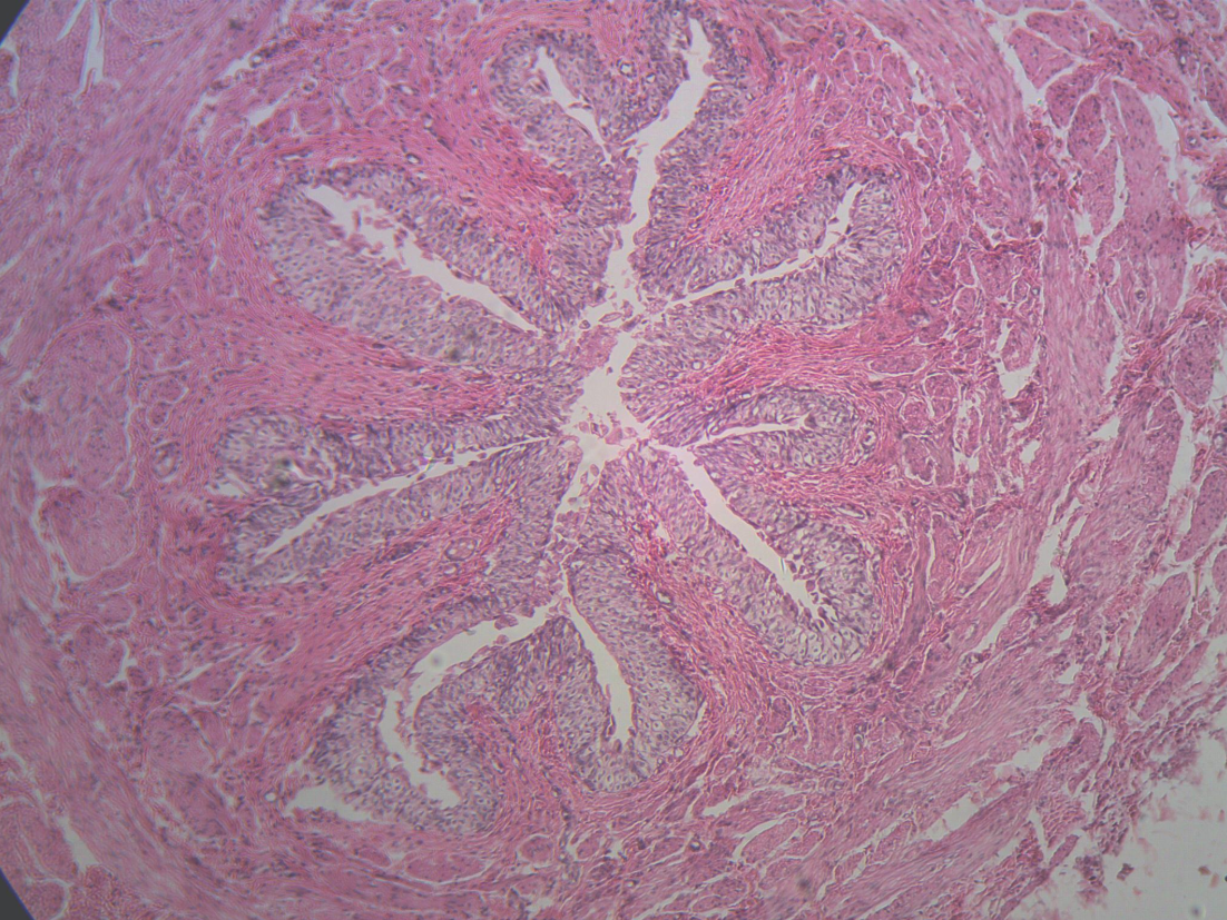
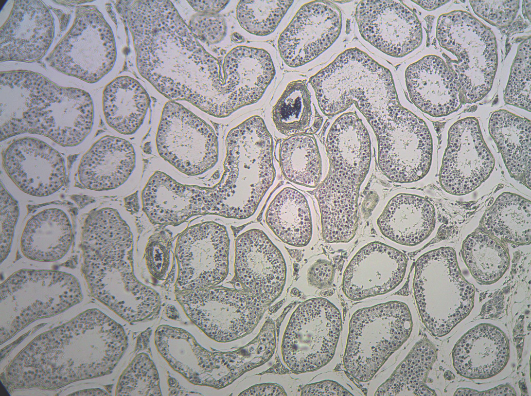
Which structures are seen here?
Seminiferous tubules
What are these structures?
seminiferous tubules
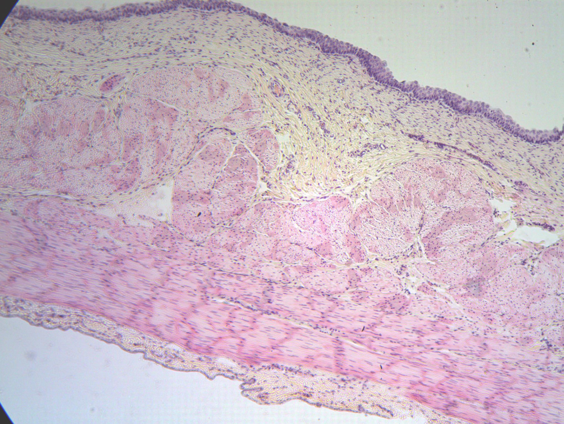
Which organ is this?
Prostate
Which organ is this?
Prostate
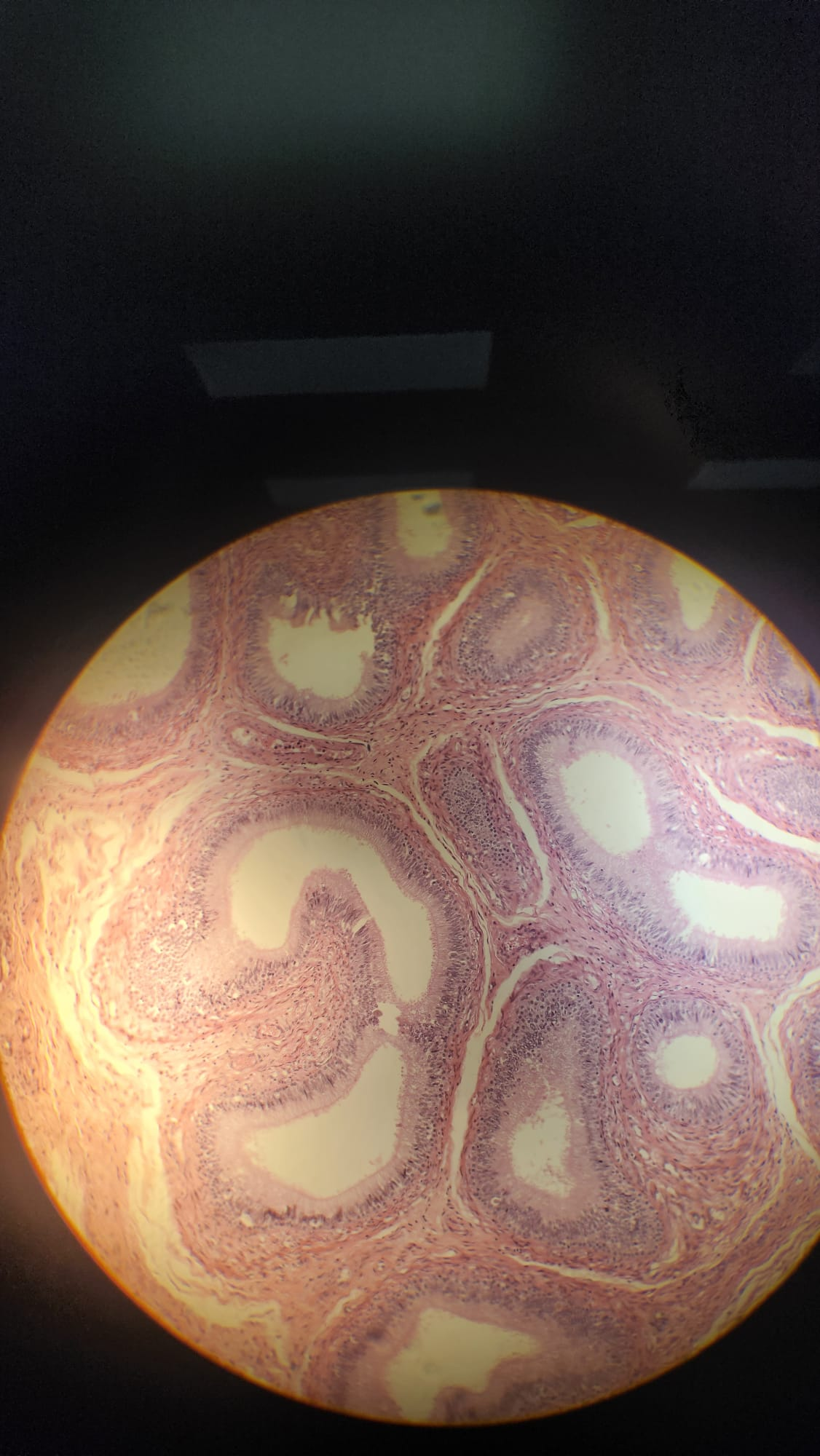
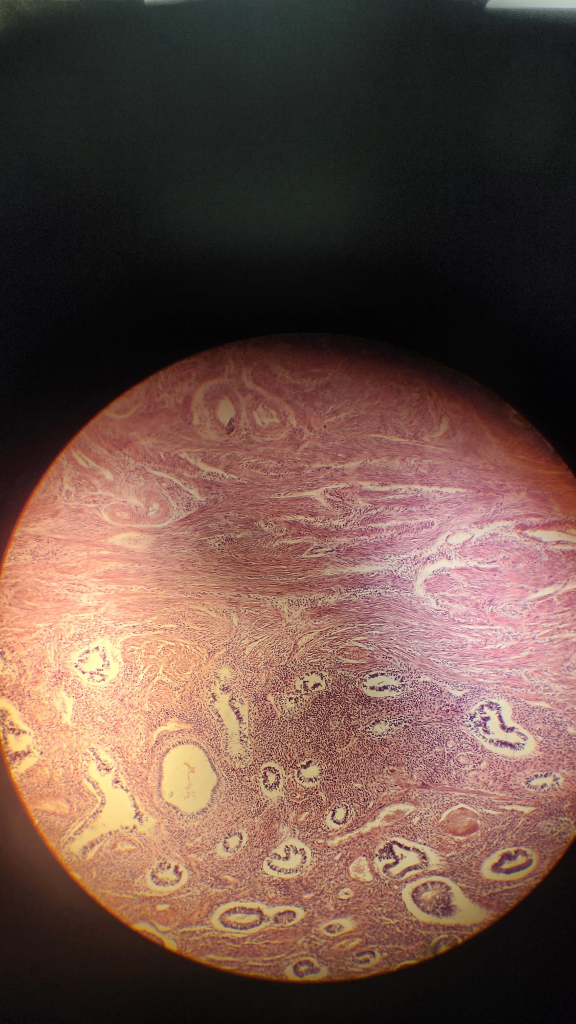
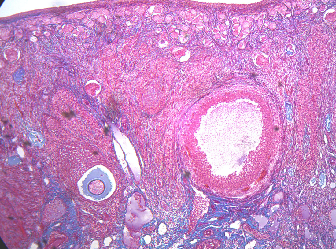
Which organ is this?
Ovary
Which organ is this?
Ovary
Which organ is this?
Ovary
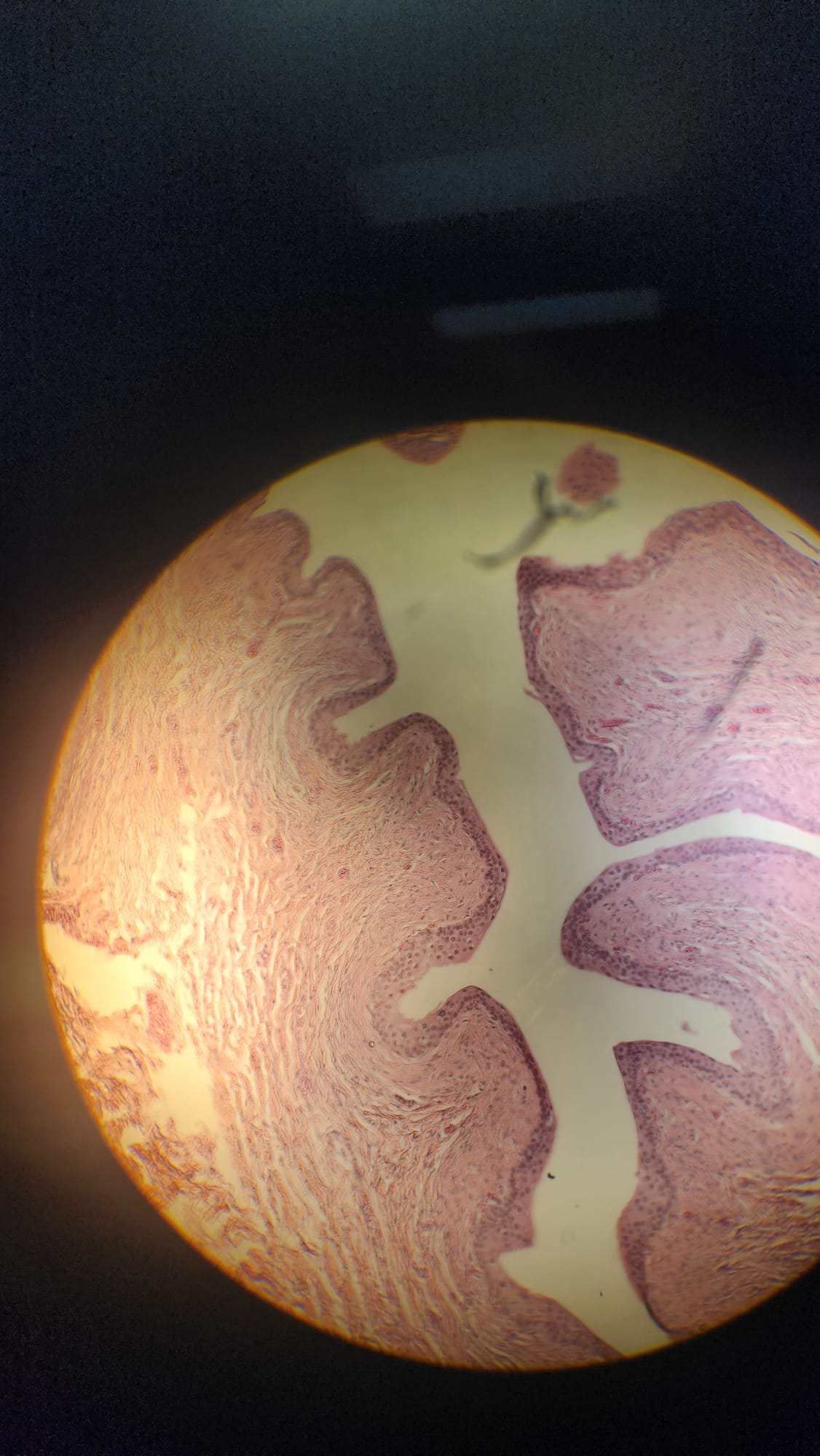
Which organ is this? Name the layers?
Uterus; myometrium (top), endometrium (bottom)
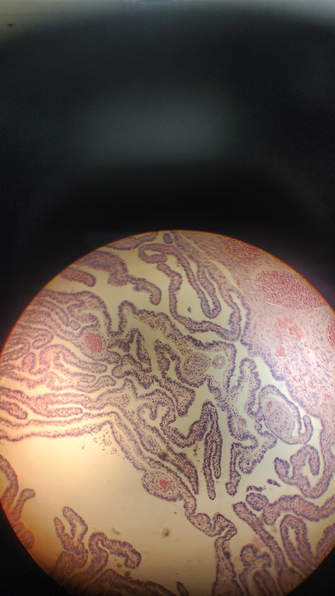
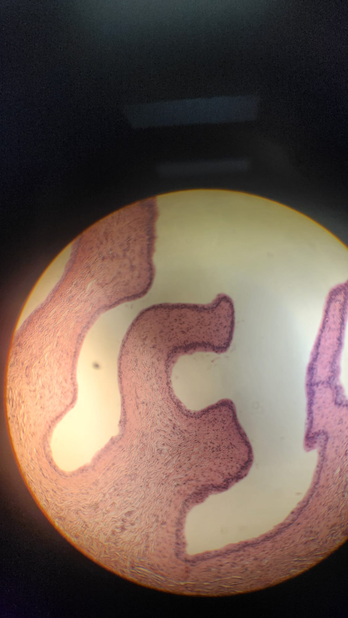
Which organ is this?
Fallopian tube
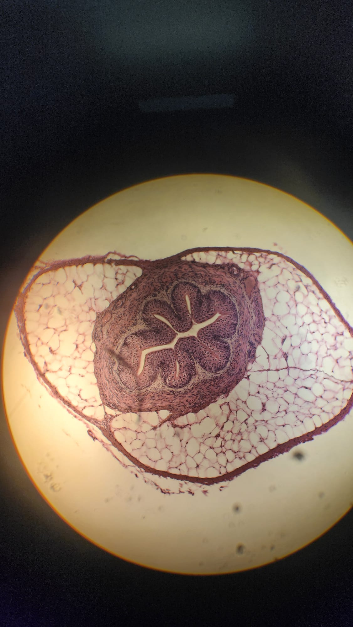
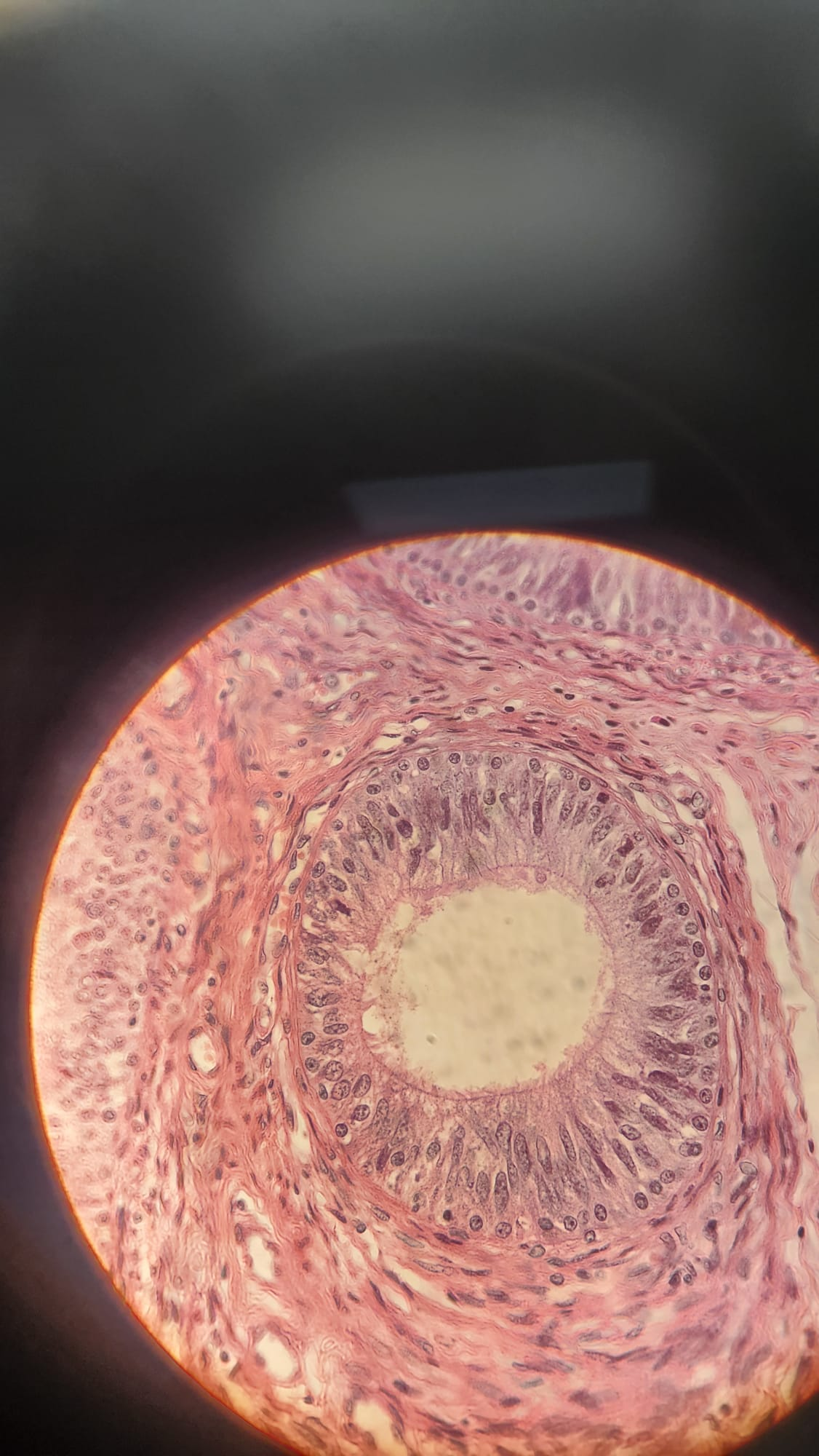
What part of the body is seen in this image?
Ureter
Where in the body is the image from?
Bladder
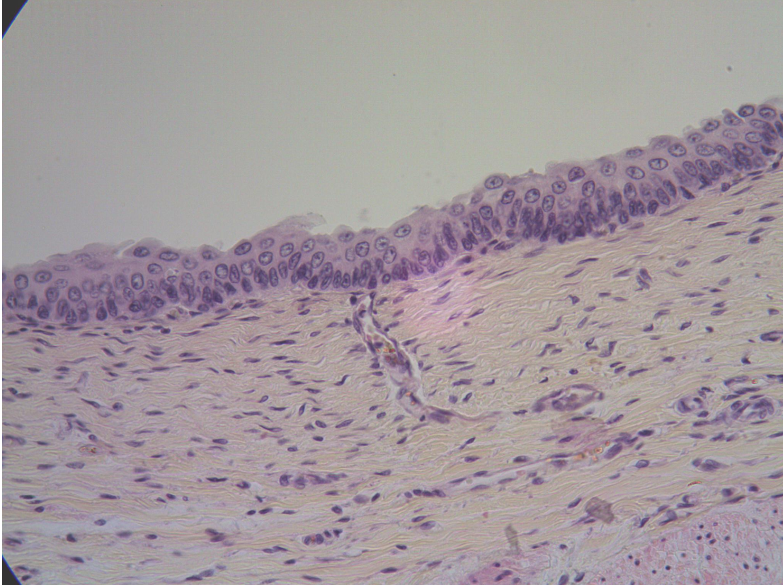
What type of cells are seen lining the lumen in this image
transitional epithelium/urothelium
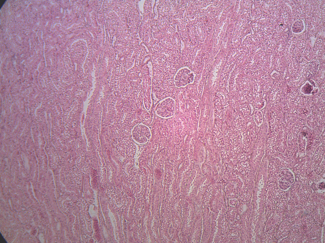
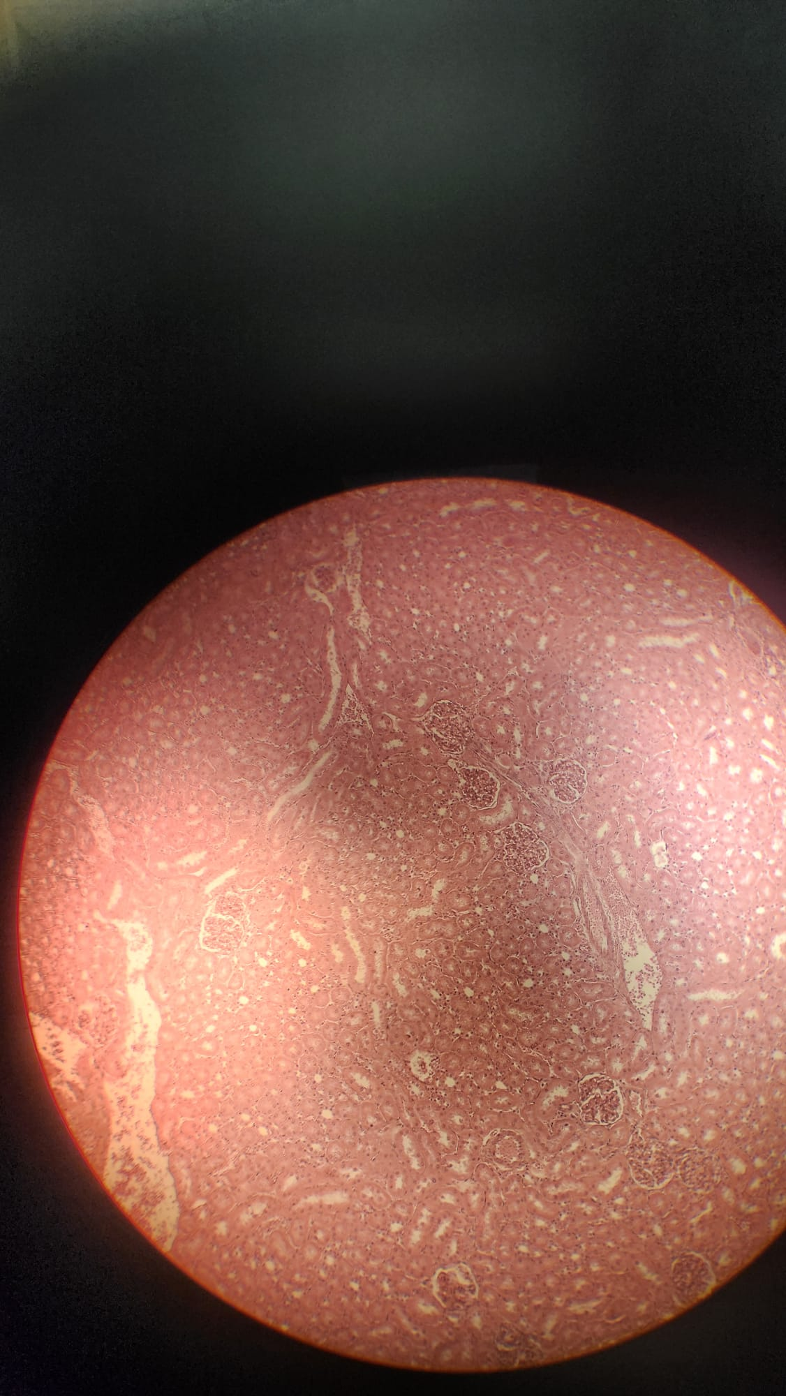
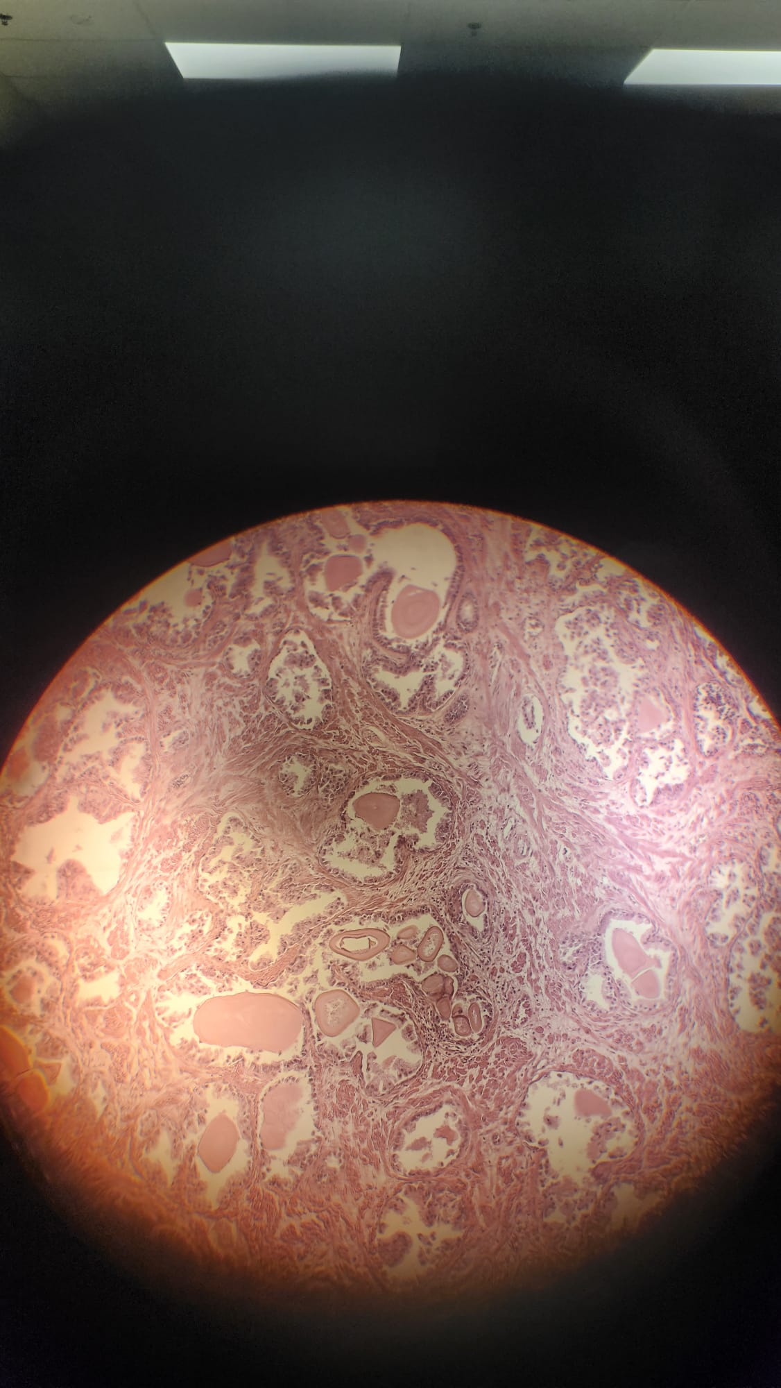
What organ is seen in this image?
Kidney
Gerota’s fascia
tough fibrous connective tissue capsule surrounding the kidney
the ___ is the functional unit of the kidney
nephron
the ___ ___ is
responsible for filtration of plasma
renal corpuscle
The ___ ___ is a single layer of flattened cells resting on a basement membrane
Bowman’s capsule
The ___ is a globular network of capillaries
glomerulus
The ___ ___ ___ is reponsible for the reabsorption of the majority of ions and water
proximal convoluted tubule
the ___ ___ ___ generates high osmotic pressure in the extracellular fluid of the medulla
loop of Henle
The ___ ___ ___ is the area of reabsorption of sodium ions and secretion of hydrogen or potassium ions
distal convoluted tubule
The ___ ___ becomes permeable to water only in the presence of antidiurectic hormone (ADH)
collecting duct
the ___ ___ is the dilated proximal part of the ureter
renal pelvis
Where is a "brush border" found?
proximal convoluted tubules
he juxtaglomerular apparatus is made up of which three components?
distal convoluted tubule, afferent arteriole and lacis cells
What term describes the transitional epithelium of the urinary tract?
urothelium
What is NOT found in the cortex of the kidney?
loop of henle
The thin descending and ascending limbs of the loop of Henle are lined with by what type of epithelium?
simple squamous epithelium
The surface transitional epithelium of the bladder are called what? (the innermost epithelial lining that are on the luminal surface)
umbrella/dome cells
The upper segment of the ureter wall has ___ layers of muscle. The lower third of the ureter and the bladder have ___ layers of muscle.
2, 3
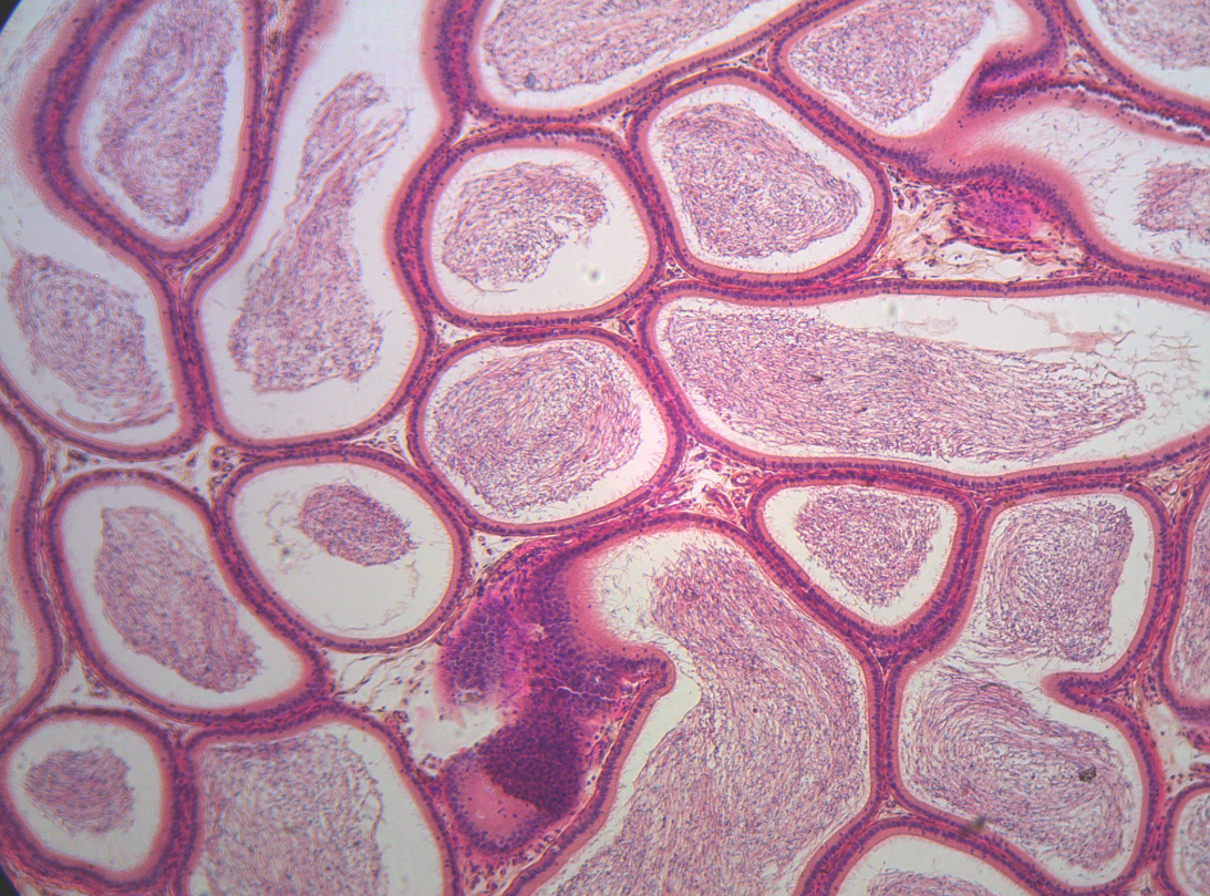
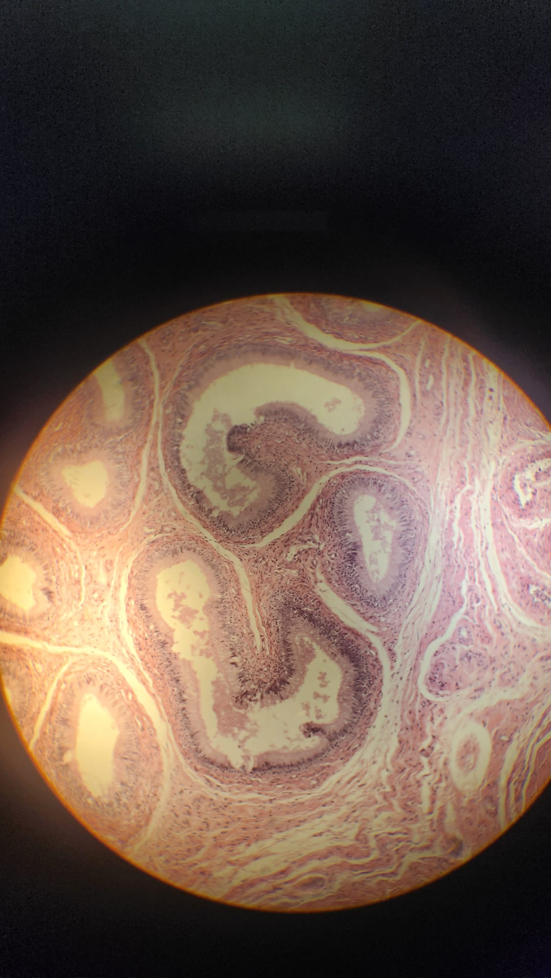
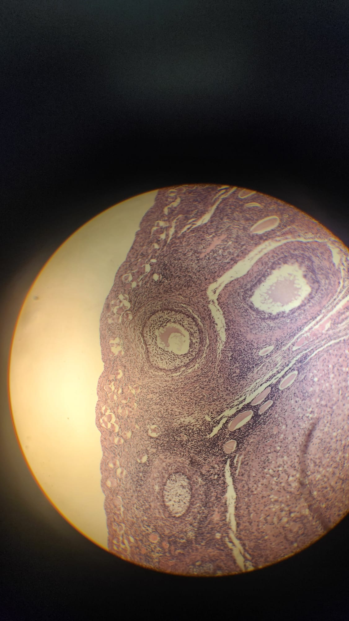
Where part of the body is seen in this image?
epididymis
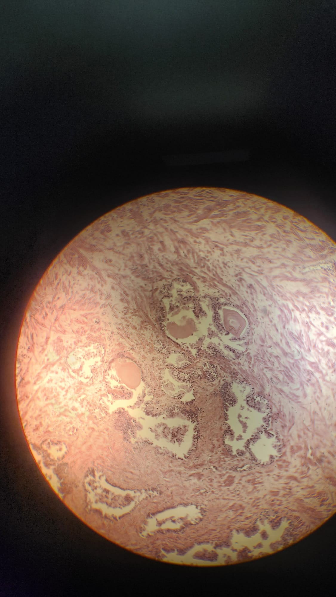
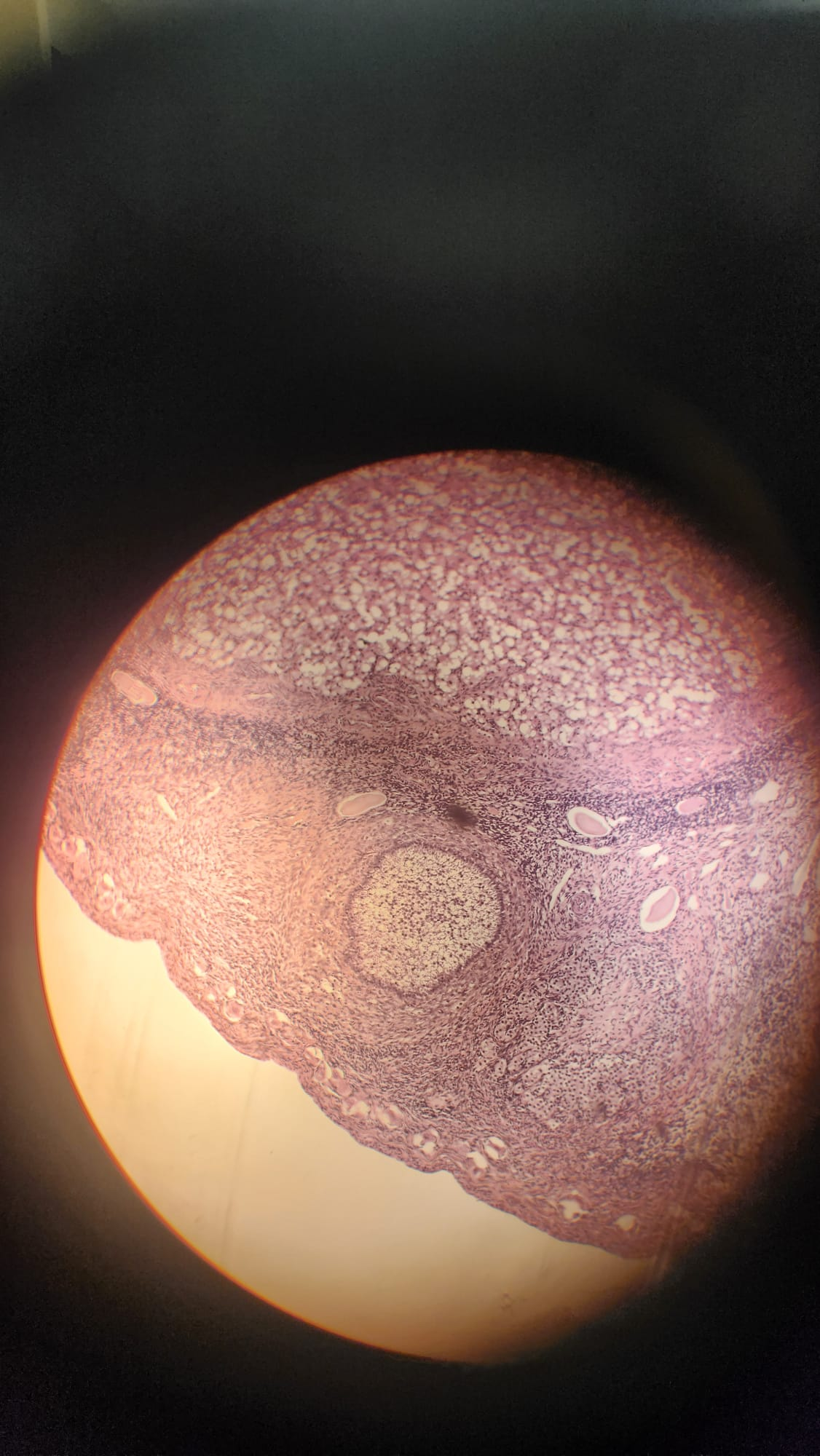
What part of the body is seen in this image?
prostate gland
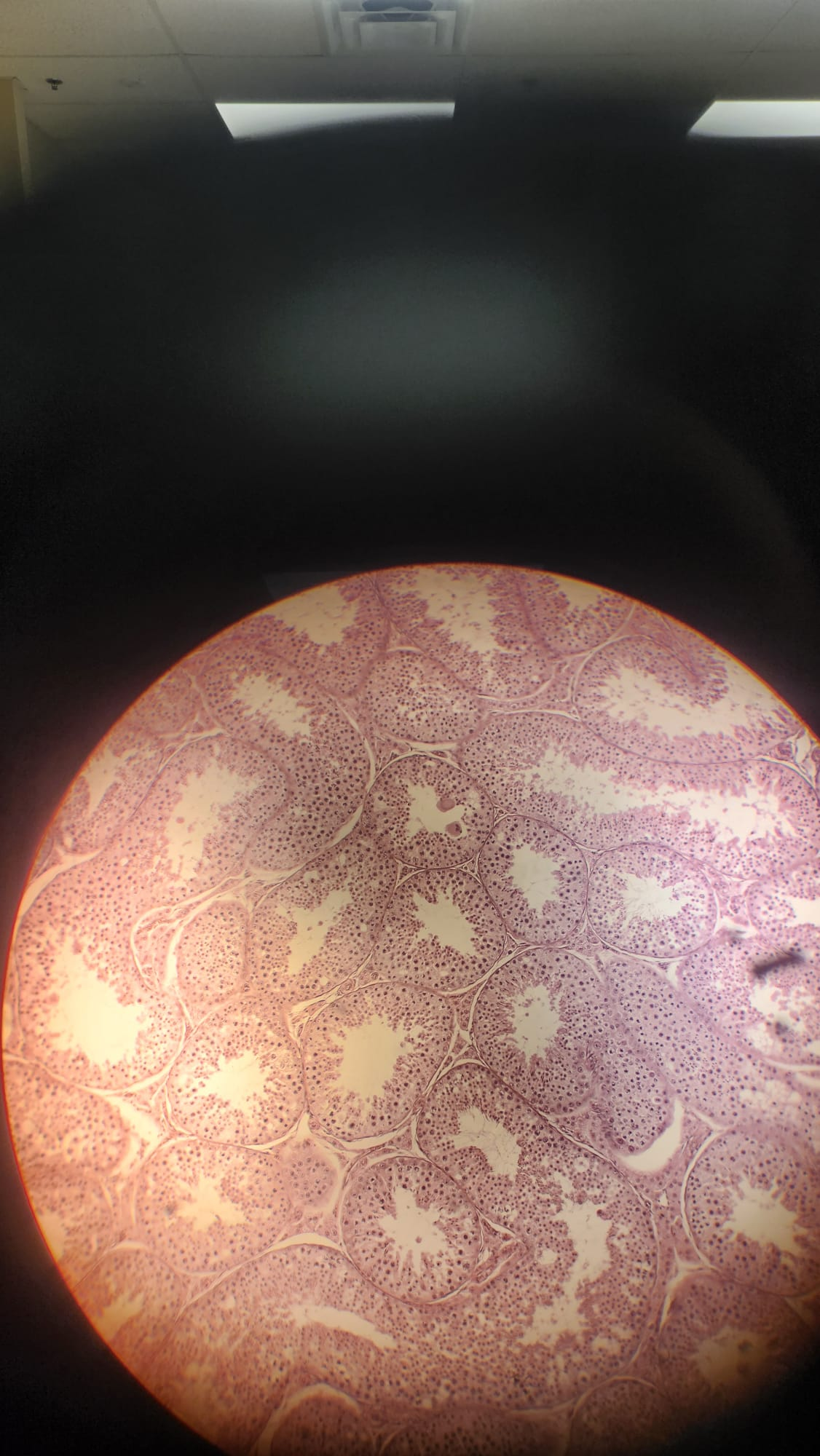
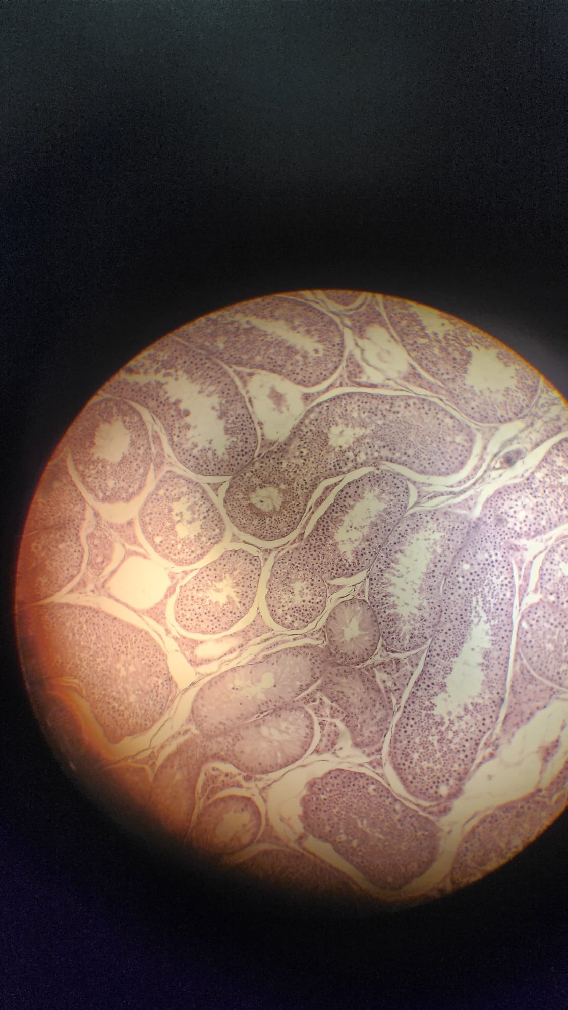
What part of the body is seen in this image?
seminiferous tubules of the testes
What type of cells are found in the seminiferous tubules and what are their functions?
1. Germ cells: give rise to sperm cells
2. Sertoli cells: support and nourish developing spermatozoa
What are the interstitial cells of the seminiferous tubules and what do they secrete?
Leydig cells secrete testosterone
The epidiymis is a tube of ___ muscle lined by ___ epithelium. The epithelial cells have the surface modificaton of ___ which are thought to be involved the ___ of ___ ___.
smooth, pseudostratified columnar, sterocilia, absorption, excess fluid
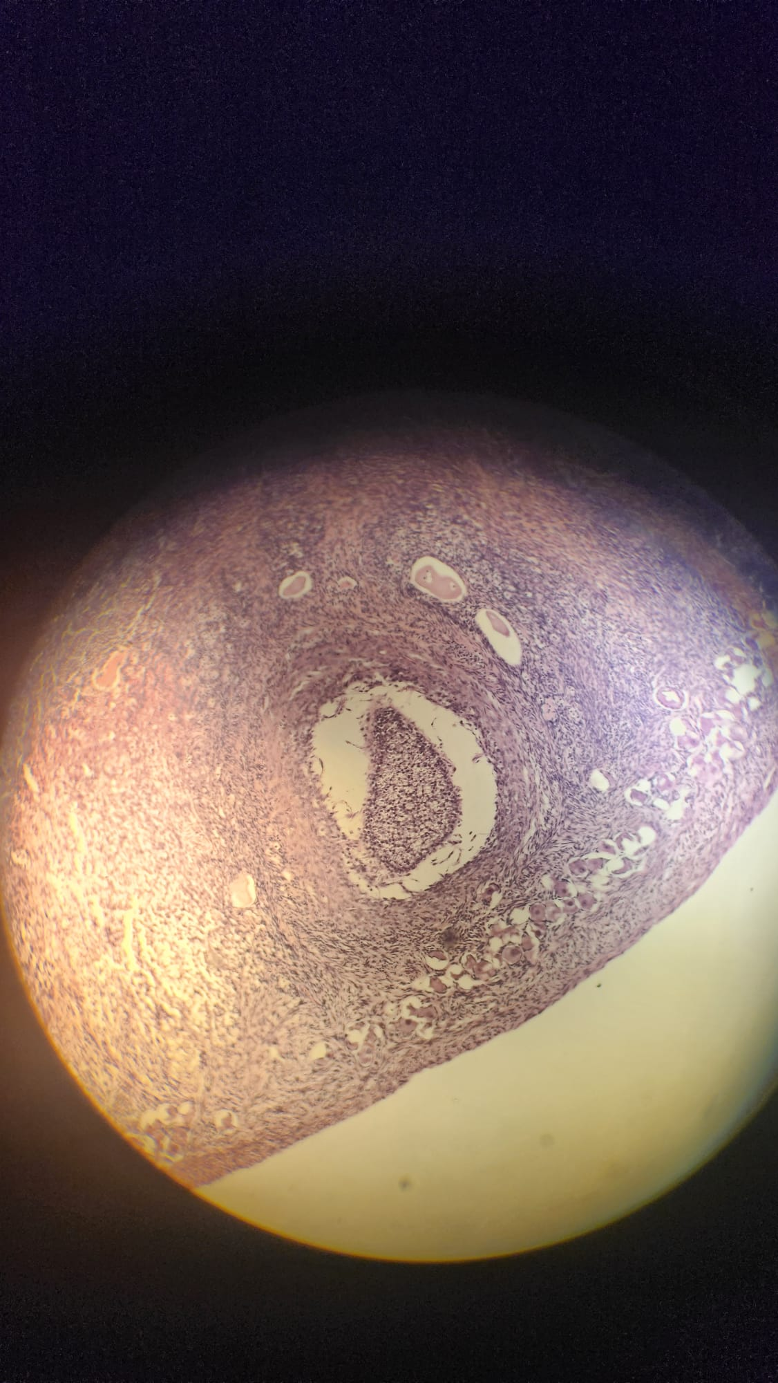
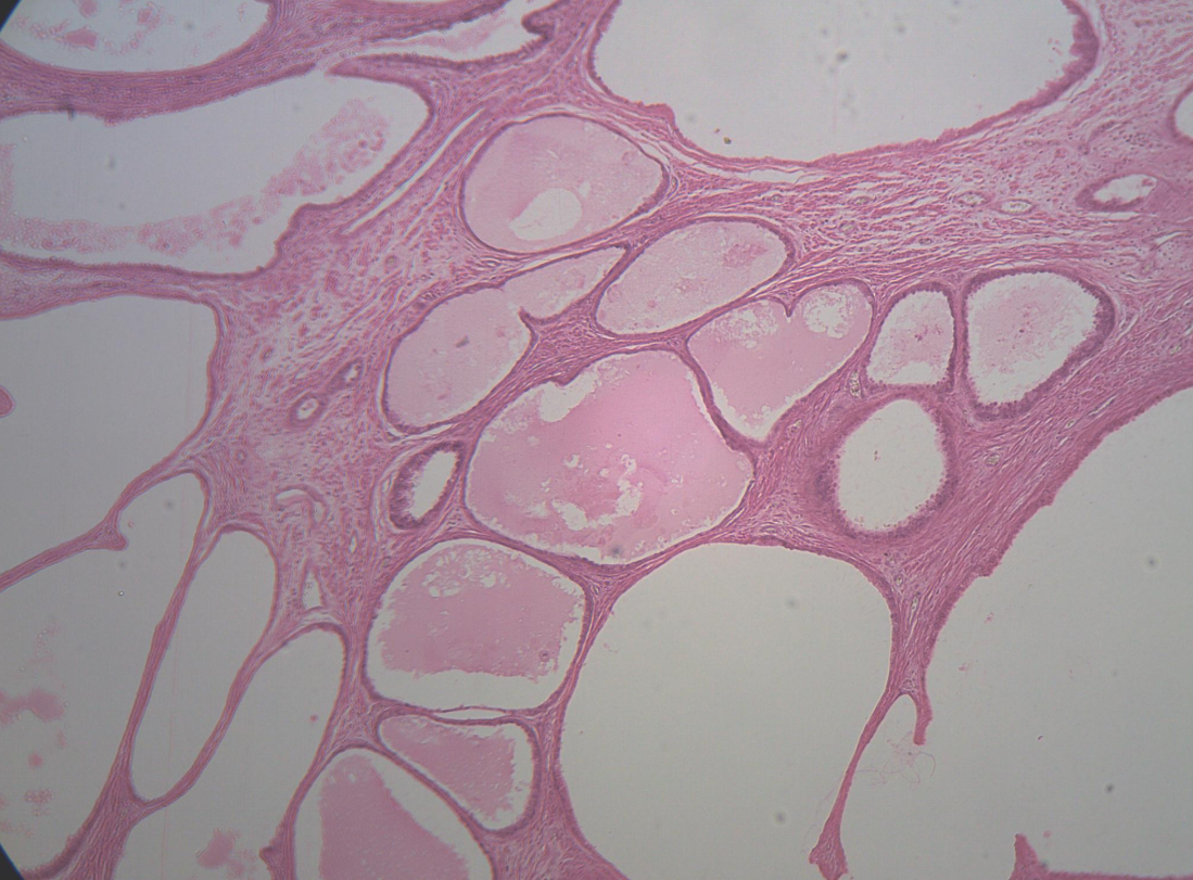
What part of the body is seen in this image?
ovary
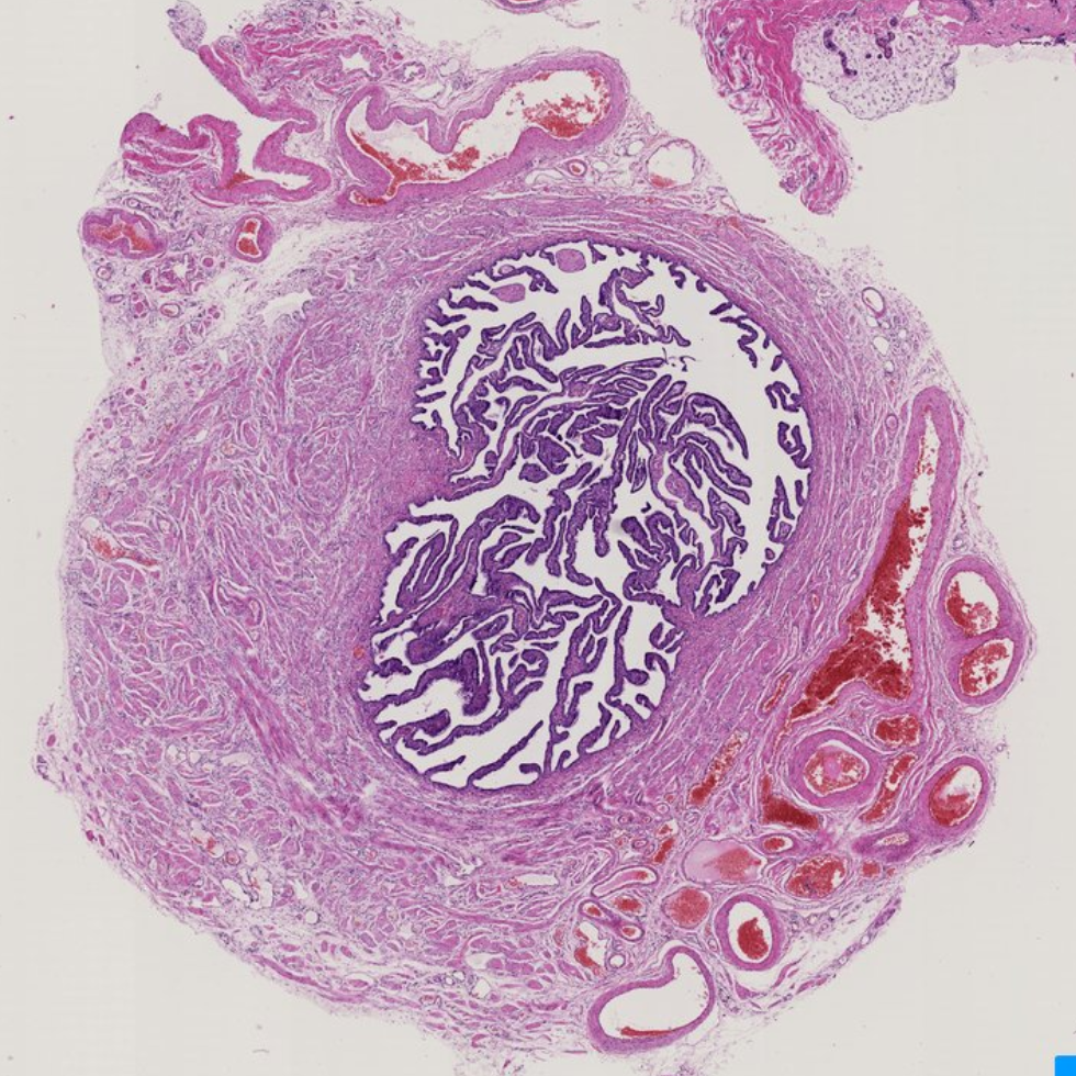
What part of the body is seen in this image?
Fallopian tube.
The wall of the fallopian tubes has ___ layers of smooth muscle.
2
What type of epithelial cells line the lumen of the fallopian tubes?
three types of tall columnar epithelial cells: ciliated, non-ciliated, intercalated cells
List the ovarian follicles in order of maturity from least mature to most mature.
primordial follicle, early primary follicle, primary follicle, secondary follicle, Graafian follicle
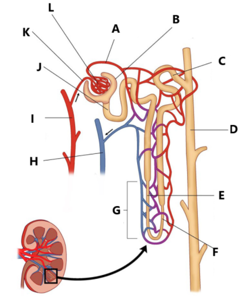
Label the following diagram
A. efferent arteriole
B. Bowman’s capsule
C. Distal convoluted tubule
D. collecting duct
E. descending limb of the loop of Henle
F. ascending limb of loop of Henle
G. loop of Henle
I. renal artery
J. proximal convoluted tubule
K. afferent arteriole
L. glomerulus