Diag/Inter Acute Lab - Week 2 - Pulmonary and Cardiovascular Assessments
1/73
There's no tags or description
Looks like no tags are added yet.
Name | Mastery | Learn | Test | Matching | Spaced | Call with Kai |
|---|
No analytics yet
Send a link to your students to track their progress
74 Terms
Where does the trachea bifurcate?
At the level of the sternal angle
What are the differences between the right and left lungs?
There are differences in size and the number of the lobes. The right lung is larger and has three lobes divided by two fissures. The left lung is smaller than the right and only has two lobes divided by one fissure.
What is the difference between the right and left mainstem bronchi?
The right main bronchus is wider, shorter than the left main bronchus, which is thinner and longer.
What are the superior and inferior limits of the lungs with normal inspiration?
-Superior border: 1-1.5 inches above the clavicle
-Inferior border: T6/6th rib
What is the inferior limits of the lungs with deep inspiration?
Border: T10-T12
What is the inferior limits of the lungs with deep expiration?
Border: T3
Where is the left ventricular apex located?
Inferior border in the 5th intercostal space 6 cm left of the sternum (Point of maximal impulse)
Where should you auscultate to listen to the aortic valve?
2nd intercostal space at the right sternal border
Where should you auscultate to listen to the pulmonic valve?
2nd intercostal space at the left sternal border
Where should you auscultate to listen to Erb's Point? And what is the significance of listening to Erb's Point
3rd intercostal space at the left sternal border.
This is the area of the chest where heart murmurs can best be heard.
Where should you auscultate to listen to the bicuspid/mitral valve?
5th intercostal space, medial to the left midclavicular line; 6mm left of the sternum
Where should you auscultate to listen to the tricuspid valve?
4th intercostal space at the left sternal border
Are there noticeable differences in left and right heart sounds?
Yes, depending on where you auscultate you will better be able to hear each individual valve.
What are normal heart sounds?
S1 (lub) is made by the closure of the mitral and tricuspid valve at the onset of ventricular systole. It is a high frequency sound with lower pitch and longer duration than during S2.
S2 (dub) is made by the closure of the aortic and pulmonary (semilunar) valves at the onset of ventricular diastole. It is a high freequency sound with higher pitch and shorter duration than S1.
What are abnormal heart sounds?
-S3
-S4
-Gallop Rhythem (S3 and S4)
-Pericardial friction rub
-Muffled heart sounds
What is S3? and what might it suggest?
Low-frequency sound heard after S2 during rapid ventricular filling.
Associated Pathologies:
-Heart Failure: Volume overload and increased ventricular filling pressures can lead to an S3 sound.
What is S4? and what might it suggest?
Low-frequency sound heard just before S1 during atrial contraction.
Associated Pathologies:
-Hypertension: Stiffening of the ventricles, especially the left ventricle, can lead to an S4 sound.
What is a pericardial friction rub? and what might it suggest?
Grating or scratching sound heard during both systole and diastole.
Associated Pathologies:
-Pericarditis: Inflammation of the pericardium, causing friction between pericardial layers.
What are muffled heart sounds? and what might it suggest?
Diminished or indistinct heart sounds.
Associated Pathologies:
-Pericardial Effusion: Accumulation of fluid in the pericardial sac, dampening heart sounds.
What is a gallop rhythm? and what might it suggest?
Presence of both S3 and S4 sounds in rapid succession.
Associated Pathologies:
-Heart Failure: The combination of S3 and S4 sounds may be heard in advanced heart failure.
What is the proceadure for listening to lung sounds?
-Postition the patient in sitting
-Listen directly on skin if possible
-Have patient take deep breaths in and out through the mouth
-Listen to both the anterior and posterior chest wall
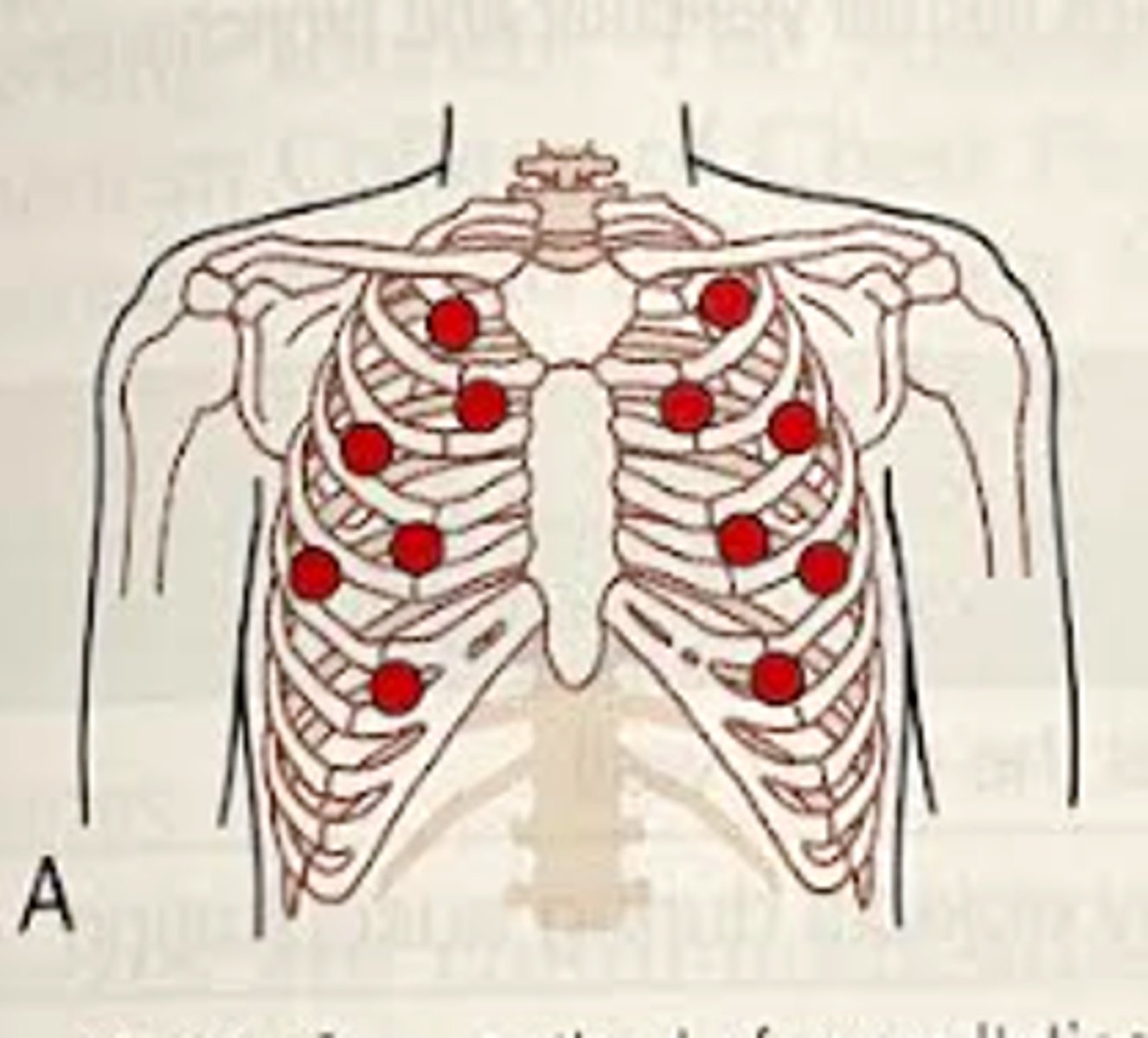
What is the proceadure for ausculating the anterior chest wall for lung sounds?
Start at one side and then move to the opposite side
-Start at the apex of the lungs (slightly above the clavicle)
-2nd intercostal space midclavicular (assesses upper lobes of right and left lung)
-3rd intercostal space midclavicular (upper lobes of right and left lung)
-4th intercostal space midclavicular (right middle lobe, left upper lobe)
-5th intercostal space (right middle lobe, left upper lobe)
-6th intercostal space midaxillary (lower lobes of right and left lungs)
-7th intercostal space (lower lobes of right and left lungs)
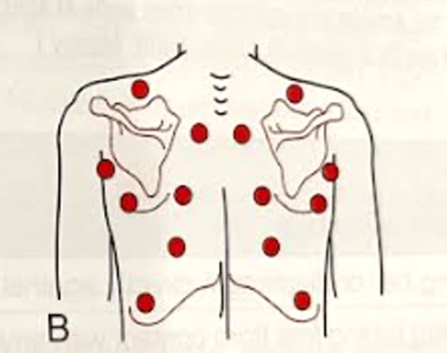
What is the proceadure for ausculating the posterior chest wall for lung sounds?
Start above the scapula at the apex of the lungs
-Intercostal spaces between C7 - T3 between spine and scapula (upper lobes of right and left lungs)
-Intercostal spaces between T3 - T10 between spine and scapula (lower lobes of right and left lungs)
-Midaxillary when below scapula (lower lobes of right and left lungs)
What are the three normal breath sounds?
-Tracheal/bronchial
-Bronchovesicular
-Vesicular
Where can tracheal and bronhial sounds be ascultated?
Tracheal and bronchial sounds can be heard anteriorly directly over the trachea and manubrium
Where can Bronchovesicular sounds be ascultated?
-anteriorly at the 1st and 2nd intercostal spaces
-posteriorly between the scapulae at the level of T3/T4.
Where can vesicular sounds be ascultated?
anteriorly and posteriorly throughout the peripheral lung field (anteriorly, 3rd intercostal space and below, posteriorly everywhere but between the scapulae at the level of T3/T4?
Describe the pitch and intensity, cycle, and duration for tracheal and bronchial breath sounds?
-loud and tubular,
-equal inhalation and exhalation,
-short pause between inhalation and exhalation.
Describe the pitch and intensity, cycle, and duration for bronchovesicular breath sounds?
-softer version of bronchial sounds,
-both inhalation and exhalation
-no pause, it is continuous from inhalation to exhalation.
Describe the pitch and intensity, cycle, and duration for vesicular breath sounds?
-soft and low pitched,
-inhalation and first third of exhalation
-no pause between inhalation and exhalation sounds, they are continuous
What are the abnormal breath sounds? (9)
-Adventitious beath sounds
-Crackle (formerly Rales)
-Pleural friction rub
-Rhonchi
-Stridor
-Wheeze
-Bronchial
-decreased or diminished breath sounds
-absent breath sounds
What is adventtitous breath sounds?
Abnormal breath sounds or normal breath sounds that are heard outside of their normal location/phase of respiration.
What does a crackle sound like?
an abnormal, discontinuous high pitched popping sound heard more often during inspiration.
What doees plueral friction rub sound like?
Dry, crackling sound heard during both inspiration and expiration
What does rhonchi sound like?
Continuous low-pitched sounds described as having a "snoring" or "gurgling"
What does stridor sound like?
Continous high pitched wheeze heard with inspiration or expiration.
What does a wheeze sound like?
continuous "musical" or whistling sound composed of a variety of pitches
What is a diminished breath sound? and what might it indicate?
A less audible sound that may indicate severe congestion, emphysema, or hypoventilation.
What might an absent breath sound indicate?
absent lung sound may indicate pneumothorax or lung collapse.
When would bronchial sounds be abnormal?
When they are heard in locations where vesicular sounds are normally present.
What are two pathologies that may be associated with wheezing?
Asthma and COPD
What are three pathologies that may be associated with crackles?
-Pneumonia (inflammation and fluid accumulation in the alveoli)
-Pulmonary embolism
-Interstitial lung disease (scarring or fibrosis in lung tissue)
What are two pathologies that may be associated with Rhonchi?
-Bronchitis
-COPD
What is a pathology that may be associated with stridor?
Upper airway obstruction (blockadge in the trachea or larynx, such as in croup or foreign body aspiration).
What are two pathologies associated with absent breath sounds?
-Pleural effusion (fluid in the pleural space limits lung expansion)
-Pneumothorax (air in pleural space causing lung collapse).
What is a pathology that is associated with bronchial breath sounds?
Consolidation (i.e. pneumonia). Solidification of lung tissue leads to enhanced transmission of bronchial sounds.
What is a pathology associated with pleural friction rub?
Pleuritis (aka pleurisy), inflammation of the pleura causing friction between the pleural layers.
What is mediate percussion?
The act of tapping the surface of the body to identify areas of altered density
What position should the patient be in for mediate percussion?
Anterior - sitting or supine
Posterior - sitting
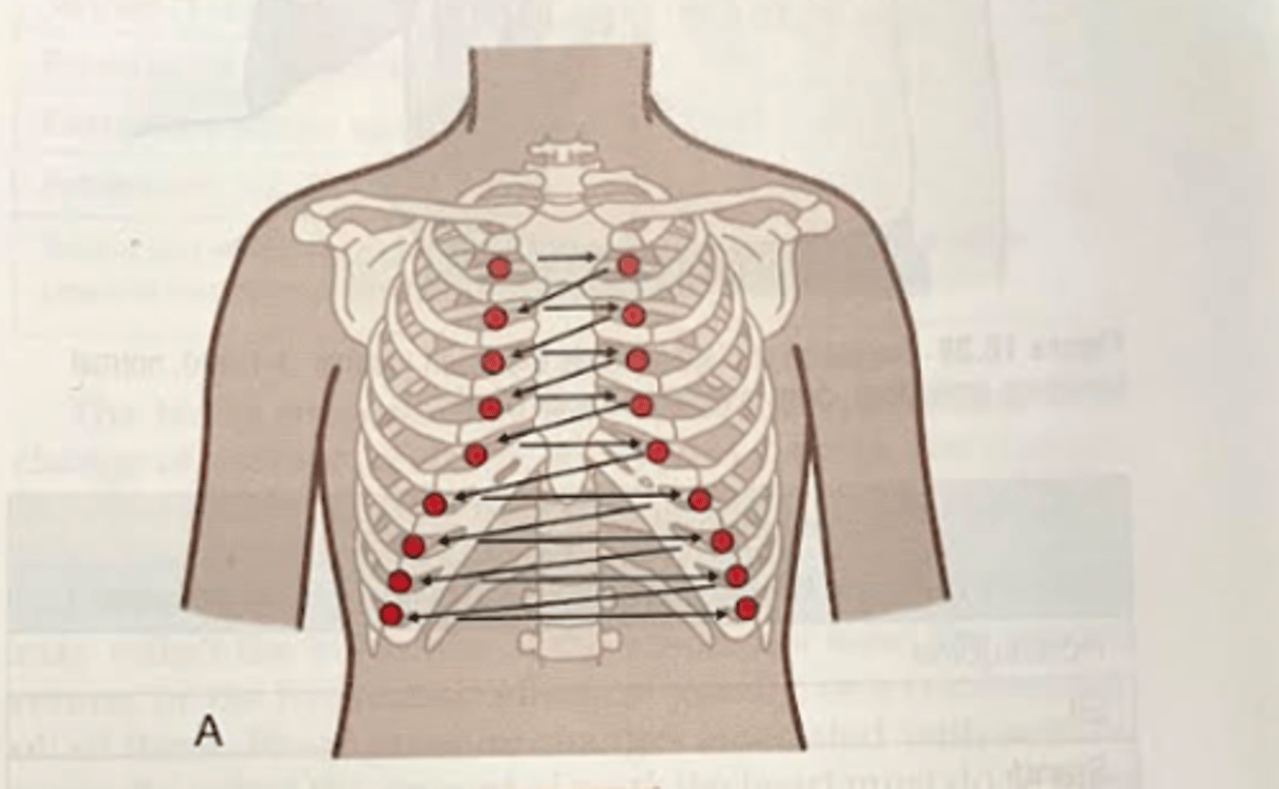
What is the proceadure for mediate percussion of the anterior chest?
Systematic approach, starting at the clavicle and progressing down through each intercostal space.
-Tap 3 consecutive blows before moving on and always compare side to side.
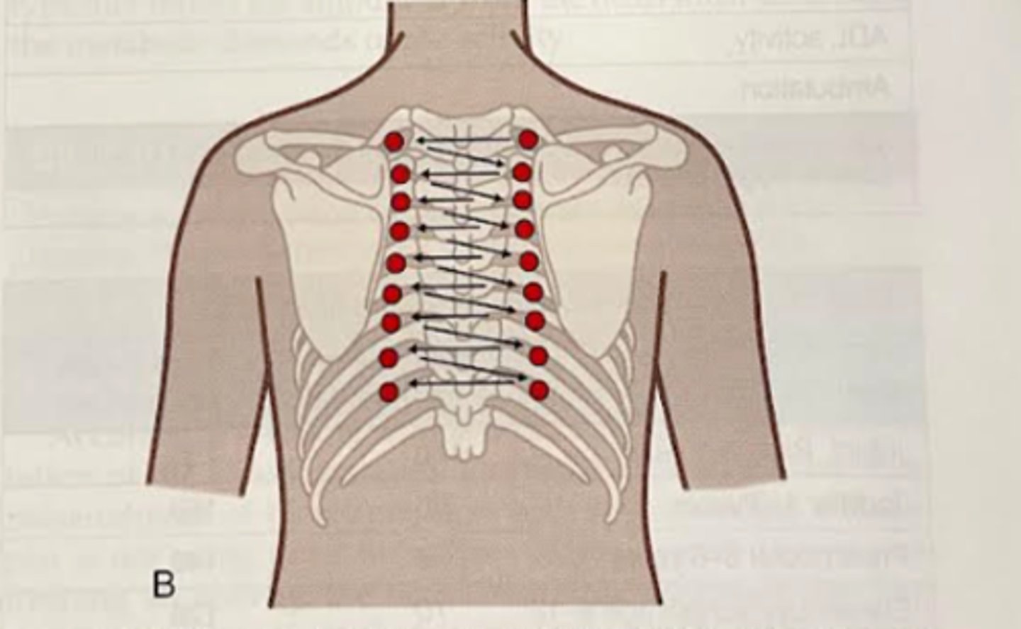
What is the proceadure for mediate percussion of the posterior chest?
Systematic approach, starting at the level of T1 and progressing down through each intercostal space.
-Tap 3 consecutive blows before moving on and always compare side to side.
How do percussion sounds work?
The percussion sounds progress from tissues of high density to tissues of low density.
What is a flat or dull percussion sound? And what may it suggest if heard in the upper lung?
The sound elicited by percussion of the thigh muscle. In the upper lung, it would suggest a neoplasm, atelectasis, or consolidation of the lung.
What is the normal sound that should be heard when percussing the lungs?
A resonance
What is hyperresonance? And what might it suggest?
Immediate sound between resonance and tympanny.
-It is a percussion not emitted by the emphysematous lung.
-It would suggest pulmonary emphysema or pneumothorax.
What is tympany? and what might it suggest?
Hollow sound vaguely resembling a drumbeat.
-Occurs almost exclusively with a large pneumothorax.
How do you palpate the abdominal aorta? and what would be a concerning finding that may suggest an aneurysm?
Assess the width of the aorta by placing your hands on each side of the aorta just above the umbilicus.
Abdominal aortic aneurysm - width >4cm with lateral pulsations
What are heart murmurs?
Heart murmurs are vibrations of longer duration than the heart sounds and are often due to disruption of blood flow past a stenotic or regurgitant valve; the sounds are variably described as soft, blowing, or swishing.
What is a systolic murmur?
Abnormal sounds heard during systole (when the heart is contracting).
Associated Pathologies:
-Aortic Stenosis: Narrowing of the aortic valve, causing turbulence during ejection of blood.
-Mitral Regurgitation: Backflow of blood from the left ventricle into the left atrium during systole.
What is a diastolic murmor?
Description: Abnormal sounds heard during diastole (when the heart is relaxing and filling with blood).
Associated Pathologies:
-Aortic Regurgitation: Backflow of blood from the aorta into the left ventricle during diastole.
-Mitral Stenosis: Narrowing of the mitral valve, impeding blood flow from the left atrium to the left ventricle.
Is the diaphragm symetrical?
No. It comes further down on left side because of the liver.
How do you palpate the mediastinum?
Tip of the index finger being placed in the suprasternal notch
-Move to the SC joint to the left and push inward to check for shift to the left
-Move to the SC joint on the right and push inward to check for shift to the right
Is a tracheal shift significant?
Yes. The observation of tracheal deviation is a critical finding that often prompts further investigation, such as imaging studies (chest X-ray, CT scan) and other diagnostic tests, to identify the underlying cause and guide appropriate management.
It's important to note that tracheal deviation is a serious sign, and prompt medical attention is warranted for a thorough evaluation and diagnosis.
What is atelectasis? and how would it affect the trachea as felt through palpation?
Collapse or incomplete expansion of a lung or a portion of it.
Atelectasis can cause tracheal deviation towards the affected side.
What is a large pulmonary abscess? and how would it affect the trachea as felt through palpation?
Localized collection of pus within the lung.
It can cause deviation of the trachea toward the affected side.
What is hemothorax? and how would it affect the trachea as felt through palpation?
Accumulation of blood in the pleural cavity.
It can cause lung compression, resulting in tracheal deviation away from the affected side.
What is pleural effusion? and how would it affect the trachea as felt through palpation?
Accumulation of fluid in the pleural space.
It may cause the lung on one side to collapse, leading to tracheal deviation away from the affected side.
What is pneumothorax? and how would it affect the trachea as felt through palpation?
Presence of air in the pleural space, leading to lung collapse.
It can cause deviation of the trachea away from the affected side.
How would a tumor or mass affect the trachea as felt through palpation?
Growing masses can displace structures, including the trachea.
It can cause deviation of the trachea toward or away from the affected side, depending on the location and size of the mass.
If a patient has a lobectomy, which direction will the trachea deviate
Toward the affected side.
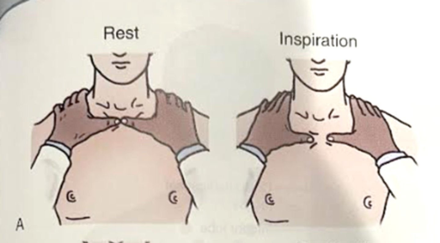
How do you palpate the upper lobes of the lungs?
-Palms of hands anteriorly over the chest wall from the 4th rib upward. Fingers should be stretched upward and over the trapezius. Thumbs should be placed together along the midline of the chest at about the sternal notch
-Patient to take a maximal inspiration and therapist's hands should be relaxed so they move with the chest wall
-Noting: symmetry and extent of movement
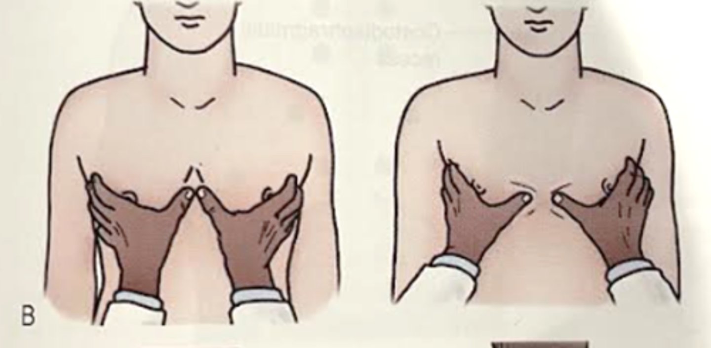
How do you palpate the right middle and left lingula?
-Placing fingers laterally and over the posterior axillary folds. Palms pressed firmly on the anterior chest wall. Skin in drawn medially until thumbs meet at the midline. Patient to take a maximal inspiration and therapist's hands should glide with the movement of the lobes underneath
-Noting: symmetry and extent of movement
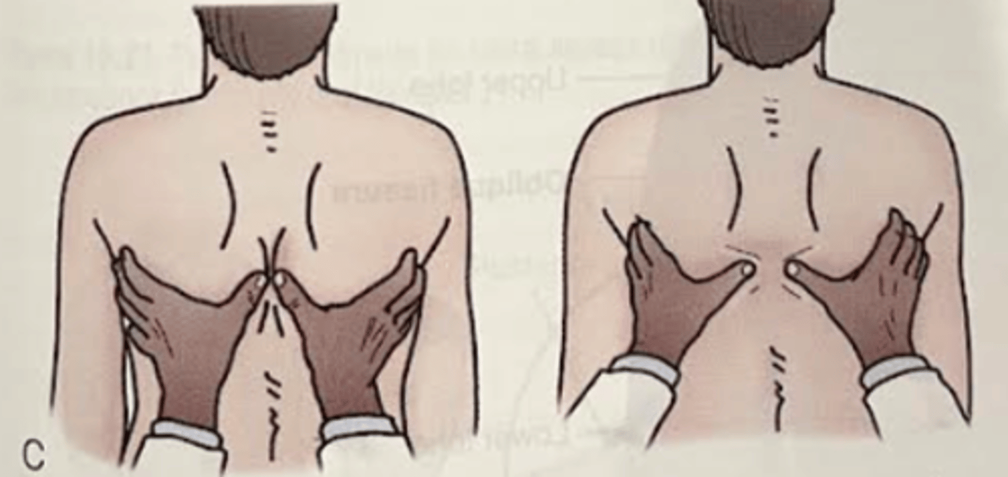
How do you palpate the lower lobes?
Patient's back toward the therapist. Therapist's fingers wrapped around the anterior axillary fold. Skin is drawn medially until the tips of the thumbs meet the spinal column. Patient is to take a maximal inspiration and the therapist's hands should allow hands to glide with the movement of the rib cage
Noting: symmetry and extent of movement
How do you palpate/assess the diaphragm?
Have patient in supine position. Palpation of the anterior chest wall with the thumbs over the costal margins and thumb tips meeting at the xiphoid process. With a deep inspiration, the hands should travel equally apart, total circumferential diameter increasing by at least 2-3 inches