Bio nervous system
1/107
There's no tags or description
Looks like no tags are added yet.
Name | Mastery | Learn | Test | Matching | Spaced | Call with Kai |
|---|
No analytics yet
Send a link to your students to track their progress
108 Terms
Neuron definition
what is true about the shape of a neuron
a specialized cell capable of transmitting electrical impulses and then translating those electrical impulses into chemical signals.
*each neuron has a shape that matches its function
1.Where is the nucleus located and what is this also called
2. what is located within the nucleus
The nucleus is located in the cell body which is also called the soma
ER and ribosomes are located there
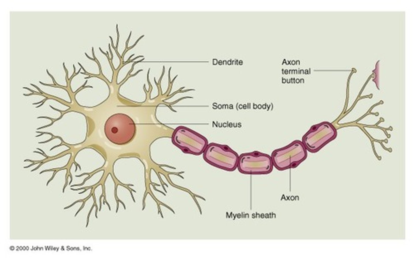
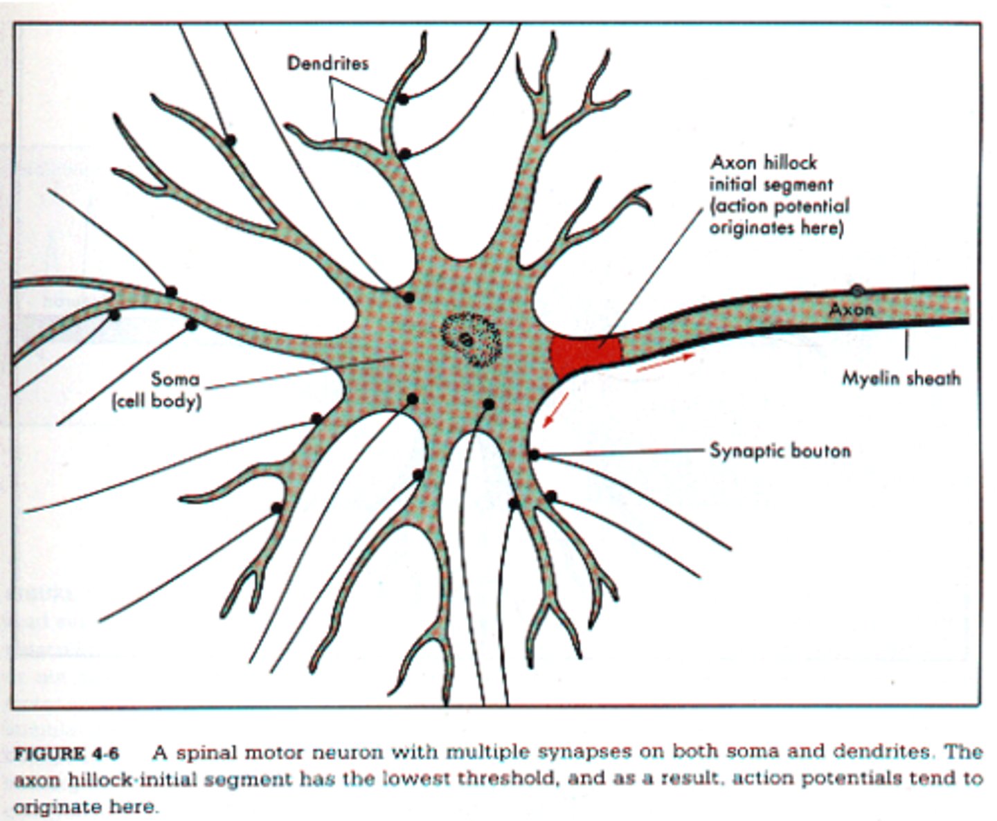
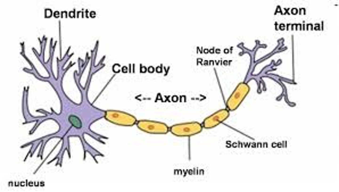
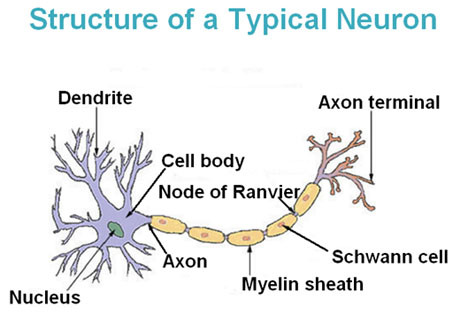
Soma
neuron cell body
Dendrites
The portion of a neuron that receives stimuli from other cells and conveys them toward the cell body
Axon Hillock what happens there? and what is it
Transition point between the cell body (soma) and the axon of a neuron; the site of action potential initiation**
Action Potentials
Is when an abrupt change in the membrane potential of a nerve or muscle caused by changes in membrane ionic permeability; results in conduction of an impulse in nerves or contraction in muscles
or the transmission of electrical impulses down the axon.
Axon
The long fiber of a neuron; it conducts impulses away from the cell body toward the synapse

Myelin
what most mammalian nerve fibers are insulated by
a fatty membrane
myelin sheath definition and function
The white, lipid-containing material surrounding the axons of the many neurons in the central and peripheral nervous systems.
-Maintains the electric signal within one neuron and increases the speed of conduction in the axon.
Oligodendrocytes
Memory device
Myelin Producing Cells in the Central Nervous System
Oligodendrocyte is oligarchy which is all about ME
My from “myelin” as in me me me so think centering yourself equals the central nervous system
Schwann Cells
Myelin Producing cells in the Peripheral nervous system
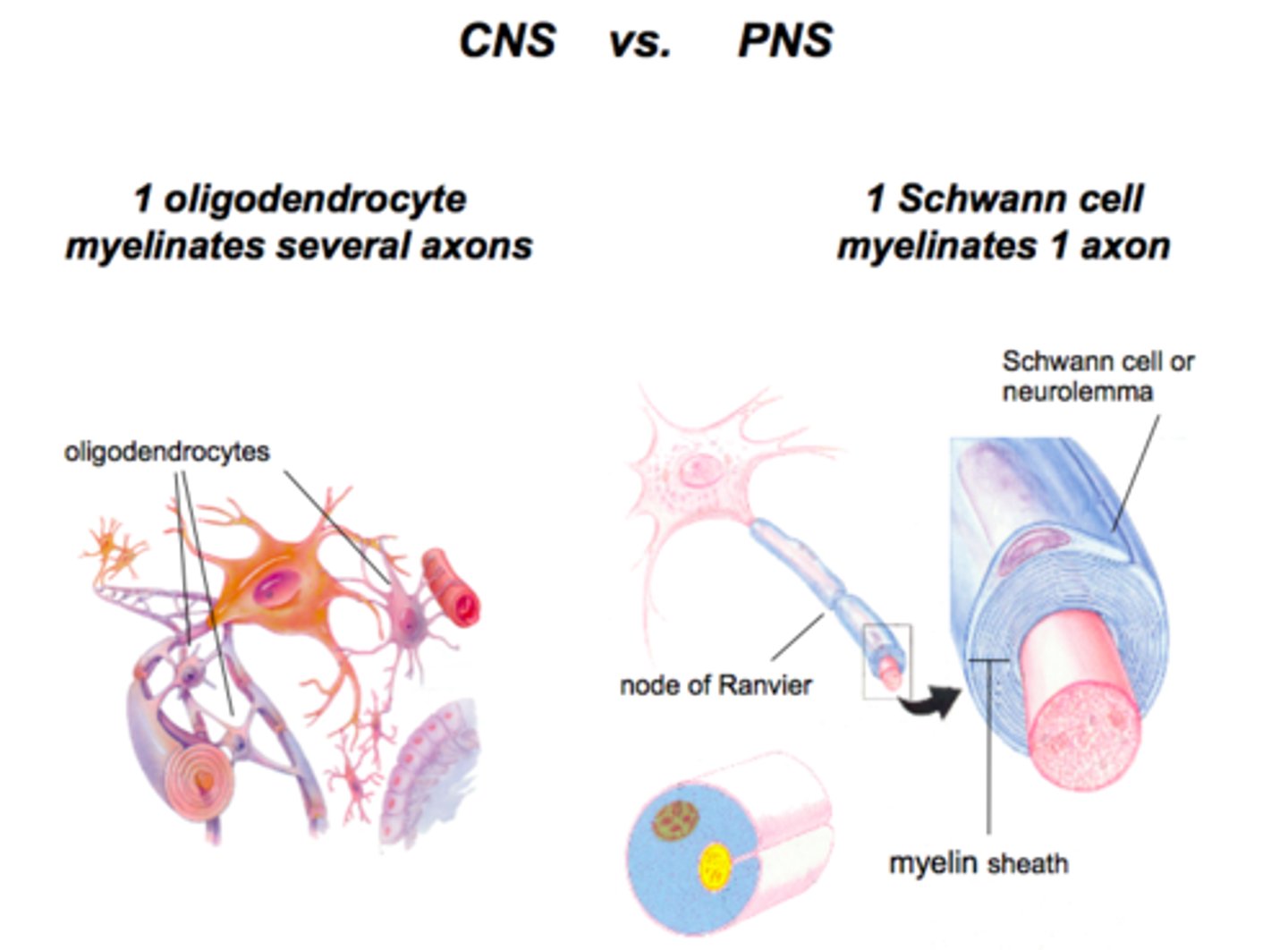
Nodes of Ranvier
What is their role?
Points on a myelinated axon that are not covered by myelin
They are necessary for rapid conduction
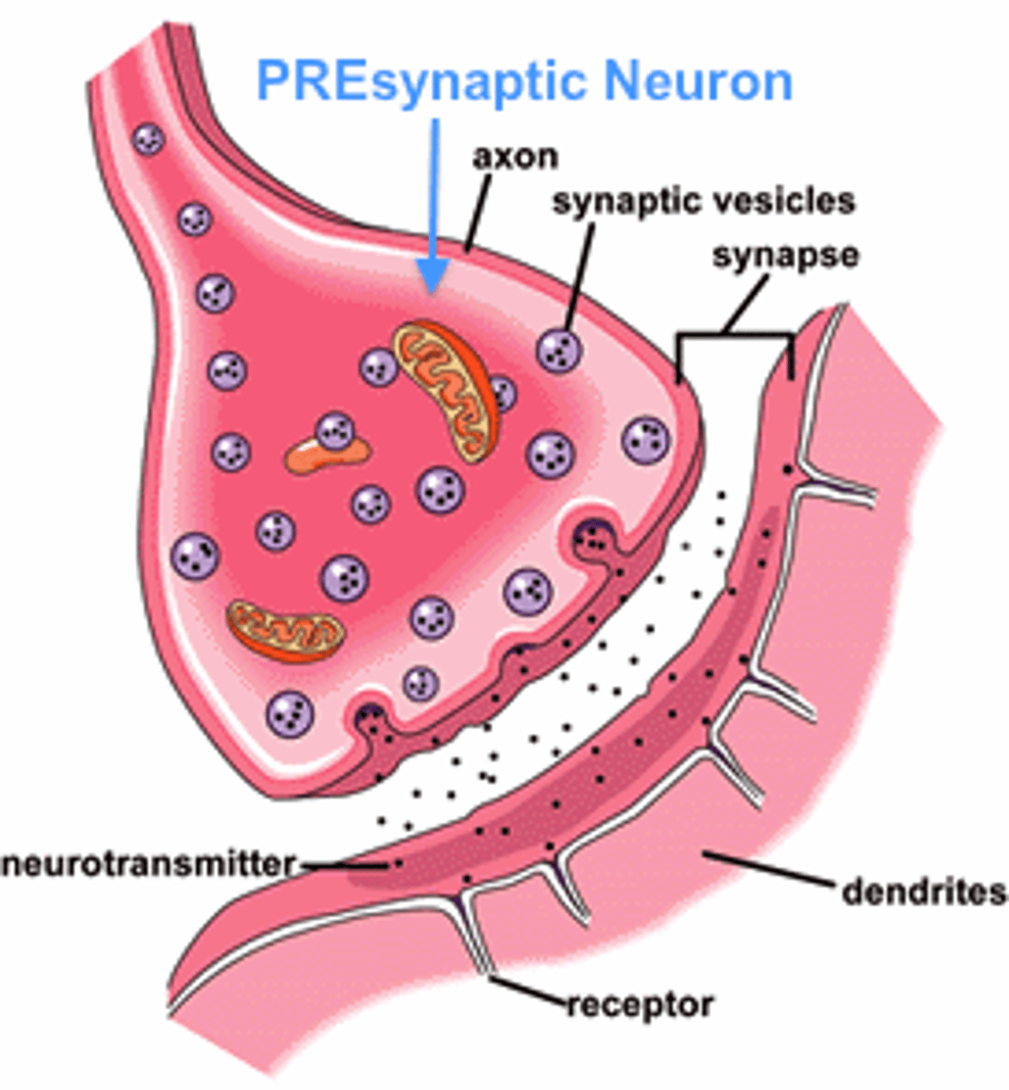
Nerve Terminal and what is it also called and what is its role
End of the axon from which neurotransmitter molecules are released; also called a "synaptic bouton"

Synaptic Bouton (Knob)
nerve terminal
also called axon terminal— not to be confused with axon hillock which is at the start of the cell. Remember, terminal means END like terminal cancer
Neurotransmitters
The chemicals that transmit information from one neuron to another.
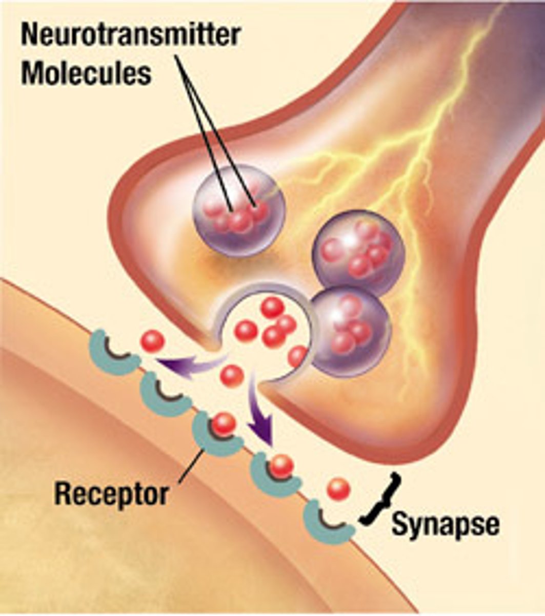
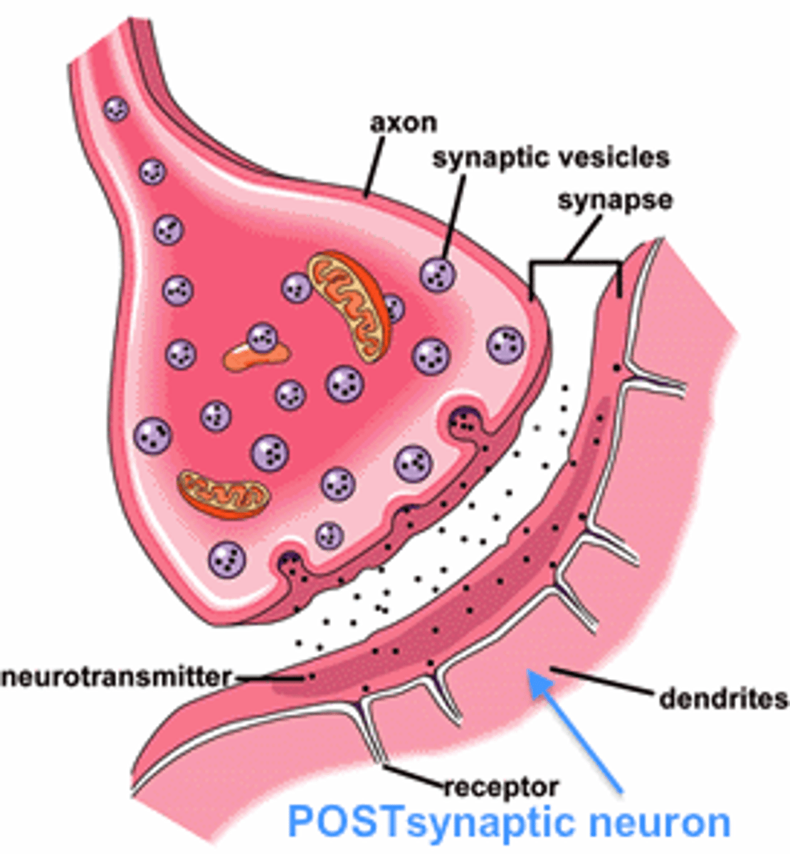
Synaptic Cleft
Space between the neurons into which the terminal portion of an axon releases neurotransmitters which bind to the dendrites of the postsynaptic neuron
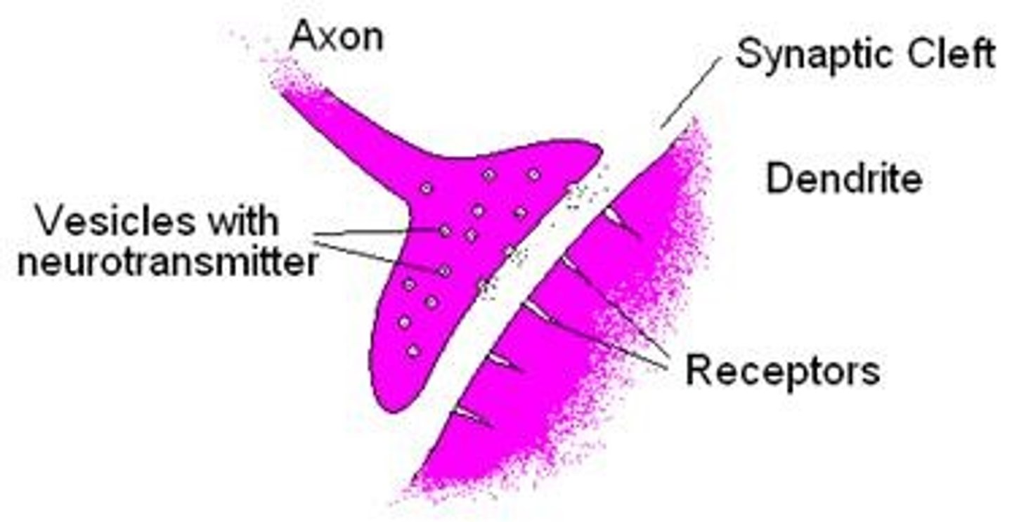
Synapse
together, the nerve terminal, synaptic cleft, and postsynaptic membrane are known as a synapse.
What is a nerve? (memory devices) and the three types
A bundle of neurons in the PNS
you’re getting on my nerve— think PMSing
1)sensory
2)motor
3)mixed
Sensory Nerve
Nerves that carry sensory information (afferent) to the brain and spinal cord
Motor Nerve
Nerves that carry motor information
(efferent) from the brain and spinal cord to the mucslces/glands
Mixed Nerve
Nerves that carry both motor and sensory information
Tracts and differentiate them from nerves
AXONS bundled together in the CNS
*unlike nerves, tracts only carry ONE type of information. Nerves carry multiple information sensory, motor, or both.
Multiple NEURONS bundle together to form a_______.
nerve in the PNS
Axons bundled together form_________.
tracts in the CNS
Cell bodies of neurons of the same type within a NERVE cluster together in_____________.
ganglia in the PNS.
gangs get on my nerves. nerves equals pmsing. pms sounds like pns
Cell bodies of the individual neurons within a TRACT cluster together in _______________.
nuclei in the CNS.
Axon bundles in CNS:
Multiple neuron bundles PNS:
Tracts
nerves
Nuclei
The cell bodies of neurons in the SAME TRACT in CNS
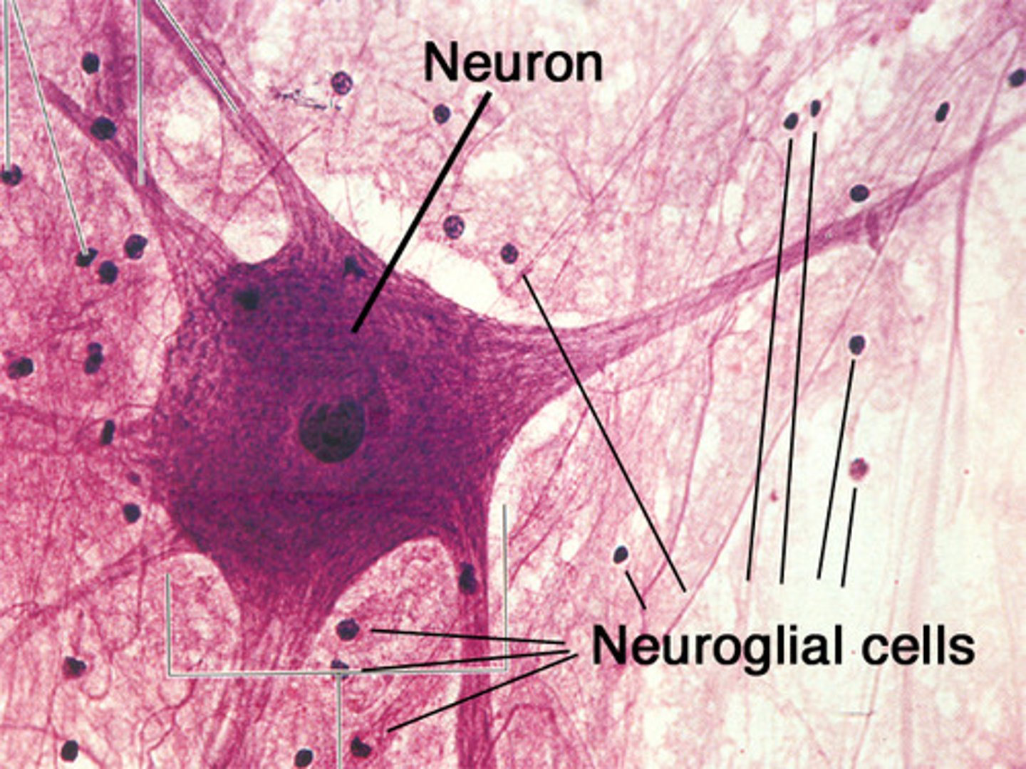
Glial Cells also called and what is their function
Neuroglia
cells in the nervous system outside of neurons that play a role in structure and support
Neuroglia cells
(Glial Cells) play supportive and structural roles for neurons; responsible for functions such as holding neurons in place, supplying neurons with oxygen and nutrients, insulating neurons from other neurons, destroying pathogens, and removing dead neurons
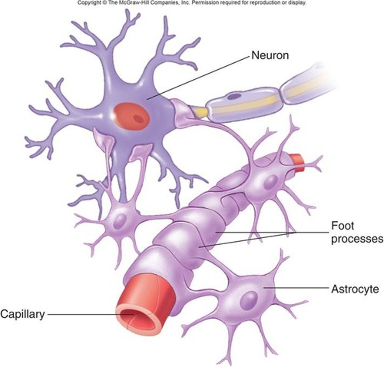
Astrocytes
type of glial cell
nourish neurons and form the blood-brain barrier, which controls the transmission of solutes from the bloodstream into nervous tissue
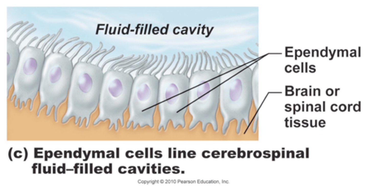
Ependymal Cells
type of glial cells
line the ventricles of the brain and produce cerebrospinal fluid, which physically supports the brain and serves as a shock absorber
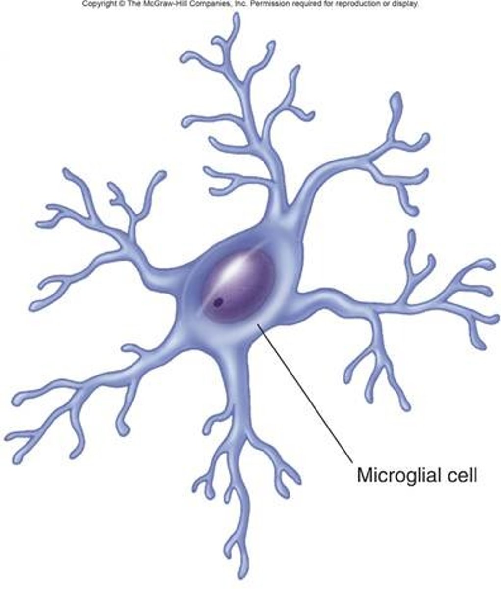
Microglia
are phagocytic glial cells that ingest and breakdown waste products and pathogens in the CNS
Mylenating glial cells in CNS and PNS?
In CNS=oligodendrocytes
IN PNS=schwann cells
*both produce myelin around axons
Resting Membrane Potential and what is it for neurons
is the net electric potential difference that exists across the cell membrane, created by movement of charged molecules across that membrane.
-for neurons this potential is about -70mV with the inside of neuron being (-) and outside being (+).
What are the two most important ions involved in generating maintaining the resting potential?
potassium (K+) and sodium (Na+)
what is true about the concentration of potassium inside and outside the cell
it is higher inside the cell
what does this concentration difference cause
causes potassium to move outside the cell
what enables the movement of potassium outside the cell
potassium leak channels
what happens as potassium leaks out of the cell
causes a build of negative charge inside the cell and positive charge outside
therefore, some potassium will begin to move back into the cell
eventually, there will be one potassium coming into the cell for every one potassium that is pushed out the cell
this means, no net movement
what is it called when there is no net movement of potassium
potassium equilibrium
what is the potential difference that represents potassium equillibrium
equilibrium potential of potassium
what is true about the concentration of sodium
it is higher outside the cell
in what direction does sodium move and how is this movement enabled
it moves from outside to inside
its movement is facilitated by sodium leak channels
what is it called when there is no net movement and what potential difference represents this state
sodium equillibrium
equillibrium potential of sodium
what is true about the movement of sodium and potassium
it occurs at the same time
describe the tug of war between sodium and potassium
Potassium’s movement pulls the cell potential toward –90mv
sodium’s movement pulls the cell potential the opposite way, toward +60 mV.
a balance of these two effects is reached at around –70 mV for the average nerve cell,
This balance, this net effect of sodium and potassium’s equilibrium potentials, is the resting membrane potential.
The resting potential is closer to potassium’s equilibrium potential because the cell is more permeable to potassium.
Neither ion is ever able to establish its own equilibrium, so both ions continue leaking across the cell membrane.
what is necessary given the continual leaking of sodium and potassium in and out of the cell and what accomplishes this
a movement of sodium and potassium back against their gradients
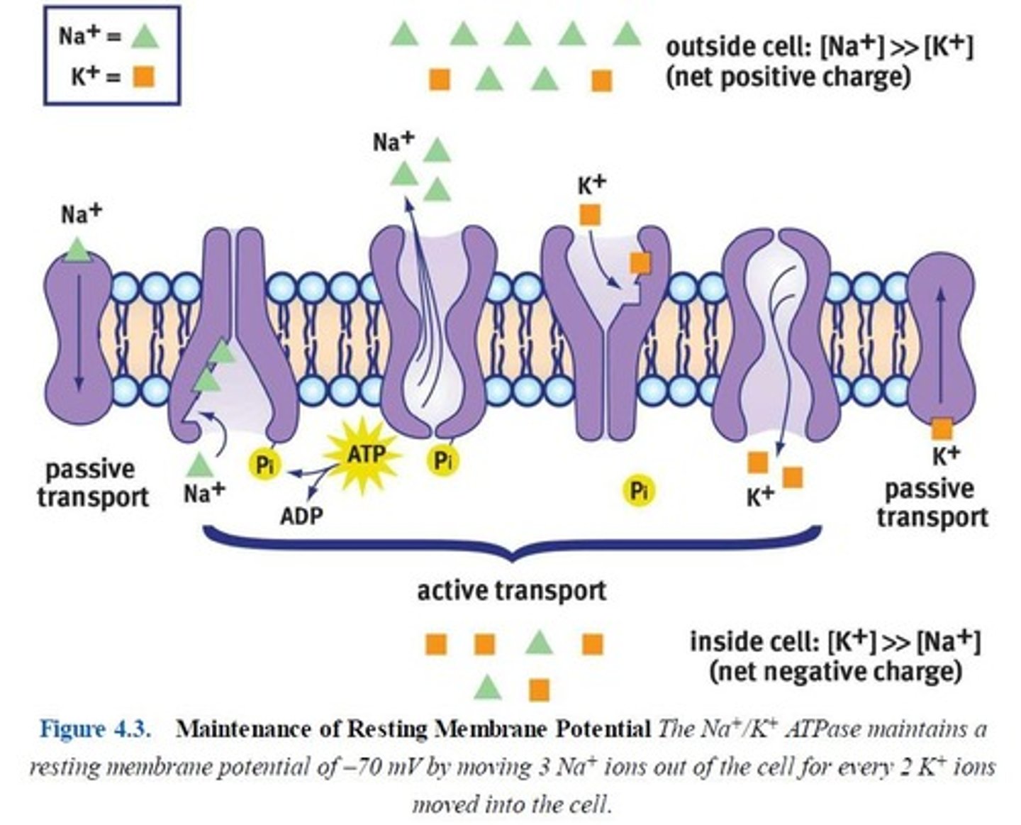
Na+/K+ATPase
Na+/K+ ATPase function and memory device
Sodium-Potassium Pump.
Pumps out 3 sodium for every 2 potassium pumped in. Maintains gradient resting membrane potential.
memory devices: 3 out, 2 in because the membrane potential is negative, it makes sense that it pumps out more positive particles than it takes in
A greater concentration of Na+ is found outside neuron and a greater concentration of K+ found inside a neuron at the resting potential
-ATP required for each transport since both are moved against the gradient.
think PumpKin (K in and Na out— reverse of their gradients)
mnemonic of the direction of ion movement by Na/K ATPase
pumpKin
- K=potassium into the cell
-therefore, sodium out of the cell
Sodium and Potassium concentration inside and outside the cell
outside the cell (net positive): sodium>>potassium
inside the cell (net negative charge):
potassium>> sodium
THE PUMP RESTORES THEM BACK TO ORIGNAL POSITIONS SO THE POSITION AT EQUILLIBRIUM IS POTASSIUM IN AND SODIUM OUT
potassium leak channels
facilitate the outward movement of potassium, which allows the slow leak of potassium out of the cell. ( leaks K+ out)
What types of input can neurons receive and what does this cause
-excitatory input causes depolarization
-inhibitory input causes hyperpolarization
Depolarization
Raising the membrane potential (Vm) from its resting potential; more positive and likely to fire an action potential

Hyperpolarization
Lowering the membrane potential from its resting potential--more negative on inside
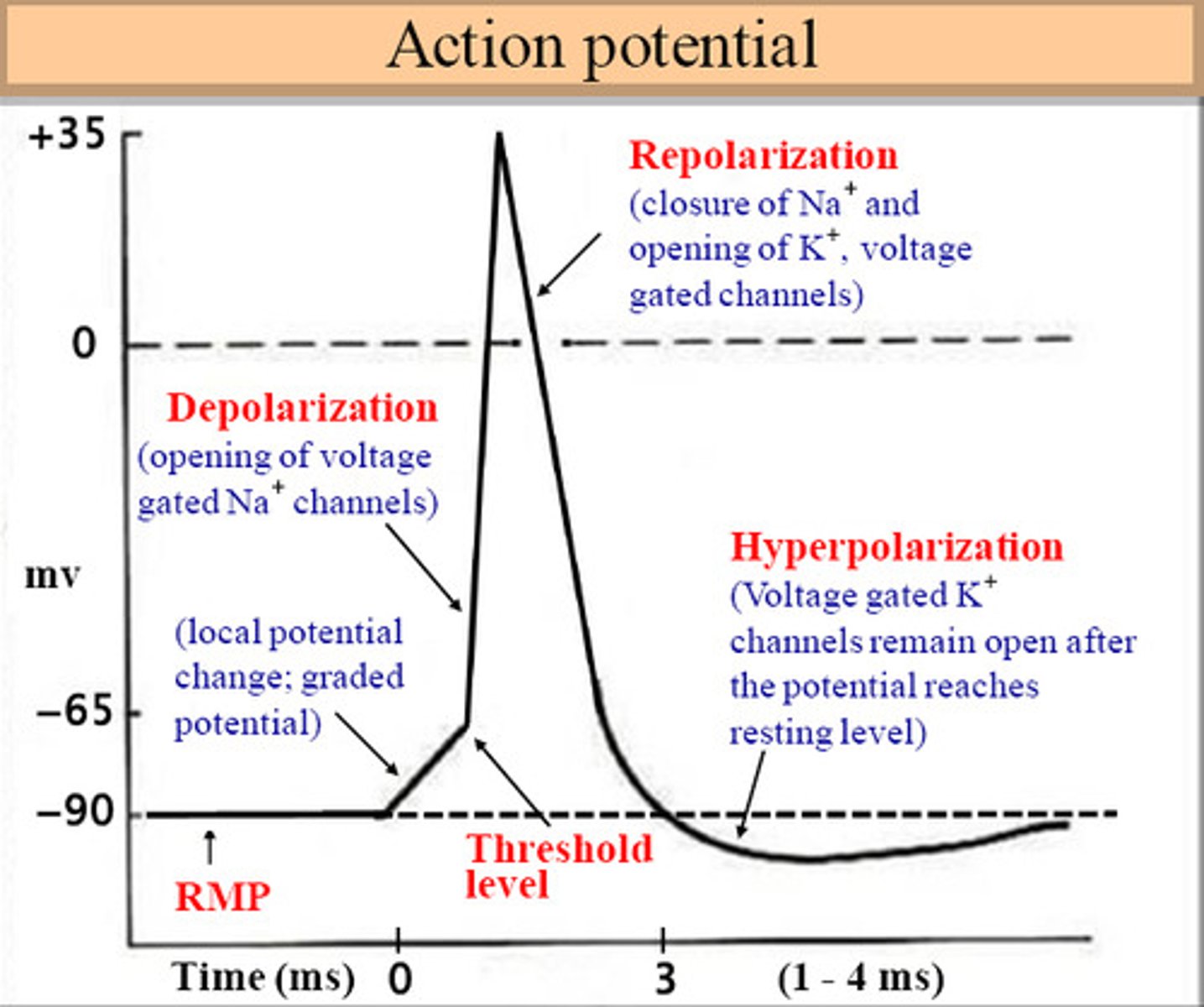
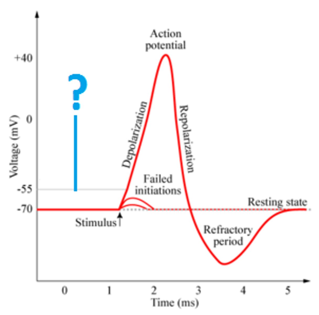
Threshold
The lowest magnitude of stimulus strength that will induce a response-- -55 to -40 mV
Summation and the two types
the additive effect of multiple signals.
A small excitatory signal may not be sufficient to bring the axon hillock to threshold.
a postsynaptic neuron may receive information from several different presynaptic neurons, some of which are excitatory and some of which are inhibitory. The additive effect of multiple signals is known as summation
two types of summations:
-spatial
-temporal.
Temporal Summation
In temporal summation, multiple signals are integrated over a small period of time. A number of small excitatory signals firing at nearly the same moment could bring a postsynaptic cell to threshold, enabling an action potential.
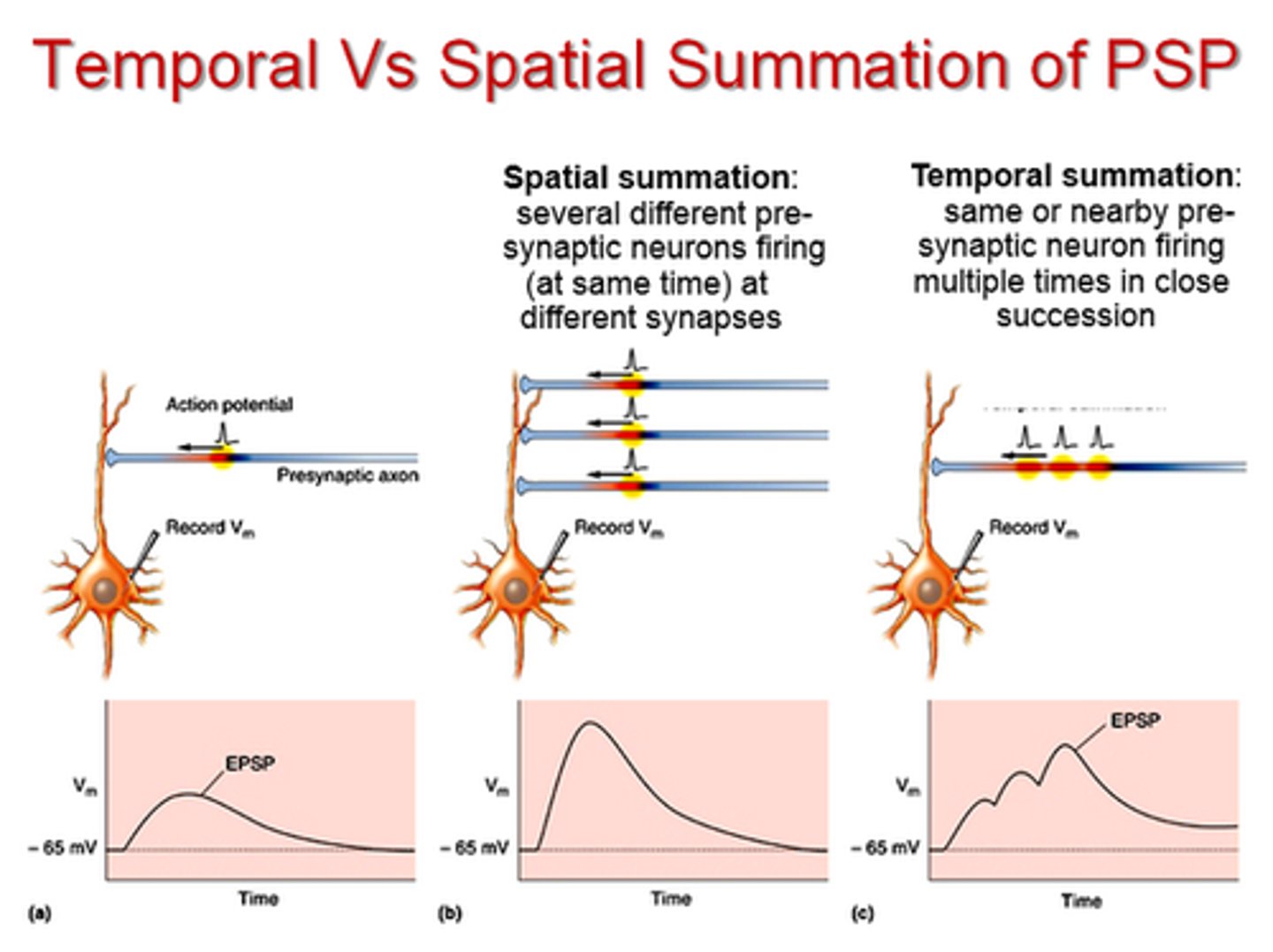
Spatial Summation
the additive effects are based on the number and location of the incoming signals.
A large number of inhibitory signals firing directly on the soma will cause more profound hyperpolarization of the axon hillock than the depolarization caused by a few excitatory signals firing on the dendrites of a neuron.

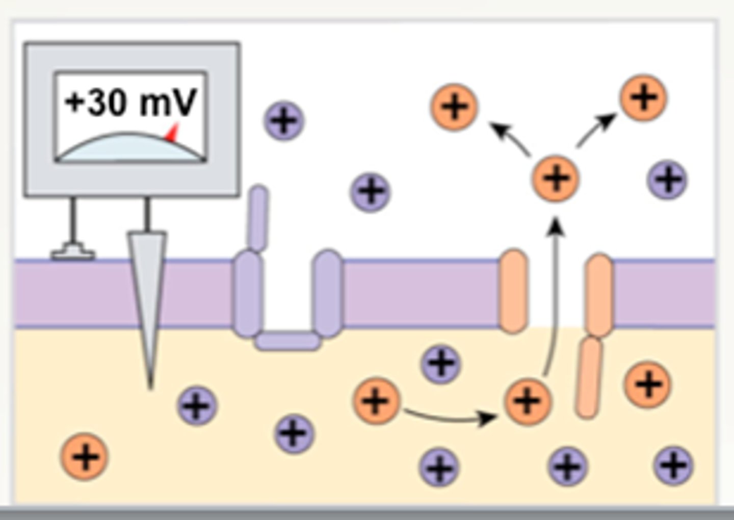
What happens when the threshold potential is reached and what does this rely on and memory devices
(basically the opposite of what happens initially where potassium was moving out)
voltage gated sodium channels open
sodium rushes inside
A gradient consisting of a difference in electrical potential as well as chemical concentration
relies on the electrochemical gradient because the inside of the cell is more negative than the outside
Na+ wants to go into the cell because the cell is more negative inside (electrical gradient) and has a lower concentration of Na+ inside (chemical gradient).
What happens first to sodium channels as membrane potential becomes more positive
Inactivated Sodium Channels
When Vm approached +35mV (peak of AP), potassium channels open.
What happens next to sodium channels as membrane potential becomes more negative
Deinactivated Sodium Channels
When Vm is brought back near -70mV after sodium channels have been inactivated
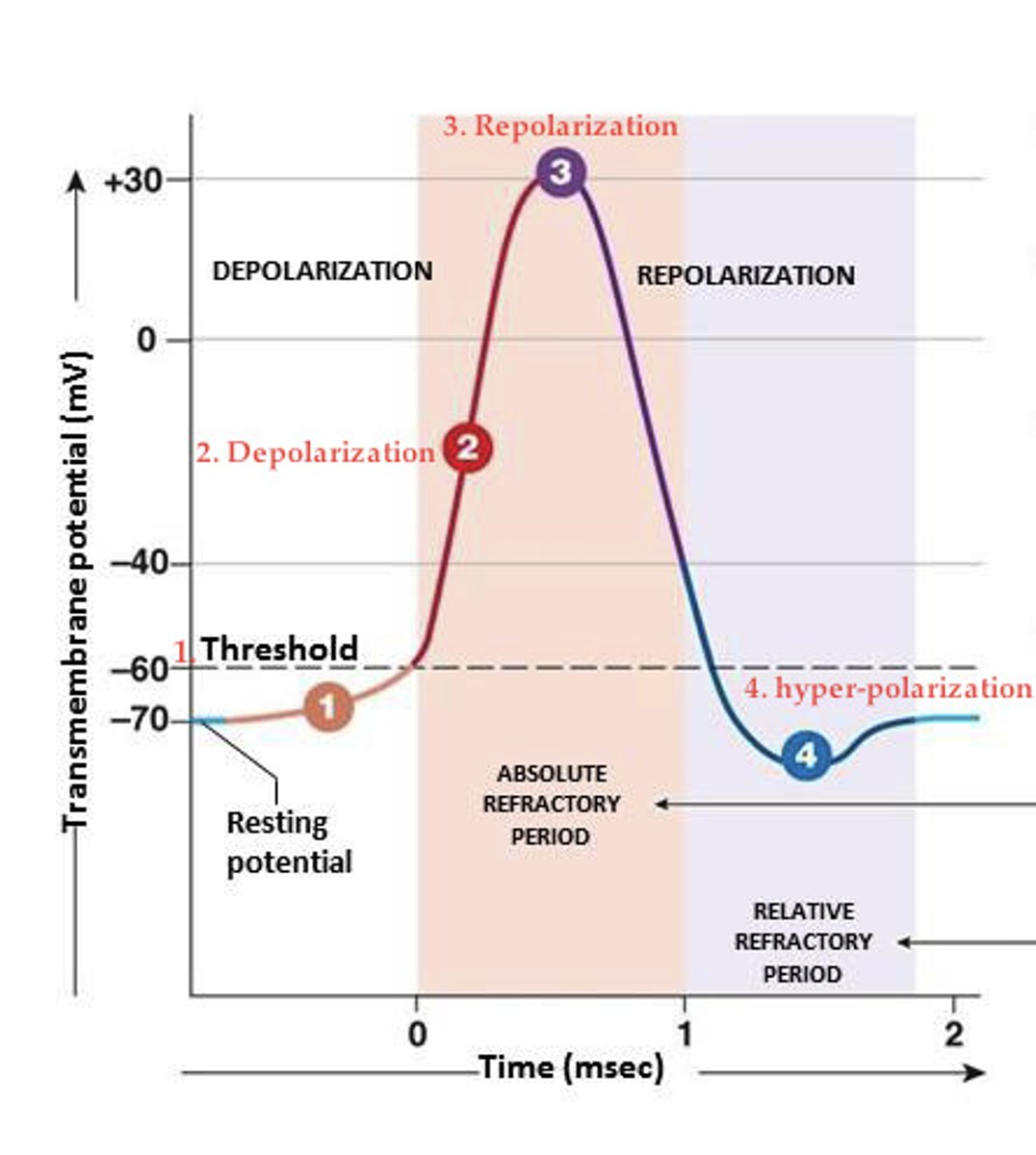
three states of sodium channels
open closed inactive
Closed Sodium Channels
Sodium Channels before the cell reaches threshold and after inactivation has been reversed

Open Sodium Channels
Sodium Channels from threshold -55mV to approximately +35mV

Inactive Sodium Channels
Sodium Channels from approximately +35mV to the resting potential -70mV aka the repolarization/refractory period
The key concept of Action Potential Process
Action potential relies on both electrical and chemical gradients. The neuron starts at the resting potential around -70mV. At the resting potential, potassium is high inside the cell and sodium is high outside the cell. Once the cell reaches the threshold, sodium channels open and sodium floods the cell, making it more positive inside (depolarization). Then, sodium channels are inactivated and the potassium channels open. This allows potassium to flow out of the cell, bringing the potential into the negative range (repolarization, and then actually overshooting the resting potential (hyperpolarization). Na+/ K+ ATPase then work to restore the resting potential.
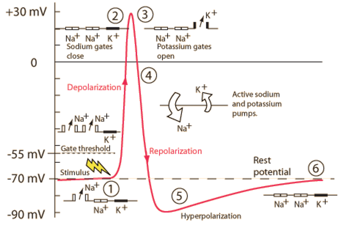
What is repolarization
What happens during repolarization
restoration of the resting membrane potential in neurons from
potassium floods out to make up for the increase positive charge of sodium ions flooding into the cell
the outflow of potassium actually overshoots and causes excess negative charge (hyperpolarization) and induces a refractory state
What are the two Refractory Periods?
absolute and relative
Absolute Refractory Period
no amount of stimulation can cause another action potential to occur
-absolutely can't occur
Relative Refractory Period
Greater than normal stimulation can cause an action potential to occur because the membrane is starting from a potential that is more negative than its resting value
Impulse Propogation- how does it happen
Movement of an action potential down an axon, resulting in neurotransmitter release at the synaptic bouton and transmission of the impulse to the target neuron or organ.
As sodium rushes into one axon segment of the axon, it will cause depolarization in surrounding regions of the axon. This depolarization results in other regions to reach threshold, opening the Na+ channels and allowing action potential to continue in wavelike fashion. Flow in one direction because after the action potential has been fired in one area of the axon, it becomes refractory
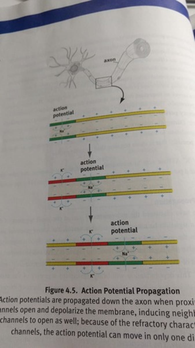
Increased length of axon increases/decreases conduction rate?
Longer axons would decrease conduction conduction rate because it increases the resistance
Greater cross-sectional area of axon increases or decreases rate of transmission?
Increases transmission due to decreased resistance.
How does mylen speed up conduction rate?
Myelin is an extraordinarily good insulator, preventing the dissipation of the electric signal.
Saltatory Conduction
electric signal hops from node to node
mnemonic: Spanish word (saltar)
Presynaptic Neuron
neuron before the synaptic cleft
Postsynaptic Neuron
The neuron after the synaptic cleft
Effector
if a neuron signals to a gland or muscle, rather than a neuron, the postsynaptic cell is termed an effector.
mnemonic: Effector Exits neuron
How does an action potential result in neurotransmitter release?
When an action potential reaches the nerve terminal, the voltage-gated calcium channels open, allowing Calcium to flow into the cell. This sudden increase in intracellular CALCIUM triggers fusion of the membrane-bound vesicles with cell membrane at the synapse, CAUSING EXOCYTOSIS of the neurotransmitter into the synapse.
Difference between electrical and chemical transmission (key concept)
-Within a single neuron, ELECTRICITY is used to pass signals down the length of the axon.
-Between other neurons, CHEMICALS (neurotransmitters) are used to pass signals to the subsequent neuron (or gland or muscle).
Types of neurotransmitter receptors and results?
-Ligand-gated ion channels=results in either a stimulation (depolarize) or inhibitory (hyperpolarization) response
-G-protein coupled receptors (GPCR)=change levels of cAMP or influx of calcium
What are 3 ways neurotransmitter is removed from synapse?
1. broken down by enzymes (i.e. acetylcholinesterase)
2. reuptake carriers back into the pre-synaptic neuron
3. Diffusion out of synapse
Acetylcholine (ACh)
A neurotransmitter found throughout the nervous system; broken down by the enzyme acetylcholinesterase
Reuptake Carriers
Brings neurotransmitters back into presynaptic neuron. An autoreceptor will signal the presynaptic cell to stop releasing and start the reuptake process
ex. serotonin (5-HT), Dopamine, Norepinephrine
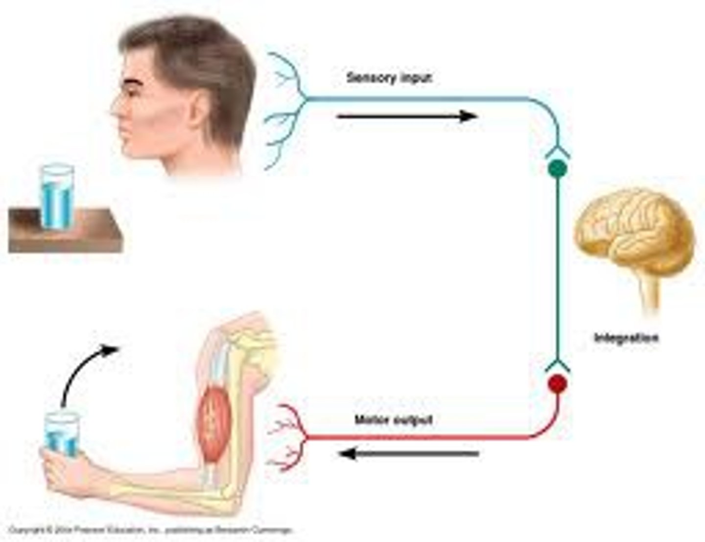
Sensory Neurons= (Afferent Neurons)
A neuron that picks up impulses from sensory receptors and transmits them toward the central nervous system (spinal cord and brain)
Motor Neurons= (Efferent Neurons)
A neuron that that transmits nervous impulses from the CNS to an effector
Mnemonic: Afferent and efferent neurons
(A)fferent neurons (A)scend in the spinal cord toward the brain; (E)fferent neurons (E)xit the spinal cord on their way to the rest of the body
Interneurons
memory device
are found between other neurons and are the most numerous of the three types. Interneurons are located predominantly in the brain and spinal cord.
Linked to reflexive behavior.
SIM like SIM card: interneuron is between sensory and motor
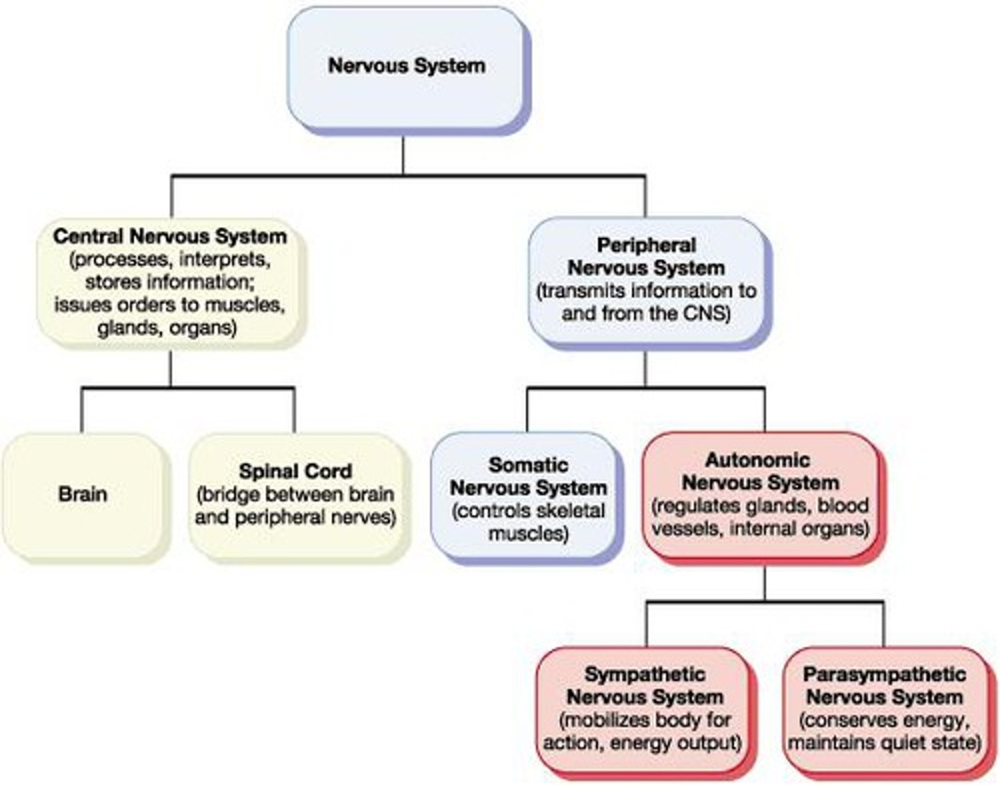
Central Nervous System (CNS) breaks down into the
The brain and spinal cord
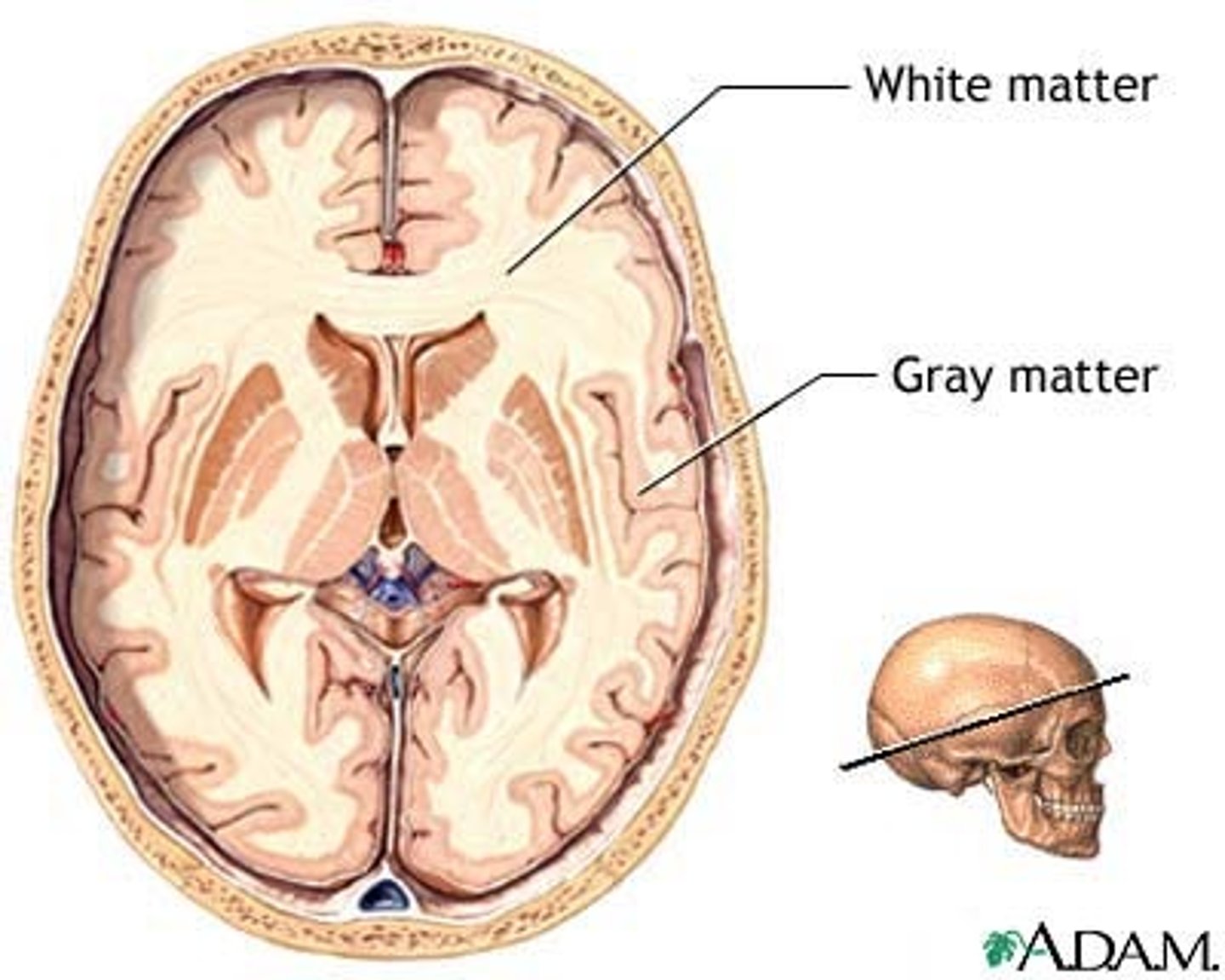
What does the brain consist of?
memory device
white matter is deeper than grey matter in the brain. Opposite in the spine.
white matter deeper in the brain, gray matter ahead of it because when you have brain fog, you cant think deep, so grey matter is not deep but white matter is
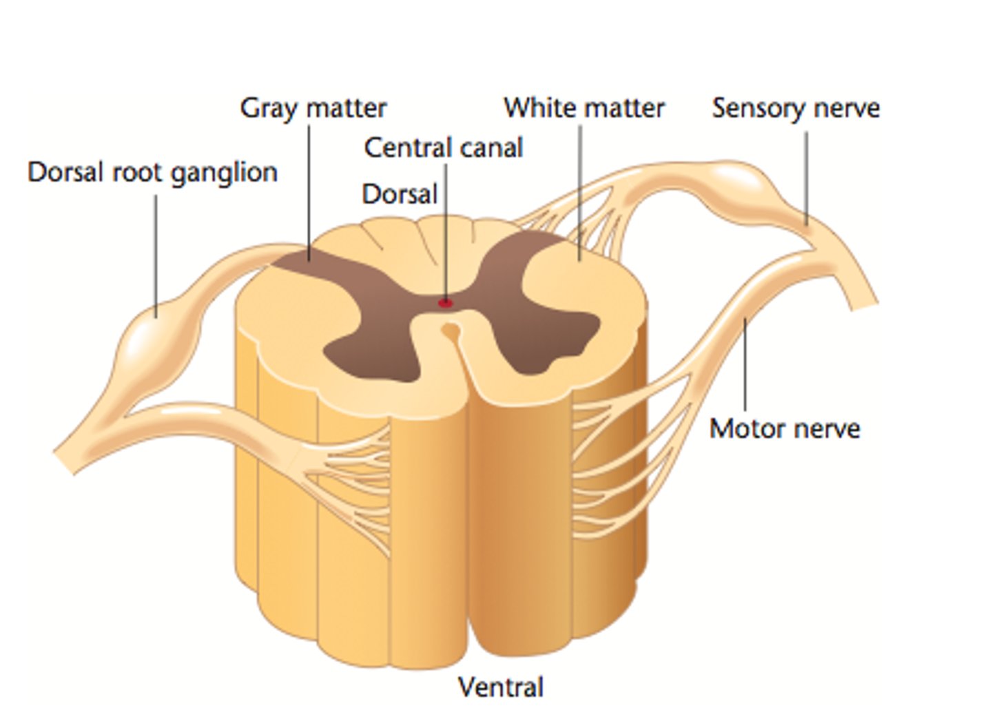
White Matter
consists of axons encased in myelin sheaths
- in the brain, the white matter lies deeper than the grey matter.
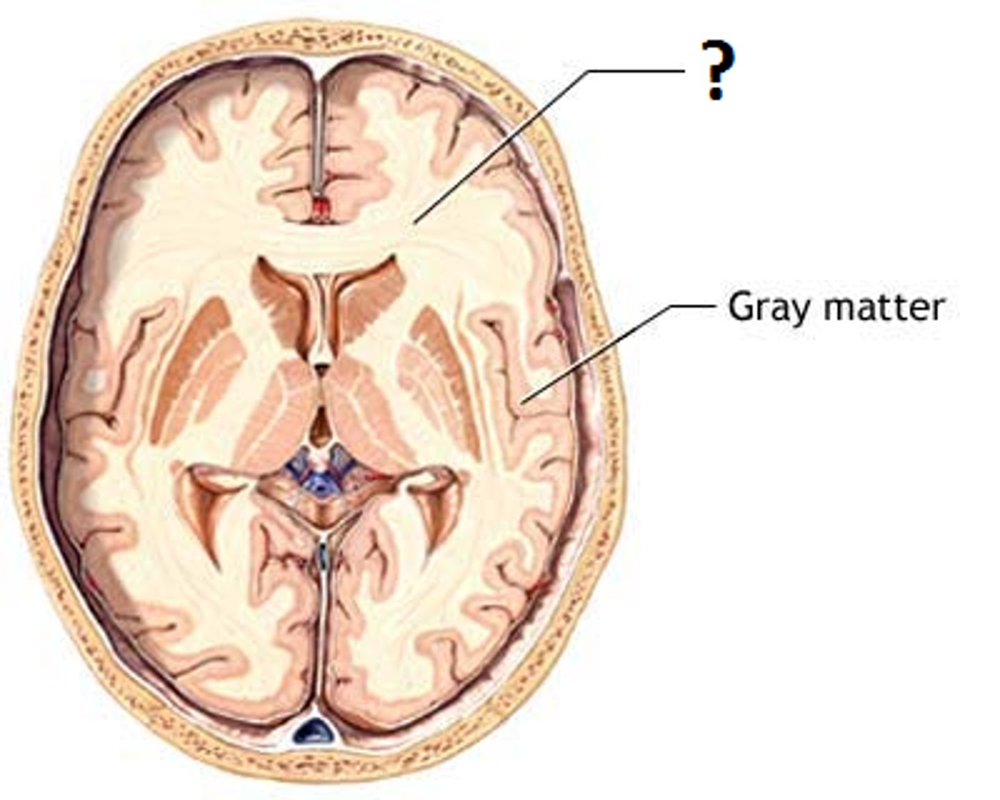
Grey Matter
Any region in the central nervous system that consists of unmyelinated cell bodies, dendrites, and synapses. Gray matter is on the outside of the brain.
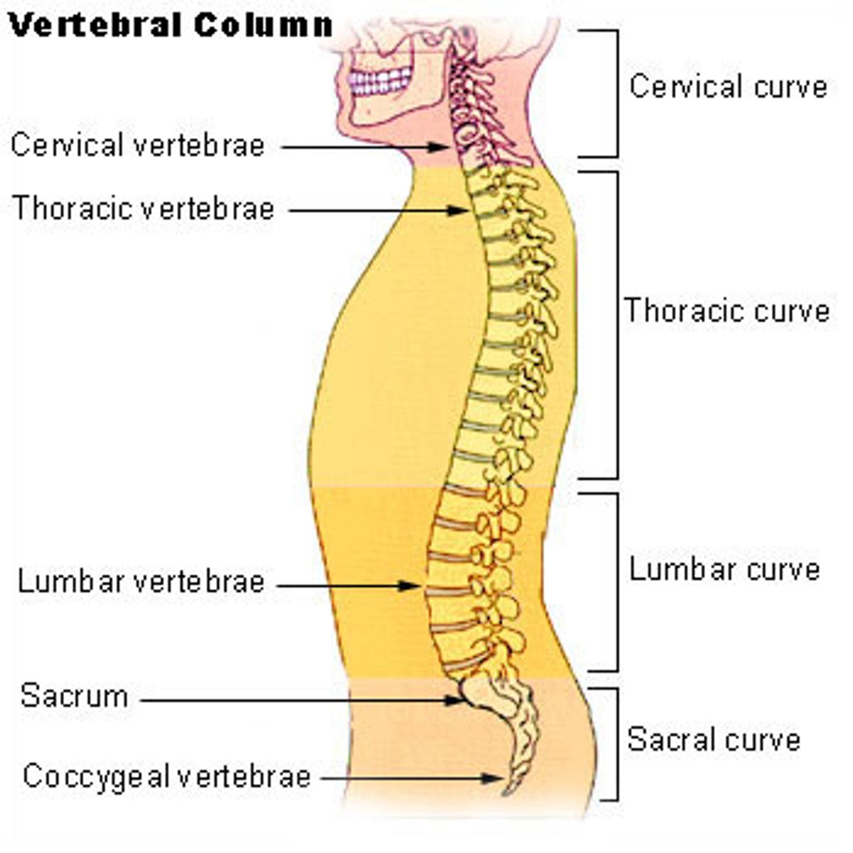
4 parts of spinal cord and memory device
From head to toe:
1)Cervical
2)Thoracic
3)Lumba
4)Sacral
(Can They Leave? Shit!)
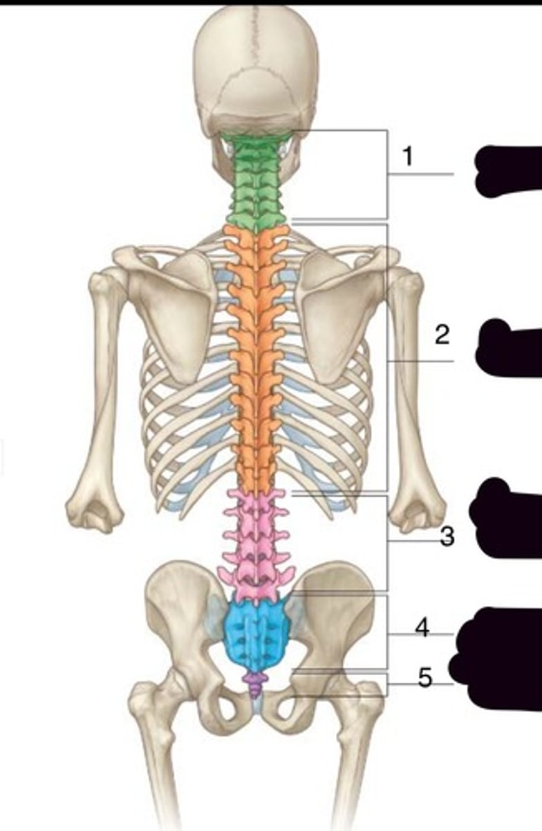
what protects the spinal cord
vertebral column protects spinal cord
Note important spinal structure
dorsal root ganglia
the axons of motor and sensory neurons are in the spinal cord.
The sensory neurons bring information in from the periphery and enter on the dorsal (back) side of the spinal cord.
The cell bodies of these sensory neurons are found in the dorsal root ganglia.
Motor neurons exit the spinal cord ventrally, or on the side closest to the front of the body
Peripheral Nervous System (PNS)
what is it subdivided into
what is its primary role
Includes all neurons outside the central nervous system, including sensory and motor neurons; it is subdivided into the somatic and autonomic nervous system.
-transmits info to and from CNS
Somatic Nervous System
Subdivision of the peripheral nervous system that governs all voluntary action.
Divisions of NERVOUS SYSTEM
Central Nervous System (CNS) and Peripheral Nervous System (PNS)