Benign Neoplastic Diseases - Module 2
1/274
There's no tags or description
Looks like no tags are added yet.
Name | Mastery | Learn | Test | Matching | Spaced | Call with Kai |
|---|
No analytics yet
Send a link to your students to track their progress
275 Terms
Tumour/lesion of abnormal cells that proliferate at a high rate
What is a neoplasm
Either
Are neoplasms diffuse or focal
Either (not all neoplasms are cancer)
Are neoplasms benign or malignant
Primary or metastases
What are the the two types of malignancy
Primary
If you see a solitary malignant mass is it most likely primary or a metastases
Metastases
If you see multiple malignant masses are they most likely primary or metastases
Typically asymptomatic, no altered lab test, well defined, encapsulate, slow growing, do not metastasize, hypovascular or avascular
Describe the the typical benign neoplasm
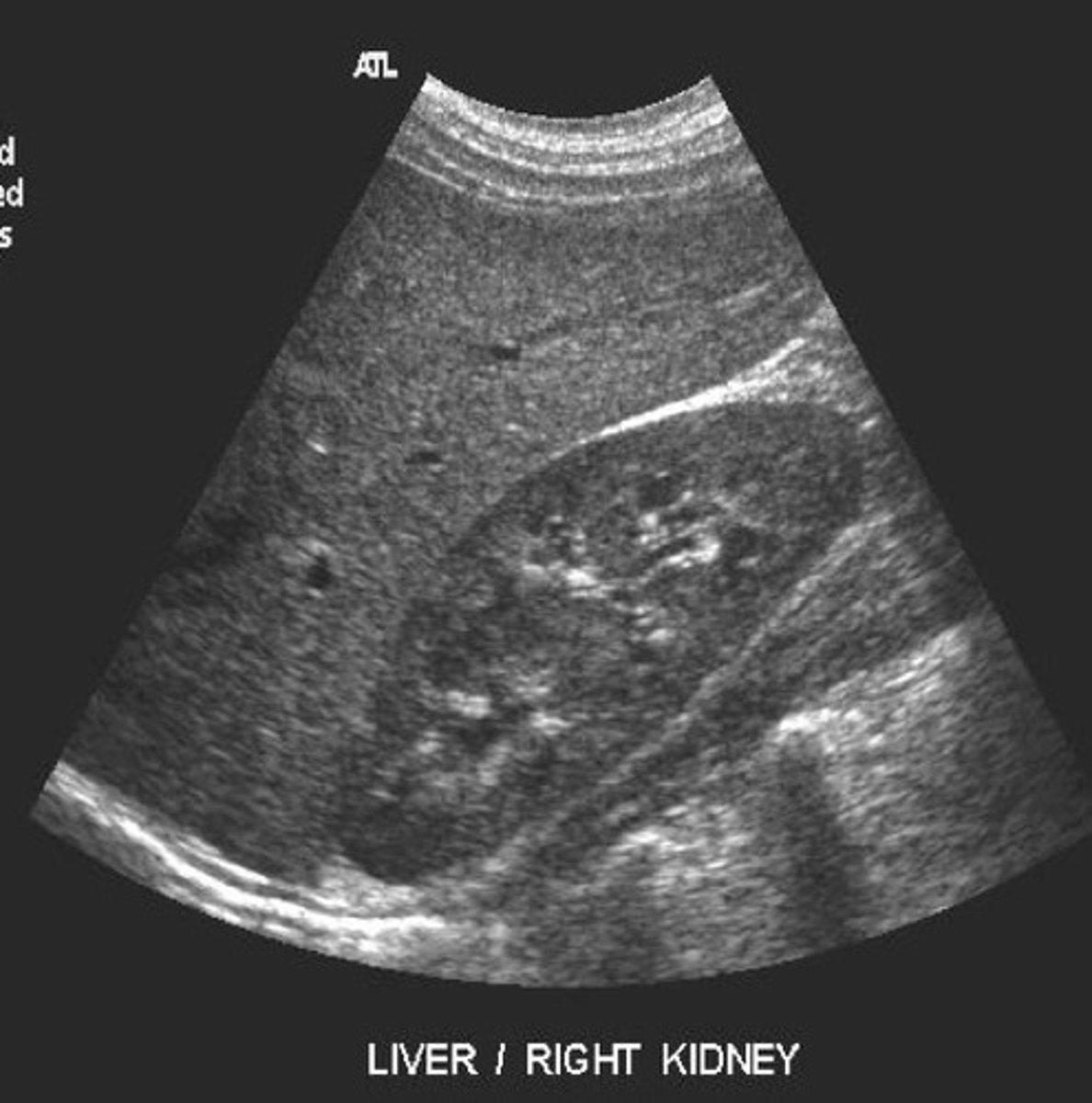
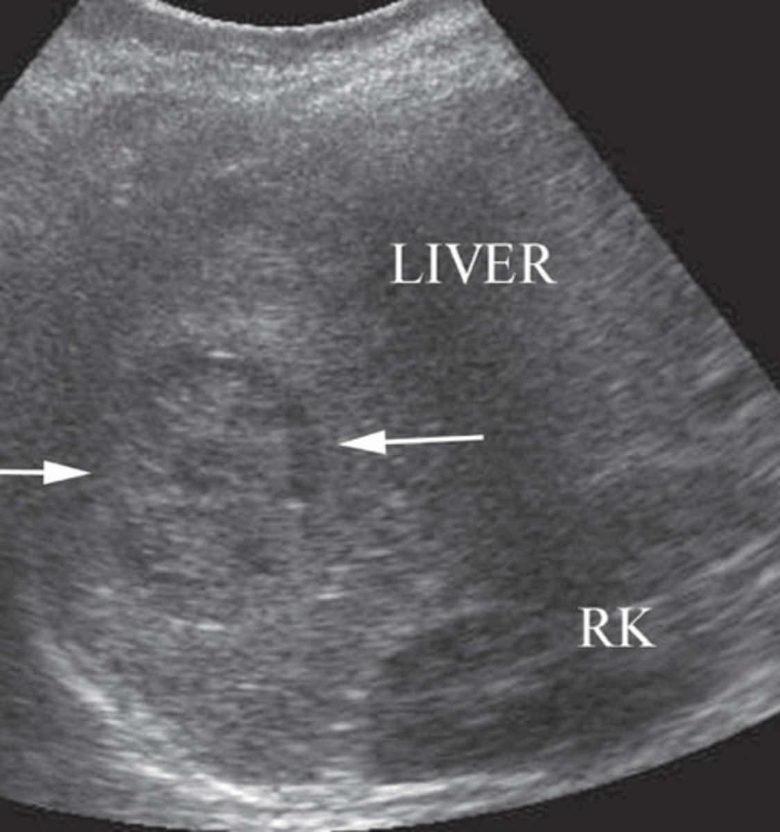
Homogeneous, slightly more echogenic or isoechoic to the kidney, smooth borders, interspersed vessels, diaphragm well seen
Describe the normal sonographic appearance of the liver
Kidney
What should you compare the echogenicity of the liver to
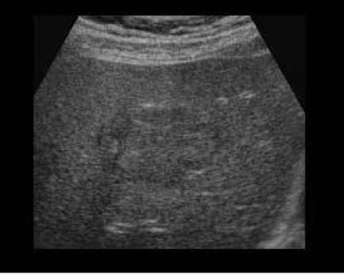
Normal sonographic appearance of liver and kidney
What does this image represent
Hemangioma
What is the most common benign liver tumour
Blood vessel mass
Describe the break down of the word hemangioma
Women (5:1)
Do hemangiomas occur more often in men or women
Capillaries
What are hemangiomas made up of
Asymptomatic
What are the symptoms of a hemangioma
Cavernous hemangioma
What is another term for hemangioma
Typically small (<3cm), well define, homogenous, and hyperechoic
Describe the sonographic appearance of a hemangioma
May include a heterogenous central area with hypoechoic components
Describe a different sonographic appearance that a hemangiomas may show
No
Do hemangiomas demonstrate any colour flow on Doppler
Bc the blood vessels are too small
Explain why hemangiomas don't show up on colour Doppler
Pregnancy or estrogen therapy (HRT)
What may cause a hemangioma to increase in size
No
Do hemangiomas increase in size with birth control use
Thought to be a link between hemangiomas and estrogen but not fully proved
What is the link between hemangiomas and estrogen
6 months to document change
How long after the initial discovery of an hemangioma would the patient need a follow up g
<3cm
What is the typical size of a hemangioma
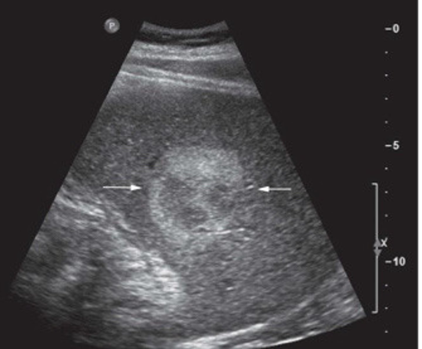
Hemangioma
What does this image show
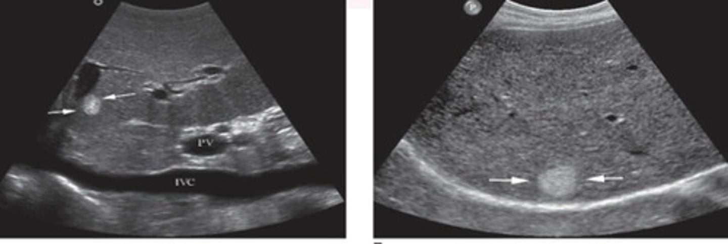
Hemangioma (would need follow up bc bigger)
What does this image show
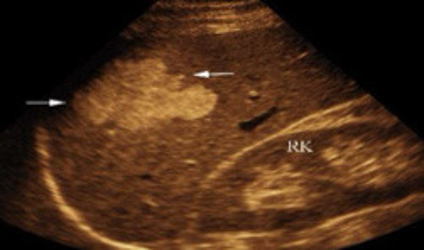
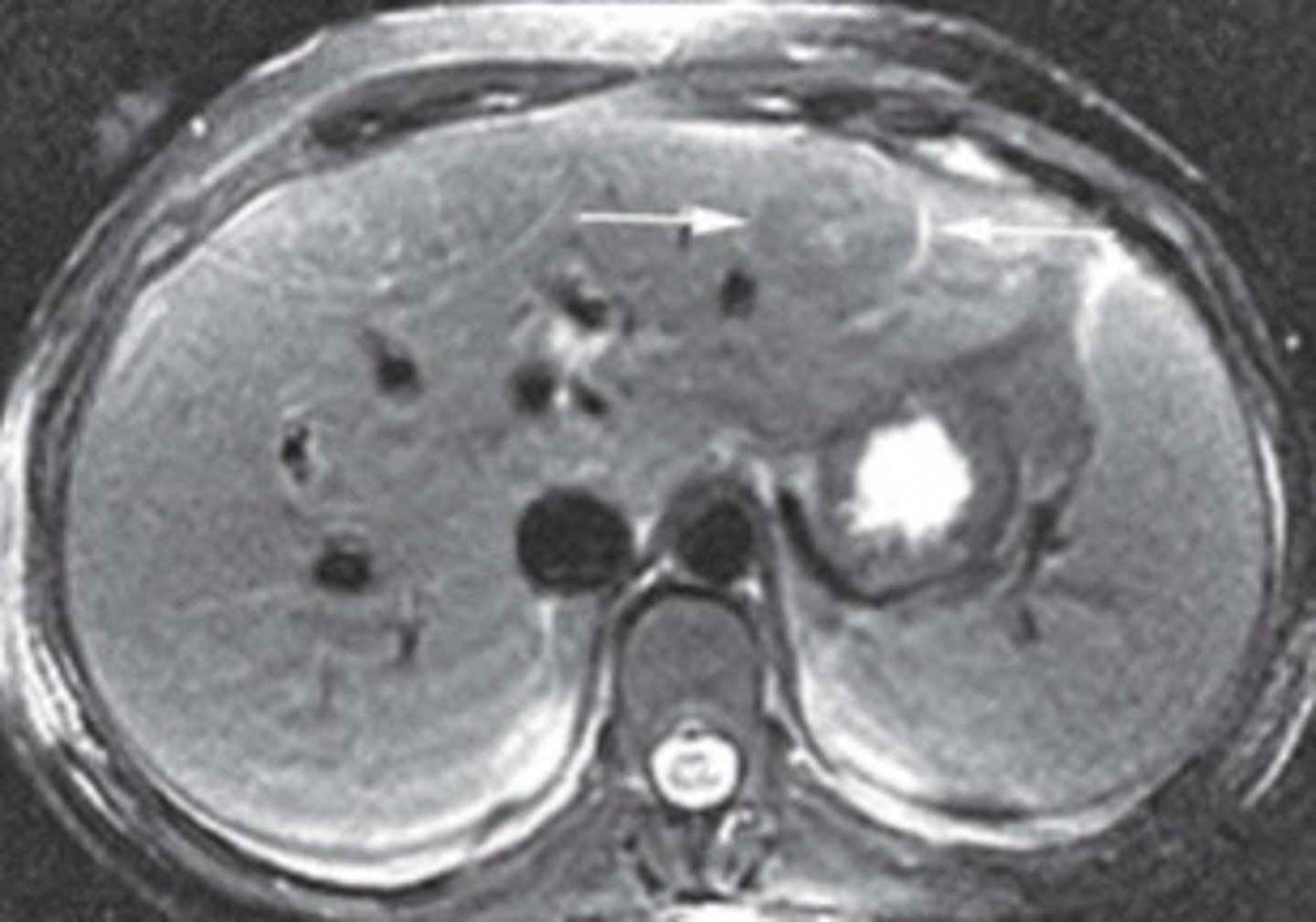
Atypical appearance of hemangioma
What does this image show
Atypical appearance of hemangioma
What does this image show
Focal nodular hyperplasia
What does FNH stand for
Normal liver tissue, but abnormal arrangement (hyperplastic lesions)
What is a FNH made of
Women
Are FNH's more common in women or women
Hormones (estrogen or FSH)
What is FNH possible influenced by
Asymptomatic
What are the symptoms of FNH
Stealth lesion
What is the nickname of FNH
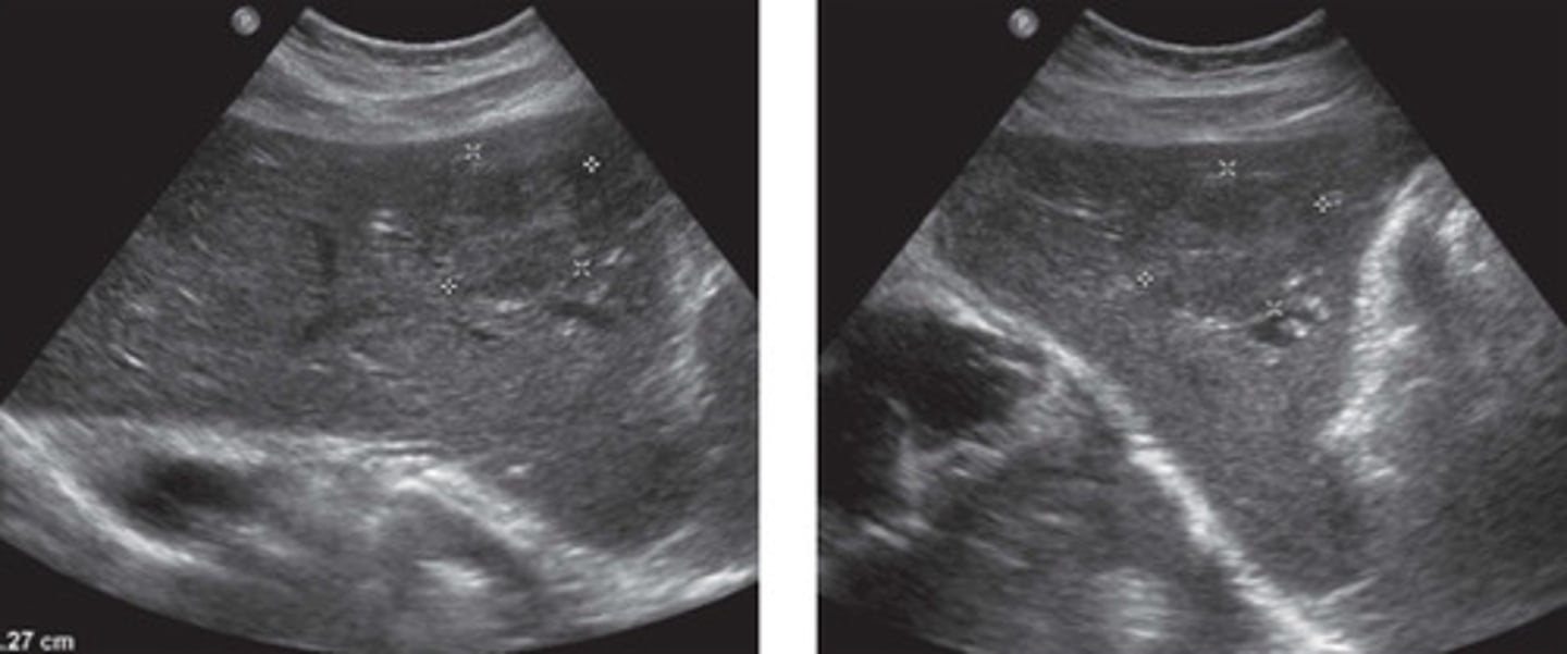
FNH
What does this image show
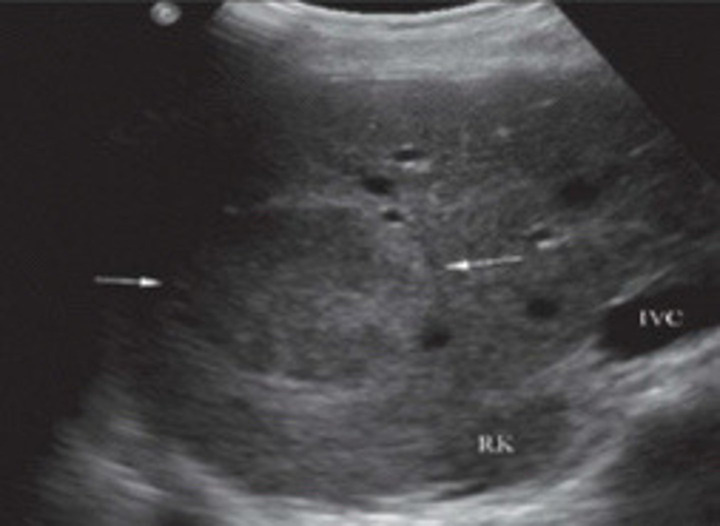
Subtle, less than 8cm, central area of decreases echogenicity, Doppler flow centrally
Describe the sonographic appearance of FNH
Look for contour abnormalities, displaced vessels in the liver, central scar (decreased echogencity in the center)
What are some tips to help you find a FNH
Hepatic veins main be randomly displaced or take a curve
Explain an example how a FNH may cause a liver vessel to be displaced
Sulphur colloid scan
What extra image modality is needed to diagnose a FNH
Hot/warm
How does a FNH appear on a sulphur colloid scan
FNH
Are hemangiomas or FNH's usually bigger
Bc has some features that are suggestive of malignancy (decreased echogencity centrally, Doppler flow centrally)
Why are FNH's a diagnostic challenge
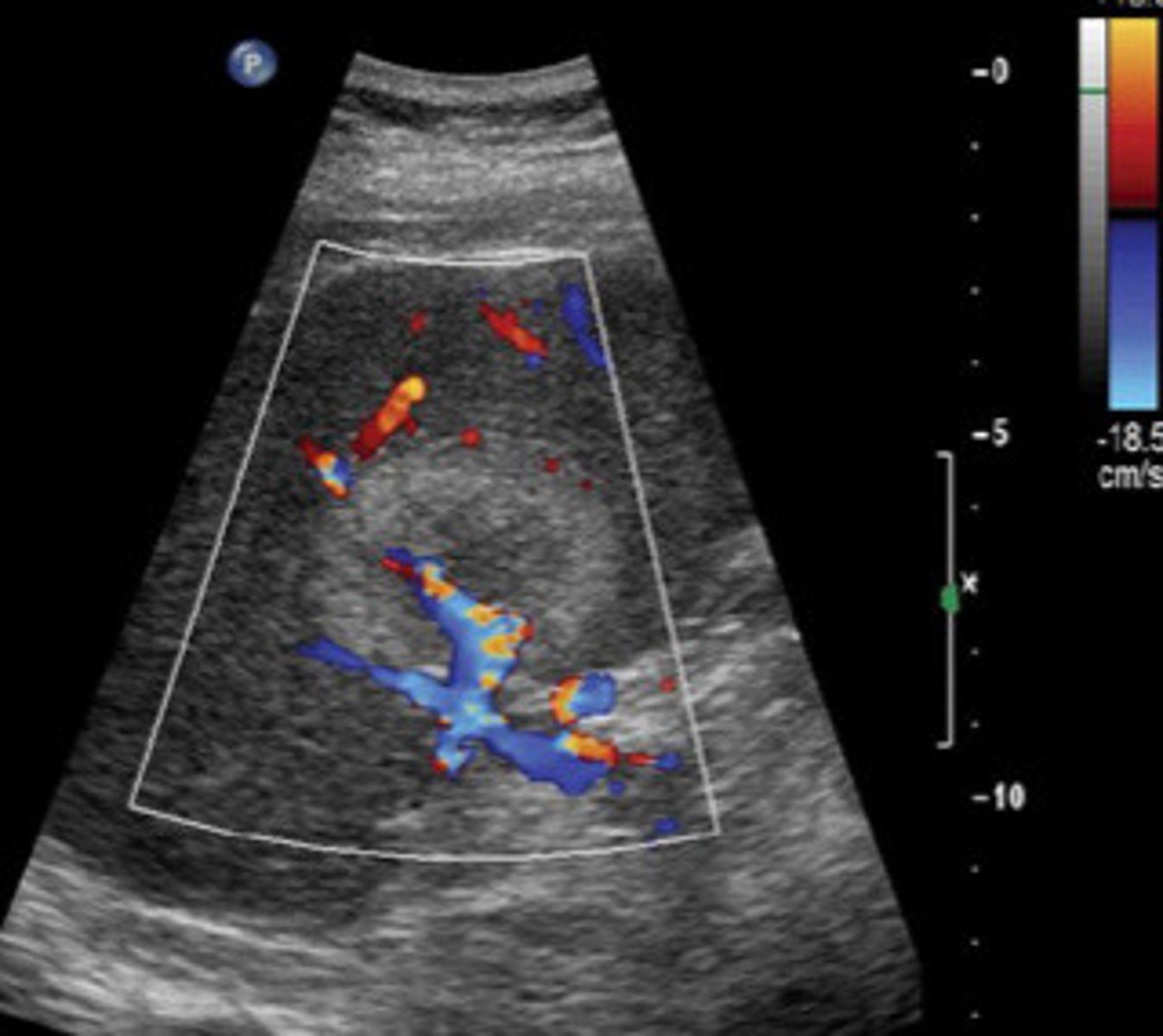
Use colour map
What is another tip to help see a FNH
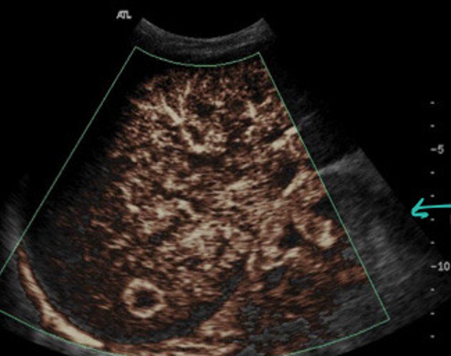
FNH
What does this image show
FNH (seen better with colour map)
What does this image show
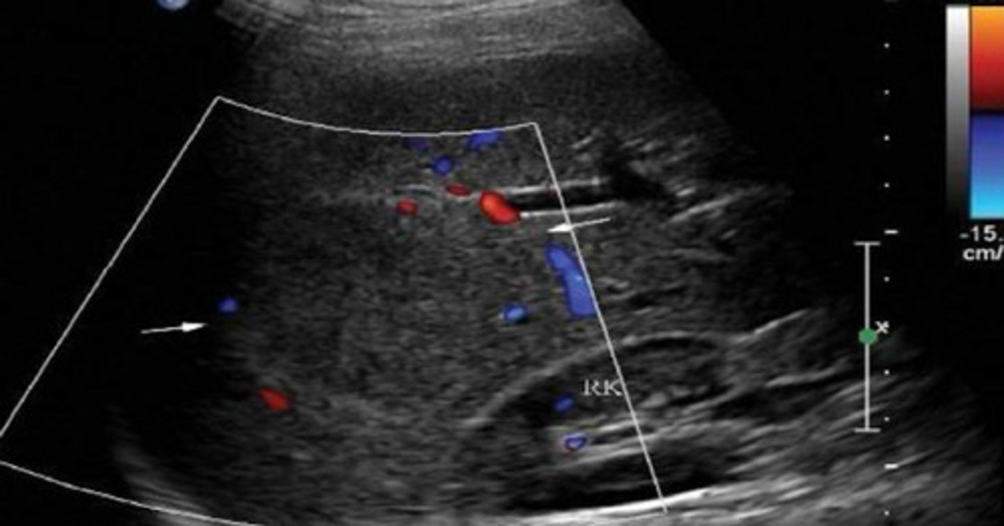
FNH
What are the arrows pointing to
False, less common
T/F: adenomas are most common than FNH
Oral contraceptives
What has adenomas been proven to be linked to
Type 1 Glycogen Storage disease (GSD)
Disease is GSD
Glycogen storage disease
What does GSD stand for
Von Gierke's disease
What is another term for GSD
inherited disorder caused by deficiencies of specific enzymes in the glycogen metabolism pathway. Leads to excess glycogen in pt's liver and kidneys
Describe what GSD is
Asymptomatic
What is the usual symptoms of adenomas
Can hemorrhage or infraction, which will cause pain (RUQ?)
What is a complication of adenomas what what symptom would this cause
Non specific (usually hyperechoic but variable, solid, solitary, and well encapsulated, Doppler show central area of Doppler
Describe the appearance of an adenoma
FNH
What is the appearance of an adenoma often similar to
Cold
What does an adenoma appear on a sulphur colloid scan
8 to 15cm
What is the typical size of a adenoma when found on ultrasound
Hyperechoic but can have variable echogencity
What is the usually echogencity of a adenoma
Phagocytic cells
What are kupffer cells
Contains kupffer cells which gobble up the sulphur which makes them appear warm on the scan
Explain why a FNH appear warm on a sulphur colloid scan
Do not have kupffer cells so do not phagocytize the sulphur and appear cold
Explain why a adenoma appears cold on a sulphur colloid scan
Adenoma
What does this image show
False, pretty common in other parts of the body but not the liver
T/F: lipomas are one of the most commonly found neoplasms in the liver
Subcutaneous tissue
What type of tissue are lipomas usually found in
Asymptomatic
What are the symptoms of a lipoma in the liver
Hyperechoic, homogenous
Describe the sonographic appearance of a lipoma
Hemangioma
What is the appearance of a lipoma similar to
Contrast enhance CT or MRI, RBC scintigraphy, surfer colloid scans, micro bubble enhanced sonography, biopsy
What are the 5 different tests often used for neoplasms in the liver
Nuclear medicine test
What is a RBC scintigraphy
Inject something that is picked up for a RBC, and would show up in a hemangioma.
Describe the RBC scintigraphy test
Used to prove an atypical hemangioma
What are RBC's used for
Nuclear medicine test, inject sulphur and see if mass absorbs it
What is a surfer colloid scan
FNH vs adenoma
What is a sulfur colloid scan used for
Ultrasound scan with bubbles
What is a micro bubble enhanced sonography
If not getting the answers with the imagining, will do biopsy to make sure
When is a biopsy used
Repeat ultrasound after 3-6 months (make sure not changing then leave it alone)
What is the treatment for a hemangioma
Conservative, depending on size (leave it alone if small, may remove if causing symptoms
What is the treatment for a FNH
Surgery recommend
What is the treatment for an adenomatoid
Bc can get so large and hemorrhage (results in more serious complications)
What is the treatment for an adenomatoid
Conservative (same as hemangioma, if it ain't broke don't fix it)
What is the treatment for a lipoma
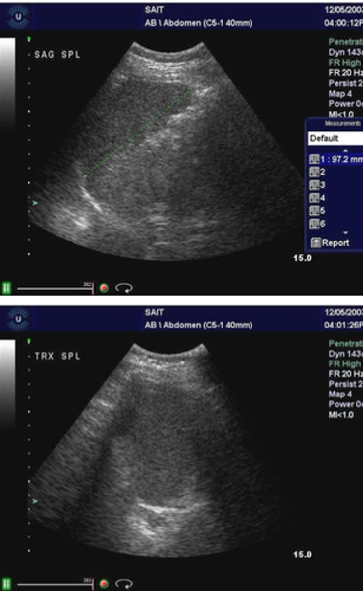
Extremely homogenous, more echogenic than the kidney, isoechoic or slightly more echogenic to the liver, hilum disrupted by vessels, inverted comma shape
Describe the normal sonographic appearance of the spleen
Splenomegaly
What is indicated when the spleen starts to loose the normal shape of an inverted comma
Cavernous hemangioma
What is the most common benign neoplasm of the spleen
Liver
Is a hemangioma in the liver or the spleen more common
True
T/F: a cavernous hemangioma in the spleen is congenital
Asymptomatic
What is the symptoms of a hemangioma in the spleen
Hyperechoic to complex with cystic degeneration (more variability in appearance than in liver)
Describe the sonographic of a hemangioma in the spleen
No, further testing is required to differentiate
Are ultrasound findings for a hemangioma in the spleen conclusive
Hemangioma in the spleen
What does this image show
Hemangioma in the spleen
What does this image show
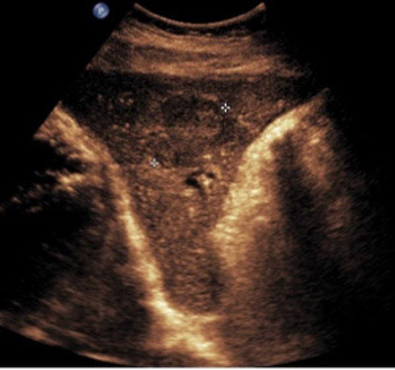
Hemangioma in the spleen (see with contrast study)
What does this image show
Larger and more complex usually than as seen in liver
Compare the sonographic appearance of a hemangioma in the spleen as compared to one in the liver
No
Do hemangiomas in the spleen usually have a lot of colour flow
Rare
Are hamartoma's rare or common
Lymphoid tissue
What tissue is hamartoma's made of
Homogeneous, solid, echogenic, not encapsulated
Describe the sonographic appearance of a Hamartoma
Rare
Are lymphangioma's rare or common
Lymphatic malformation in the spleen
What is a lymphangioma