Biology- physiology and anatomy part 1
1/96
There's no tags or description
Looks like no tags are added yet.
Name | Mastery | Learn | Test | Matching | Spaced | Call with Kai |
|---|
No analytics yet
Send a link to your students to track their progress
97 Terms
Levels of life
atom, compound or molecule, organelle, cell, tissue, organ, organ system, organism
Tissue
a group of connected cells with a similar function (4 basic types)
4 basic types of tissue
connective, epithelial, muscle, nervous
connective tissue + structure
made of cells that form the body’s structure (ex. bone and cartilage)— they support and join other tissues; structure is cells scattered throughout the extracellular matrix; PROPER CONNECTIVE, SUPPORTIVE, AND FLUID
epithelial tissue
made up of cells that line inner and outer body surfaces (skin & lining of digestive tract); protects body and internal organs, secretes substances such as hormones
muscle tissue
made up of bundles of long, thin, cylindrical cells known as muscle cells with the ability to contract due to specialized proteins; (muscles attach to bones which enable the body to move)
nervous tissue
made up of neurons, or nervous cells, that carry electrical messages. nervous tissue makes up the brain and the nerves that connect the brain to all parts of the body
Organ
a structure that consists of two or more types of tissue that work together to do the same job
3 types of connective tissue
proper connective tissue, supportive, and fluid tissue
examples in epithelial tissue
epidermis: skin cells keratinized stratified squamous cells; is relatively impermeable
endothelium: lining of blood capillaries formed by simple squamous
urothelium: epithelium lining surface of the urinary bladder called specialized stratified columnar
3 types of muscular tissue
cardiac muscle, smooth muscle, and skeletal muscle
cardiac muscle
involuntary and only covers the walls of the heart (eg. Heart muscles)
smooth muscle
involuntary and usually covers walls of internal organs (eg. digestive tract)
skeletal muscles
voluntary and usually attached to skeleton — one cell has multiple nuclei to create energy & tear and repair (eg. bicep)
physiology
the study of the function of the parts of an organism
anatomy
the study of the structure of an organism’s parts
how are organs and organ systems regulated
by the nervous and endocrine system; nervous system controls all body’s activities and endocrine system secretes hormones to regulate these activities
negative feedback
a response to stimulus that keeps a variable close to a set value (turns a system on or off)
order of homeostasis
stimulus, receptor, signal, and response
what are the 12 organ systems
cardiovascular, lymphatic, digestive, endocrine, integumentary, muscular, nervous, reproductive, respiratory, skeletal, urinary, and immune
Positive feedback
response to an event would increase the likelihood of the event continuing until reaches an endpoint (example contraction during childbirth/oxytocin release or nursing mothers)
skeleta system consists of
bones, ligaments, cartilage
cartilage
type of dense connective tissue, made of tough protein fibers, that provides a smooth surface for the movement of bones at joints
ligament
band of fibrous connective tissue that holds bones together and keeps them in place
functions of skeletal system
support, structure, protecting internal organs, attachment surfaces for muscles, producing blood cells, storing minerals, and maintaining mineral homeostasis
How are mineral levels in blood maintained?
when mineral levels are too high, bones absorb them and store them as mineral salts. When its too low, bones release some of the minerals back into the blood
2 major divisions of bones
axial- bones along central axis such as cranium, vertebral column, ribs and sternum; appendicular is everything else
How do bones produce blood
inside bones, there are chutes of bone marrow which hold stem cells (unspecialized cells)
3 types of specialized cells in human bones + collective function
collective function of bone growth and mineral homeostasis; osteoblasts, osteocytes, osteoclasts
osteoblasts
(type of specialized cell in bones) makes new bone cells and secretes collagen that mineralizes to become the bone matrix; responsible for bone growth and uptake of minerals from the blood
osteocytes
regulate mineral homeostasis; directs the mineral uptake and release as needed
osteoclasts
dissolves minerals into bone matrix and releases them into the blood; they recycle and break down unneeded mineral lattice using acids and enzymes (to make available material for bone growth)
bone marrow
found inside the pores of spongy bone — the soft connective that produces blood cells
periosteum
tough fibrous membrane that covers and protects the outer surfaces of bone
what do all bones come from/grow from
cartilage (through ossification)
Ossification
process in which mineral deposits replace cartilage and change it to bone
where does ossification begin
begins at the center and works towards the ends (growth plates at the ends of long bone is where cartilage remains in the skeleton)
Locomotor system
skeletal muscle, skeleton, and nervous system
Tendons
connect muscle to bone (facilitate movement) and are white; made of fibrous connective tissue (skeletal muscle to skeleton)
Ligament
grey-ish yellow-ish and connect bone to bone for stability; made of fibrous connective tissue; flexible and elastic
Joint
place where two or more bones of the skeleton meet; works with muscles to be mechanical levers for movement
3 types of joints
partly movable, movable (synovial), and immovable
immovable joints
allow no movement due to bones being securely held together by dense collagen (ex. skull connected by immovable joints)
movable/ synovial joints
allow the most movement. bones at these joints are connected by ligaments
partly movable
allow only limited movement; bones at these joints are held in place by cartilage (ex. ribs, sternum)
types of movable joints
ball and socket, hinge, and pivot
Do tendons or ligaments take longer to heal?
generally, ligaments take longer to heal since they have less blood supply
ball and socket joints
allows movement in all direction (like hips and shoulders)
hinge joints
only move in 1 direction like a door (bend and straighten like knee and elbow)
pivot joints
rotation
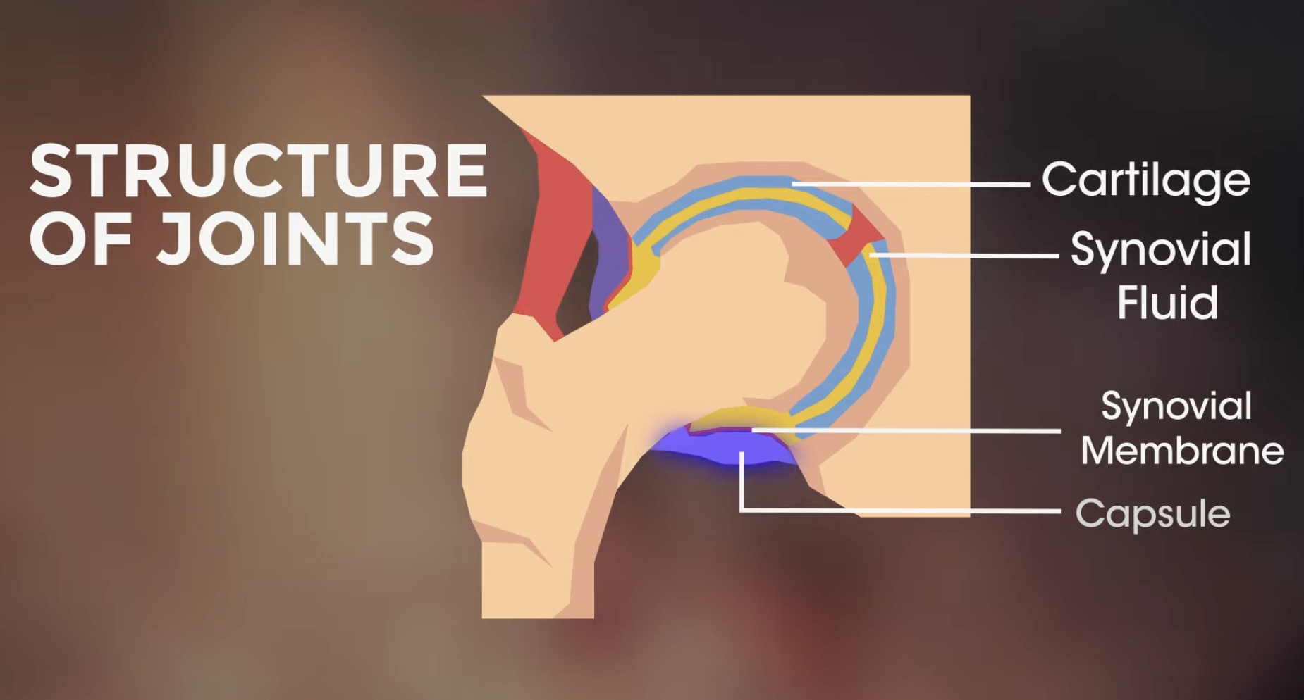
Structure of joints (components)
cartilage, synovial fluid, synovial membrane, capsule
cartilage
cushion; reduces friction and absorbs shock
synovial fluid
reduces friction by lubricating joint (oily FLUID)
synovial membrane
makes synovial fluid
capsule
surrounds joint and holds in the synovial fluid
fractures
breaks in bone by excessive stress; healing when osteoblasts form new bone and bone cells travel to the break site and takes 2-3 months before compact and spongy bone form
osteoporosis
disease where bone loses mass and becomes more fragile; prevented by healthy diet (vitamin d and calcium); common in women going through pre-menopause because the stage affects how bones absorb calcium
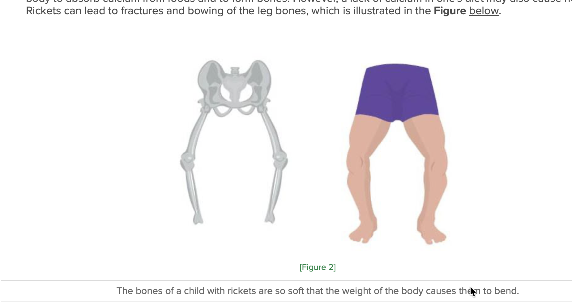
rickets
softening of bones in children (usually due to vitamin D deficiency) which impacts the absorption of calcium which can lead to fractures and bowing
osteoarthritis
condition where cartilage breaks down in joints due to wear and tear; occurs with age, too
rheumatoid arthiritis
autoimmune disease where immune cells start attacking cartilage/joint lining.
skeletal muscle
skeletal muscle fibers bundled by connective tissue and provides support and allows muscle cells to withstand contraction; this connective tissue is also a path for nerves and blood vessels to reach the muscle
individual muscle cells
muscle fibers which contain individual contractive subunits called myofibrils
skeletal muscle extremely simplified zoom-in/break down
skeletal muscle; made of 10-100 muscle fibers; muscle fibers made of myofibrils; myofibrils have sarcomeres; sarcomeres consist of actin filament, myosin filament, and z line
muscle fibers
long & thin muscle cells with the ability to contract or shorten (thanks to connective tissue and sarcomeres)
different types of connective tissue
proper connective (like adipose), supportive (ex. bone and cartilage), and fluid (blood vessels and lymph vessels)
connective tissue structure
support and join other tissues; cells scattered throughout the extracellular matrix
cardiovascular system
transports oxygen, hormones, and nutrients to the body cells, moves carbon dioxide away from cells; heart, blood, blood vessels
lymphatic system
defends against disease and infection; moves lymph between tissues and the blood stream
digestive system
digests foods and absorbs nutrients, minerals, vitamins and water; esophagus, small intestine, large intestine and stomach
endocrine system
produces hormones that communicate between cells; pituitary gland in brain, hypothalamus, adrenal glands, ovaries, and testies
integumentary
protection from injury and water loss, physical defense from infections by microorganisms and temperature control; skin, hair, nails
muscular system
involved in movement and heat production; cardiac muscle, skeletal muscle, smooth muscle, and tendons
nervous system
collects, transfers, and processes information; brain, spinal cord, and nerves
reproductive system
produces gametes and se hormones; in females uterus, vagina, fallopian tubes, ovaries; in males penis, testes, seminal vesicles
respiratory system
brings air to sites where gas exchange occurs between blood and body cells or blood and air; trachea, larynx, pharynx, lungs
skeletal system
supports and protects soft tissues of the body; produces blood cells and stores minerals; bones, cartilage, and ligaments
urinary system
removes extra water, salts, and waste products from blood and body; controls pH, water, and salt balance; kidneys, urinary bladder
immune system
defends against disease; bone marrow, spleen, and white blood cells
Balancing water level homeostasis
water level too high; kidneys respond by producing larger value of urine (diluted/lighter) means its getting rid of excess water; if water level too low, kidneys will respond with concentrated/darker water to save
how is internal temperature regulated
thermal regulatory sensor in brain and nerves in skin sense a change; hypothalamus sends command to skin and blood vessels —— when cold, muscle cells contract causing hair to stand up and shivering (blood vessels near the skin constrict); when hot, blood vessels dilate to increase blood flow
how is internal glucose level regulated top to bottom is high and low
Beta cells in pancreas | Pancreas | Pancreas beta cells release hormone insulin which helps cells absorb glucose and turns glucose into glycogen to be stored in the liver |
Alpha cells in pancreas | Pancreas | Pancreas alpha cells will produce hormone glucagon which causes the liver to turn glycogen (stored) into glucose |
How are oxygen levels regulated
Blood vessels sense the O2 decrease and send signals to brainstem Chemoreceptor cells in the carotid body detect hypoxia in bloodstream | Brainstem Nerve impulses are propagated to effector tissues | Brainstem will tell breathing muscles (such as diaphragm) to work harder and faster Kidney produces more erythropoietin |
adipose tissue
fat
cartilage (which type of connective tissue
supportive —- hard but flexible
fluid tissue example
blood vessels and lymph vessels
muscle contraction — how start (not first step its more of like a what initiates it)
signal from brain which goes through nervous system and releases acetylcholine
each muscle fiber contains how many myofibrils + what are they
each fiber contains hundreds of myofibrils — cylindrical organelles made up of two types of protein filaments (ACTIN = THIN MYOSIN= THICK)
extreme simplified sliding filament theory (sarcomeres)
myosin filaments use energy from ATP to glide along actin filaments through their cross bridges which ultimately pulls the actin filaments together and brings the z-lines closer
motor neurons
type of neuron that carries nerve impulses from central nervous system to muscles and glands
sarcoplasmic reticulum
inside of the sarcomere unit controls calcium inside muscle
nerve cells releasing acetylcholine (neurotransmitter) to muscle effect
causes calcium ions to release and binds onto the troponin site of actin filaments; this bond displaces tropomyosin and exposes the myosin binding site
muscle contraction; steps 1-3 (before the actual gliding)
nerve cells release acetylcholine (neurotransmitter) which releases calcium ions from the sarcoplasmic reticulum; this calcium binds to troponin units which displaces tropomyosin and exposes the myosin binding sites; myosin filaments attach to this site and form cross bridges (chemical reaction causes the formation)
muscle contraction (glide explained and rest)
ATP from the myosin head will break down into ADP + P which is released energy and enables myosin to glide/pull actin filaments causing muscle contraction; ATP then binds back to the myosin head and myosin detaches from actin causing the cross bridges to be broken; the glide ends when nerve stops sending acetylcholine (glide occurs when there is supply of calcium and ATP); when nerve impulse stops, calcium goes back to sarcoplasmic reticulum and muscle returns to relaxed position
osmoregulation
the process maintaining the balance of water and solute concentrations
hypotonic
when comparing two solutions, hypotonic refers to the solution with less solute (more solvent)
hypertonic
when comparing two solutions, hypertonic refers to the solution with more solute (like being salty)
isotonic
even balance of solvent and solute in a solution