Anatomical Divisions of the brain week 7
1/18
Earn XP
Description and Tags
MBB1
Name | Mastery | Learn | Test | Matching | Spaced | Call with Kai |
|---|
No analytics yet
Send a link to your students to track their progress
19 Terms
Navigating neural space
Rostral (anterior) – ‘toward the beak’
Caudal (posterior) – ‘toward the tail’ § Dorsal (superior) – ‘toward the back’
Ventral (inferior) – ‘toward the belly’
Human neural axis bends because the head is perpendicular to the back
Lateral - toward the side
Medial - toward the midline
Ipsilateral – on same side of midline
Contralateral – on opposite side of midline
Corpus callosum
Consists of large bundle of axons that connect two hemispheres
Green & Blue represent contralateral connections
Homotopic connects complementary region of other hemisphere
Heterotopic communicates to a different brain regions
Ipsilateral is on the the same side
Callosotomy - procedure to cut the corpus collosum to stop severe epileptic seizures
Prevents communication between hemispheres
Anatomical Subdivisions of the brain
Forebrain
telencephalon
cerebral cortex
basil ganglia
limbic system
diencephalon
thalamus
hypothalamus
Midbrain
mesencephalon
tectum/tegmentum
Hindbrain
metencephalon
cerebellum
pons
myelencephalon
medulla oblongata
Telencephalon – Cerebral Cortex
Telencephalon is a subdivision of the forebrain & consists of the limbic system, Basal Ganglia and Cerebral Cortex
Cerebral cortex - largest structure of human brain
Divided into two cerebral hemispheres
Inner “white matter” is pale because it has a high proportion of axon fibres covered in fatty myelin layer.
Outer “grey matter” where the neurons synapse and connect together
Telencephalon – Basal ganglia
the nuclei of the basal ganglia (including caudate nucleus & Putamen) are responsible for controlling involuntary movement, particular aspects that are highly automatised or involuntary (such as walking).
The basal ganglia are dysfunctional in patients with Parkinson’s disease which leads to weakness, tremors, limb rigidity, poor balance and difficulty initiating movements
Telencephalon – Limbic System
1937 James Papez first proposed an emotion circuit in the brain
The “limbic system” includes the hypothalamus, thalamus, cingulate gyrus, fornix, hippocampus, amygdala & orbitofrontal cortex and some nuclei of the basal ganglia and was previously thought to be the emotion circuit
Concept of Limbic system still used today, however, the understanding of the role of different brain structures has been refined.
While the amygdala plays a crucial role in emotion, it is now known the hippocampus & parts of surrounding cortex are involved in learning & memory
Diencephalon - thalamus & hypothalamus
Thalamus:
Major relay station for sensory inputs to cerebral cortex
Divided into several nuclei
Hypothalamus:
Controls autonomic nervous system and endocrine (hormone) system
Regulates survival behaviours (fighting, feeding, fleeing, mating)
Midbrain – Mesencephalon
The midbrain (and hindbrain) are located within the brainstem.
The midbrain (or mesencephalon) is at topmost region of brainstem and sits directly above the hindbrain.
It connects the pons and cerebellum with the forebrain.
plays an important role in motor movement particularly movement of the eye, and in auditory and visual processing.
Hindbrain - Metencephalon
Includes the cerebellum (‘little brain’) which receives information from visual, auditory, somatosensory & vestibular (balance) systems helps coordination of movement. Damage to the cerebellum causes problems with walking and leads to jerky, poorly coordinated movements and problems maintaining balance.
The pons lies on the ventral surface of the brainstem. It contains several nuclei important in regulating sleep and arousal; it also relays information from the cerebral cortex to the cerebellum.
Hindbrain - Myelencephalon
The myelencephalon is more commonly called the Medulla oblongata (or Medulla) and links the hindbrain to the spinal cord and contains neurons important for autonomic functions like respiration and hear rate.
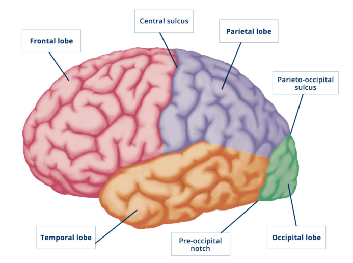
Lobes of the cerebral cortex
Primary visual cortex
Occupies medial and lateral parts of the occipital cortex/lobe at the back/posterior of the brain.
Receives sensory information from retina.
The left and right visual field are each projected to the contralateral hemisphere.
Light stimulus from the external environment from both visual fields stimulate the corresponding area of the retina within each eye.
Different regions of the retina are represented by different areas within the primary visual cortex.
areas further out in peripheral vision are processed by areas of the visual cortex that extend into the calcarine fissure
Within the primary visual cortex neurons show “orientation selectivity”
Hubel and Wiesel won the Nobel prize in 1981 for this work.
Parietal Lobe / Parietal Cortex
Involved in attention and spatial awareness.
sits on the dorsal surface of the cortex and is referred to as part of the dorsal stream and the “where” pathway – named for its role in spatial localisation
Temporal Lobe / Temporal Cortex
Important in auditory processing.
Also involved in more complex visual processing (faces & complex object recognition).
sits on the ventral surface of the cortex and is part of the ventral stream and the “what” pathway – named for its role in complex object recognition
Primary auditory cortex
Occupies superior part of the temporal cortex, as well as a patch of cortex that is buried within the Sylvian fissure. It receives auditory sensory information from the cochlea (part of the inner ear concerned with hearing).
Sounds of different frequencies (e.g., low versus high tones) are represented by different areas within the primary auditory cortex, forming a tonotopic map.
Primary somatosensory cortex
Located immediately posterior to the central sulcus (large grove between the frontal & parietal lobe)
Receives sensory information from the skin (temperature, touch & pain).
Different regions of skin surface represented by different areas along the strip of cortex, forming a somatotopic map (face & hands overrepresented)
Primary motor cortex
Located on the precentral gyrus immediately anterior to central sulcus.
Different parts of primary motor cortex send signals that control different groups of voluntary muscles (e.g., hands, feet, lips).
Like the primary sensory cortices, the primary motor cortex controls muscles on the opposite (contralateral) side of the body
Frontal Lobe
Human frontal cortex is different from other animals
relatively larger than non primates
a higher level of connectivity with rest of the brain (compared to other apes)
“higher order” functions of the frontal lobes
Voluntary, controlled behaviour
Impulse control & emotional regulation
Abstract reasoning & planning
Social cognition
Language
Phineas Gage – Classic case of altered “executive functioning” with frontal lobe damage
railway foreman in Cavendish, Virginia accident involving ‘tamping iron’ (1848)
survived profound damage to frontal lobe behavioural changes (‘no longer Gage’ )
personality changed profoundly
angry, impatient, obstinate, capricious
unable to plan for the future.
not employable in old job, (only held jobs where he did not make decisions)