Landmarks on Dental X-Rays
1/26
There's no tags or description
Looks like no tags are added yet.
Name | Mastery | Learn | Test | Matching | Spaced | Call with Kai |
|---|
No analytics yet
Send a link to your students to track their progress
27 Terms
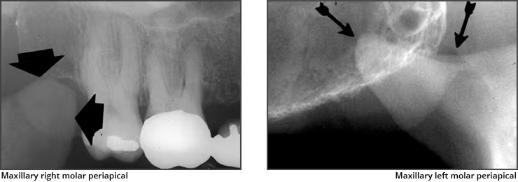
Walls of sinus
Appears radiopaque
Sinus cavity
Appears radiolucent
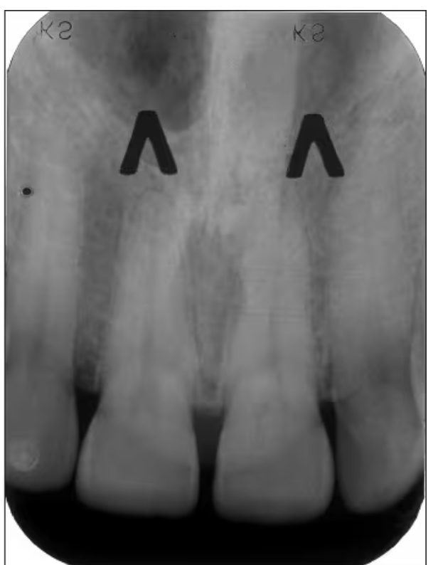
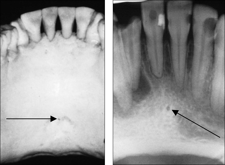
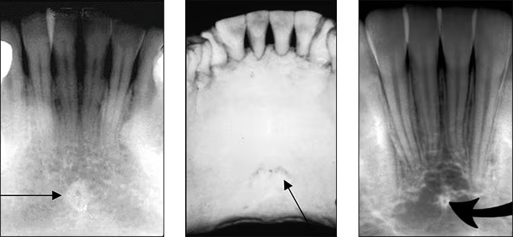
Incisive foramen / nasopalatine foramen
Appears between the roots of the central incisor & looks like a round oval less than 1 cm in diameter
Seen on maxillary central incisor PA’s
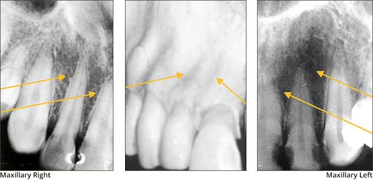
Nasal fossae
Appears radiolucent. Seen on maxillary central incision PA’s and somewhat on lateral incisor and canine PA’s

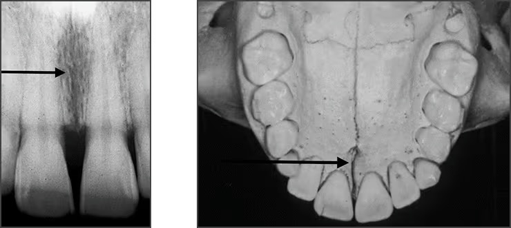
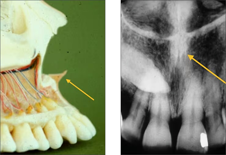
Median palatine suture
Appears as a thin vertical line in the midline on max central incisor PA’s
Is where the two halves of the maxillary come together
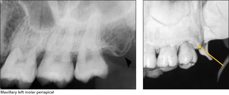
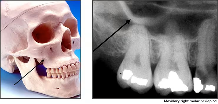
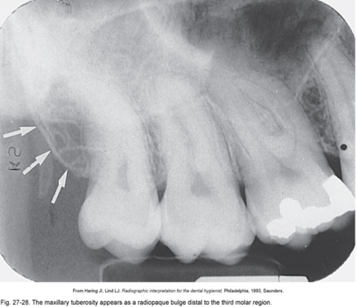
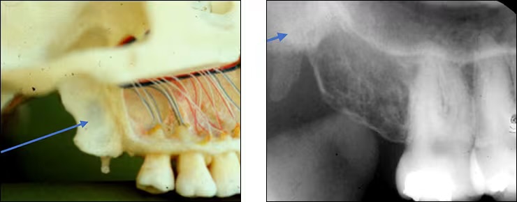
Maxillary tuberosity
Is the rounded end of the alveolar process of the maxilla
Coronoid process
Anterosuperior portion of the ramus
Seen when the patient’s mouth is open on posterior maxillary PA’s
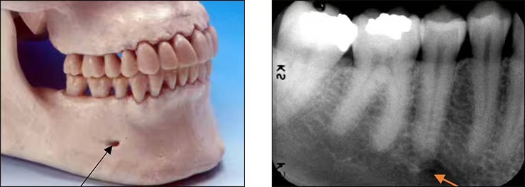
Mental foramen
Located below roots of mandibular premolars.
Is the opening for passage of the mental nerve and vessels.
Often misinterpreted as periapical lesions
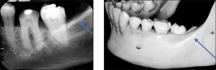
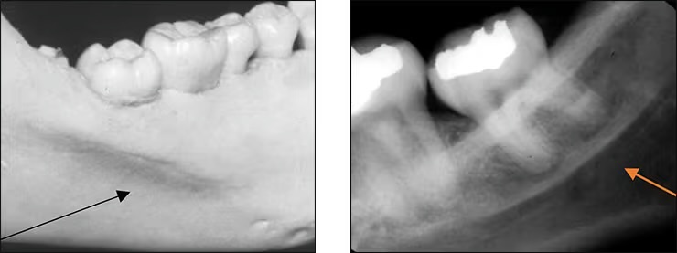
External Oblique Ridge
Bony anterior border of the ramus located n the outer aspect of the mandible

Mental Ridge
Prominence of bone on the labial surface of the anterior mandible
Some individuals display very distinct ridges while others have little or no evidence of its presence.
Seen on incisor and partially on lateral aspect of canine PA’s
Zygomatic Process (malar process)
Radiopaqe U-shaped structure representing where the zygomatic bone attaches to the maxilla
Seen on maxillary premolar and molar PA’s
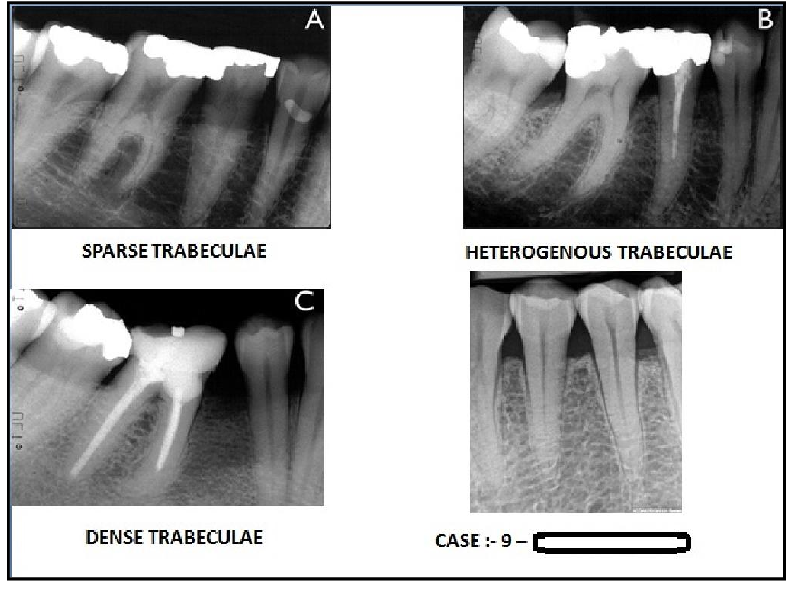
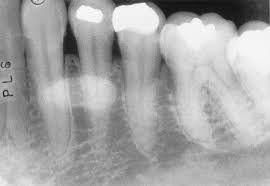
Trabelculae
Interconnected spongy bone (bony lines) that appear like fuzzy bright lines and dark spaces

Mandibular canal
Pathway in the bone where the inferior alveolar nerve and blood vessels flow through the mandible.
Extend from mandibular foramen within the ramus to the mental foramen.
Appears radiopaque
Submandibular gland fossa
Depression in the bone on the lingual aspect of the posterior mandible.
Bilateral below the maternal oblique ridge/mylohyoid line.
Is where the submandibular salivary gland rests.
Typically appears radiolucent
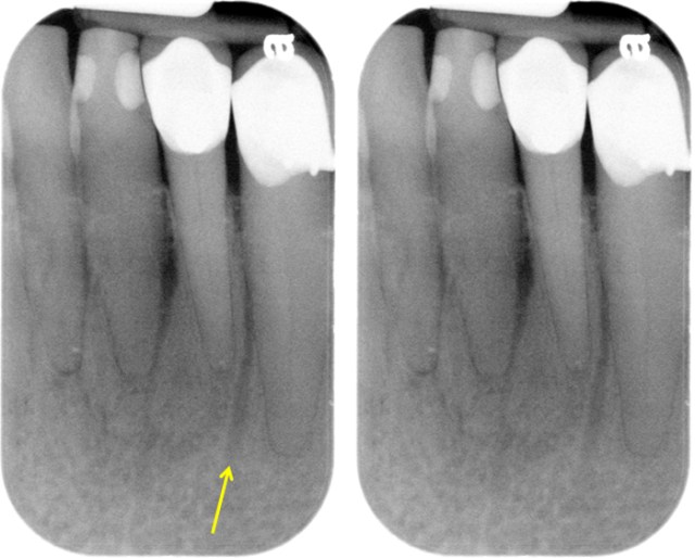
Nutrient canals
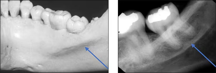
Internal Oblique Ridge / Mylohyoid line
Bony ridge found on the lingual aspect of the posterior mandible. Ranges from highly to barely visible.
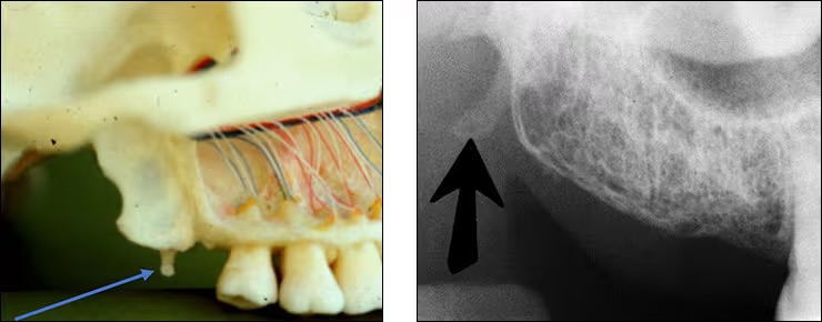
Hamular process
aka pterygoid hamulus
Tiny finger/hook like projection of bone extending from medial pterygoid plate.
Appears on maxillary molar PA’s and molar bitewings
Lingual foramen
Small pinpoint opening in bone on the lingual aspect of anterior mandible.
Surrounded by genial tubercle.
(for lingual nerve and arteries)
Mandibular Retromolar pad
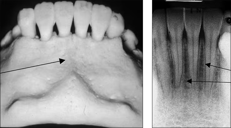
Genial tubercles
aka mental spine
Spiny protuberance (sometimes 2) of bone located in the midline on the lingual aspect of mandible below roots of incisor teeth
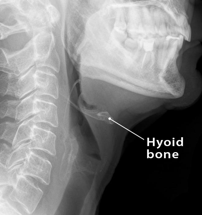
Hyoid bone
Sometimes appears as a ghost image above the mandible
Inverted Y
Depicts where the nasal fossa crosses the maxillary sinus (the boundary between them are shaped like a upside down Y)
Anterior Nasal Spine
Appears “V-shaped” or “triangular point”
Is a bony projection located at the base of the nasal septum in the maxillary midline
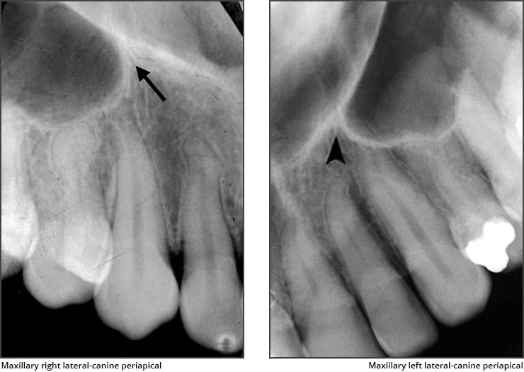
Lateral fossa / canine fossa
Appears bilaterally
Is a slight dip or depression in the bone on the labial of the maxilla near the lateral roots
Recorded don lateral and canine PA’s
Tori
Tissue appears radiopaque
Pterygoid plates
Located behind maxillary tuberosity
“thin wing bone” extending posteriorly from tuberosity
Mental fossa
Depression in the bone on the labial aspect of the mandible.
Diffuse radiolucent appearance above the mental ridge
prominence based on thickness and density of mandible