OPERATIONAL CONTROLS
1/88
There's no tags or description
Looks like no tags are added yet.
Name | Mastery | Learn | Test | Matching | Spaced | Call with Kai |
|---|
No analytics yet
Send a link to your students to track their progress
89 Terms
SUPERIOR (TOWARD HEAD)
1

INFERIOR (TOWARD FEET)
2
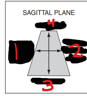
POSTERIOR
3

ANTERIOR
4

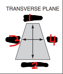
ANTERIOR
1

POSTERIOR
2

RIGHT
3

LEFT
4
Probe Indicator
The “____” on the ultrasound probe can be identified as an orientation marker (ridge, indentation, groove, or nub) on one side of the probe.
Probe Indicator
This corresponds to the indicator or orientation marker on the ultrasound image.
Left Side
What side does the orientation marker located for all standard applications and procedures?
Right Side
What side does the orientation marker located for all cardiac application
KNOBOLOGY
a terminology that describes the manipulation of ultrasound knobs and system controls in order to obtain the best image possible from diagnostic ultrasound
SINGLE OR DUAL WINDOW BUTTON
Other manufacturer it is called: Window 1 and Window 2, Or Right Window or Left Window
TRACKBALL OR TOUCHPAD
Is the mouse of the ultrasound device and the common operating instrument of the screen cursor.
TRACKBALL OR TOUCHPAD
Can be rotated freely in all axis direction.
KEYBOARD
Various capabilities as provided by the manufacturer
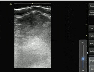
DEPTH
controls how much distance into the body the image displays in the far field.
DEPTH
used to adjust the size of the image so that organs and adjacent structures or regions of interest are equally well visualized
Increasing Ultrasound Depth
Decreasing Ultrasound Depth
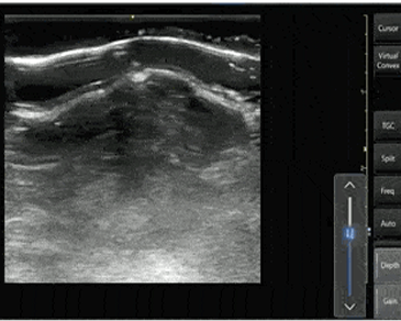
Magnified
When the depth is decreased, superficial structures will be ______
higher the frame rate
Less depth will have ____
GAIN
Is a knob that adjust the overall UTZ echo signal
Increased
As the gain is ____, the strength of the returning echoes is amplified, which produces a brighter image
Decrease
A _____ in gain will darken the image visualized on the monitor
TIME GAIN COMPENSATION
may be set up as a column of sliding knobs, or may be adjusted with knobs for “near gain” and “far gain
TIME GAIN COMPENSATION
allows selective control of gain at different depths
ZOOM
To magnify the region of interest (ROI)
READ zoom
WRITE zoom
2 types of ZOOM (by manufacturer)
READ zoom
produces the worse kind of images because it relies on stored images which enlarges the pixel density in that region.
WRITE zoom
tries to maintain the pixel density by zooming the image live which produces a better spatial resolution.
READ zoom
WRITE zoom
can produce poorer image depending on the size of the area magnified.
FOCUS
This knob, is system allows the operator to improve lateral resolution in a region of interest by adjusting the focal zone
FOCUS
It can be moved up or down by the operator and should be placed at the region of interest or posterior to that region
focal zone
The ____ normally appears at the lateral side of B mode as a triangular-shaped structure or a dot.
DYNAMIC RANGE
is a control on the ultrasound system that allows the operator to determine the range of shades of gray to be displayed on the monitor
broad dynamic range
In abdominal sonography, a ____ is the most appropriate option for assessing the echotexture of homogeneous soft-tissue structures like the liver, pancreas and spleen
Narrow dynamic range
is most appropriate for assessing anechoic structures such as the aorta and IVC
BOX STEER
Ultrasound beam steering may lead to an improved Doppler angle
Always towards the flow especially in linear probe
FREEZE
Used to pause the moving live image to be able to judge individual frames more precisely, or to save and store them.
CINE LOOP
additional control that helps with selecting the best of the image frozen image
CINE LOOP
It displays image frames acquired in the last few seconds prior to freezing.
CINE LOOP
The slow-motion (frame by frame) review capability allows the sonographer to select an appropriate image for study
CALIPER
To measure the acoustic properties of material
A Mode
Amplitude Mode
A Mode
is the simplest form of ultrasound imaging which is based on the pulse echo principle.
A Mode
A scans only give one dimensional information and not so useful for imaging
A Mode
useful for echoencephalography and echo ophthalmoscopy
B – Mode
Brightness mode (2D Mode)
B – Mode
Spatially oriented- Structures are seen as function of brightness (echoic vs anechoic), depth, and width.
Real-Time Images
If multiple B-mode images are watched in rapid sequence, they become ___
REAL TIME
Facilitates the observation of motion in any part of the subject within the cross section being scanned
REAL TIME
Essentially involves the generation of images of the same cross section repetitively and at a rate exceeding about 25 frames per second which is high enough to create the impression of continuity of events in time.
M Mode
Motion Mode
M Mode
Another way of displaying motion
M Mode
This result in a wavy line and most commonly used for cardiac ultrasound
DOPPLER MODE
It is a general term used to visualize velocities of moving tissues.
DOPPLER MODE
Evaluates blood velocity as it flows through blood vessel.
DOPPLER MODE
Blood flow through the heart and large vessels has certain characteristics that can be measured using doppler instruments.
CONTINUOUS WAVE DOPPLER (CW)
Uses different crystals to send and receive the signal
CONTINUOUS WAVE DOPPLER (CW)
One crystal constantly sends a soundwave of a single frequency the other constantly receives the reflected signal.
CONTINUOUS WAVE DOPPLER (CW)
The advantages is can accurately display flow of any velocity without aliasing.
CONTINUOUS WAVE DOPPLER (CW)
Can measure very high Doppler shift/velocities but with limited depth
SPECTRAL/PULSED WAVE (PW)
Produces short burst/pulses of sound
SPECTRAL/PULSED WAVE (PW)
Uses the same crystals to send and receive the signal.
SPECTRAL/PULSED WAVE (PW)
it follows the same pulse-echo technique used in 2D image formation.
SPECTRAL/PULSED WAVE (PW)
Advantage : specific depth and range
SPECTRAL/PULSED WAVE (PW)
Disadvantage : limited maximum detectable velocity
COLOR FLOW (CF)
produces a color-coded map of Doppler shifts superimposed onto a B-mode ultrasound image
COLOR FLOW (CF)
Assignment of color to frequency lighter shifts is usually based on and magnitude (different color hues or saturation frequency shifts).
COLOR FLOW (CF)
Best way to check for problem w/ blood flow such as deep vein “thrombosis”
POWER DOPPLER
also referred to as energy Doppler, amplitude Doppler and Doppler angiography.
POWER DOPPLER
5 times more sensitive in detecting blood flow than color doppler.
POWER DOPPLER
It is a new technique that displays the strength of the Doppler signal in color, rather than the speed and direction information
POWER DOPPLER
Provide greater detail of blood flow, especially in vessels that are located inside organs
Red
Yellow
Power Doppler uses a red-to-orange-to-yellow color scale to represent the total power in the Doppler spectrum at each sample volume. The lowest powers are displayed in ___ and the highest powers are displayed in _____
SLIDING
involves moving the entire probe in a specific direction to find a better imaging window.
SLIDING
This is usually used to find the best window, move to different areas of the body, or to follow a specific structure (such as a vessel)
TILTING/FANNING
involves moving the transducer from side to side along the short axis of the probe
TILTING/FANNING
will allow visualization of multiple cross-sectional images of a structure of interest.
TILTING/FANNING
You can apply this technique to structures such as the heart, kidney, bladder, vessels, etc
Rotating
involves turning the transducer in a clockwise or counterclockwise direction along its central axis.
Rotating
is most commonly used to switch between the long and short axis of a specific structure.
ROCKING
involves “rocking” the ultrasound probe either towards or away from the probe indicator along the long-axis
ROCKING
allows you to help center the area of interest
ROCKING
This is also referred to as “in-plane” motion because the image is kept in-plane throughout the manipulation
COMPRESSION
involves putting downward pressure on the probe to evaluate the compressibility of a structure or organ of interest.
COMPRESSION
The most common use is to evaluate for deep vein thrombosis, differentiate between artery versus vein, and evaluation for appendicitis (non-compressible)