brain anatomy and CT appearances
1/25
There's no tags or description
Looks like no tags are added yet.
Name | Mastery | Learn | Test | Matching | Spaced | Call with Kai |
|---|
No study sessions yet.
26 Terms
what are the subdivisions of the nervous system
•The central nervous system (CNS) consists of the brain and the spinal cord.
•It is covered by three ‘membranes’ (the meninges)
•Within the CNS some neurons that share similar function are grouped into aggregations called nuclei.
•The CNS can also be divided into:
Grey matter, which contains neuron cell bodies
White matter, which is rich in myelin
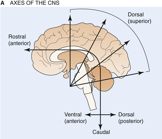
what are the axes of the CNS
nervous system bends during development
what are the major components of the CNS
•telencephalon,
•cerebellum,
•diencephalon,
•brainstem (consisting of the midbrain, pons, and medulla), and
•spinal cord.
Each of these areas has symmetrical right and left sides
what is the telencephalon: cerebral cortex
•The human cerebral cortex has a surface area of ~2200 cm2 and is estimated to contain 1.5 to 2 × 10 10 neurons. The number of synaptic contacts between these cells is ~3 × 10 14
•The cortical surface area of mammals increases massively from mouse to monkey to humans in a ratio of 1 : 100 : 1000.
There are 4 regions of the telencephalon - frontal, parietal, occipital and temporal lobe
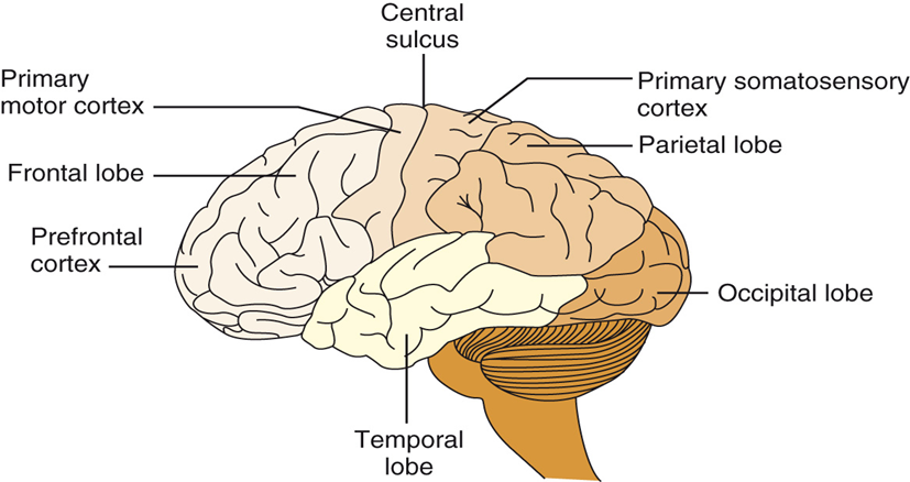
what is the role of the frontal lobe
Frontal lobe: extends anterior from the central sulcus and contains the motor and prefrontal cortices. Associated with higher cognitive functions and long-term memory storage; the posterior portion is the primary motor cortex and controls fine movements.
what is the parietal lobe
between the occipital lobe and central sulcus, associated with sensorimotor integration and processes information from touch, muscle stretch receptors, and joint receptors. This area contains the primary somatosensory cortex.
what is the temporal lobe
Temporal lobe: located laterally in each hemisphere and is the primary cortical target for information originating in the ears and vestibular organs; it is involved in vision and language
what is the occipital lobe
located in the posterior cortex - involved in visual processing. Main target for axons from thalamic nuclei that receive inputs from the visual pathways and contains the primary visual cortex.
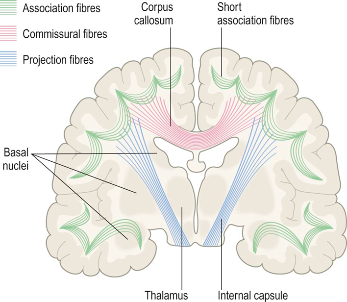
What are the connections in the telencephalon
The corpus callosum and smaller white matter tracts interconnect the two cerebral hemispheres
the basal ganglia are a functionally related group of neuron clusters consisting of the striatum (caudate nucleus and putamen), globus pallidus, amygdala, and hippocampal formation. These are all paired structures. The basal ganglia have indirect connections with motor portions of the cerebral cortex and are involved in motor control.
The amygdala participates in the expression of emotion, and the hippocampal formation is crucial in the formation of new memories. Injury to both hippocampal formations can cause a severe amnesic disorder
•There are three main types of white matter bundle in the cerebrum:
•Association fibres interconnect parts of the same hemisphere.
•Commissural fibres cross the midline to connect with matching areas of the opposite hemisphere The largest commissure is the corpus callosum, composed of some 200–300 million myelinated axons.
•Projection fibres pass to and from the cerebral cortex. The internal capsule is a massive white matter system composed of 20 million projection fibres
what is the cerebellum
•Lies immediately dorsal to the brainstem.
•Represents ~10% of the CNS by volume, but contains ~50% of all CNS neurons.
•Very large number of input connections to the cerebellum conveys information from nearly every type of receptor in the nervous system, including visual and auditory input.
•The cerebellum is associated with the regulation and coordination of movement, posture, and balance.
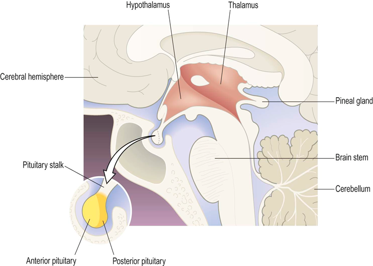
What is the diencephalon
The hypothalamus controls the and is involved in the maintenance of homeostasis. The hypothalamus lies inferior to the thalamus.
It is small but controls many critical body functions.
The pituitary gland is attached to the end of the infundibulum or pituitary stalk which extends from the hypothalamus.
The thalamus is known as the ‘gateway’ to the cerebral cortex, since most ascending sensory pathways relay in one of its nuclei in order to reach their cortical targets.
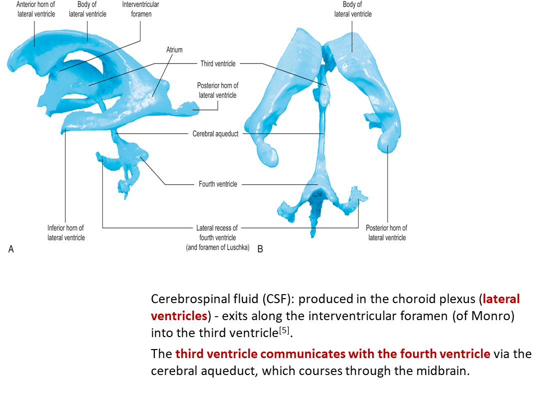
what are the ventricles
•2 (large) c-shaped lateral ventricles
•Third ventricle is narrow cavity in the midline
•Fourth ventricle located between brainstem and cerebellum
Cerebrospinal fluid (CSF): produced in the choroid plexus (lateral ventricles) - exits along the interventricular foramen (of Monro) into the third ventricle[5].
The third ventricle communicates with the fourth ventricle via the cerebral aqueduct, which courses through the midbrain.
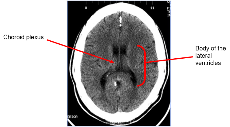
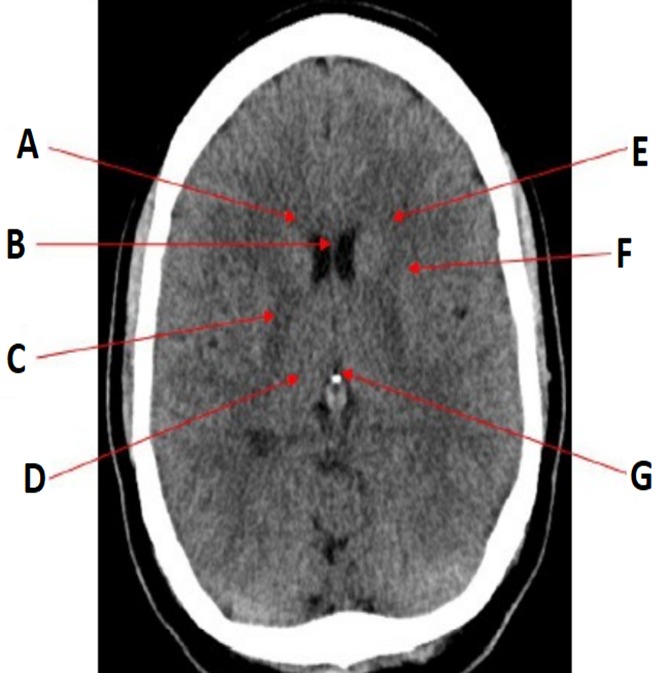
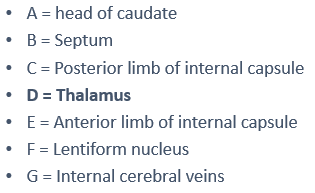
Axial CT image of the brain at the level of the body of the lateral ventricles
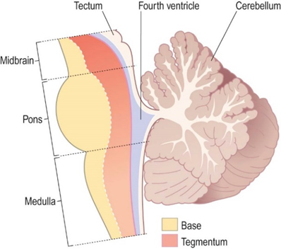
what is the brain stem?
It is composed of the midbrain, pons & medulla
Lies immediately above, or rostral to, the spinal cord
brainstem has three main functions:
1.Conduit for tracts ascending and descending through the CNS;
2.Houses cranial nerve (CN) nuclei III to XII (CN XI is in the cervical spinal cord)
3.Location for reflex centres related to respiration, cardiovascular function, and regulation of consciousness.
Surrounded by the pons and medulla anteriorly and the cerebellum posteriorly, the fourth ventricle sends CSF out of the ventricular system and into the subarachnoid space.
how is blood supplied to the brain
•The brain is supplied by two pairs of arteries that arise from branches of the aortic arch:
The internal carotid arteries (80%) ascend in the anterior part of the neck, arising from the bifurcation of the common carotid arteries.
The vertebral arteries (20%) arise from the subclavian arteries and ascend in the lateral cervical region, passing through foramina in the transverse processes of the upper six cervical vertebrae.
•The arteries at the base of the brain are linked by communicating vessels to form the circle of Willis (COW).
•This gives rise to anterior, middle and posterior cerebral arteries on each side, which supply most of the cerebral hemisphere.
•The cerebral blood supply can be divided into anterior and posterior circulations
what is the circle of willis
•Formed by a single anterior communicating vessel and two
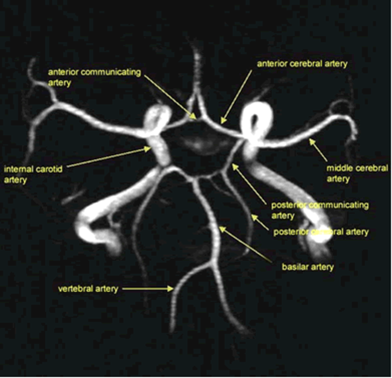
posterior communicating vessels.
what is anterior circulation
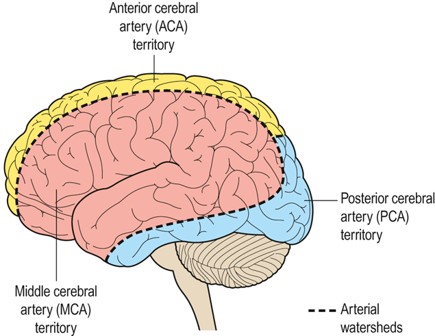
•The internal carotid artery divides at the base of the brain, giving rise to the middle cerebral artery (MCA) and the anterior cerebral artery (ACA).
•The MCA is the larger of the two and receives 80% of the internal carotid blood flow. For this reason, cardiogenic emboli are much more likely to enter the MCA than the ACA.
•The MCA continues laterally between the frontal and temporal lobes and emerges from the lateral sulcus to supply most of the hemispheric convexity.
•The ACA passes forward and medially to meet its partner between the cerebral hemispheres.
what is posterior circulation
•The posterior circulation supplies the remainder of the cerebral hemisphere and the posterior fossa contents (brain stem and cerebellum).
•The two vertebral arteries unite in front of the brain stem to become the basilar artery, which splits into the two posterior cerebral arteries (PCA).
•These vessels pass posteriorly, above the tentorium cerebelli, to supply the occipital lobe and the inferior surface of the temporal lobe.
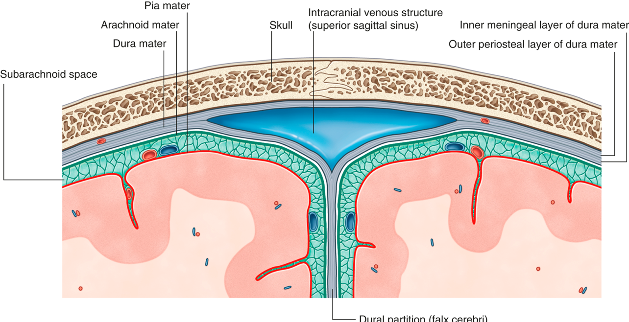
what are meninges
Meninges - connective tissue coverings which act to support and stabilize the brain and spinal cord.
Dura mater:
•Outermost covering - a tough, fibrous sheet composed of two layers.
•Outer periosteal layer is adherent to the skull, inner meningeal layer lies against the underlying arachnoid mater.
•Do separate in some regions to form dural venous sinuses, which receive cerebral venous drainage.
•Meningeal layer of dura mater folds in on itself in several areas (e.g. falx cerebri).
Arachnoid mater:
•Deep to the dura mater
•Strands of connective tissue extend from outer layer to form arachnoid trabeculae, which connect internally to the pia mater.
•The pia and arachnoid matter are separated by a subarachnoid space that contains CSF and the major blood vessels supplying the brain.
Pia mater:
•Forms a thin, veil-like layer that closely follows the gyri and sulci on brain surface.
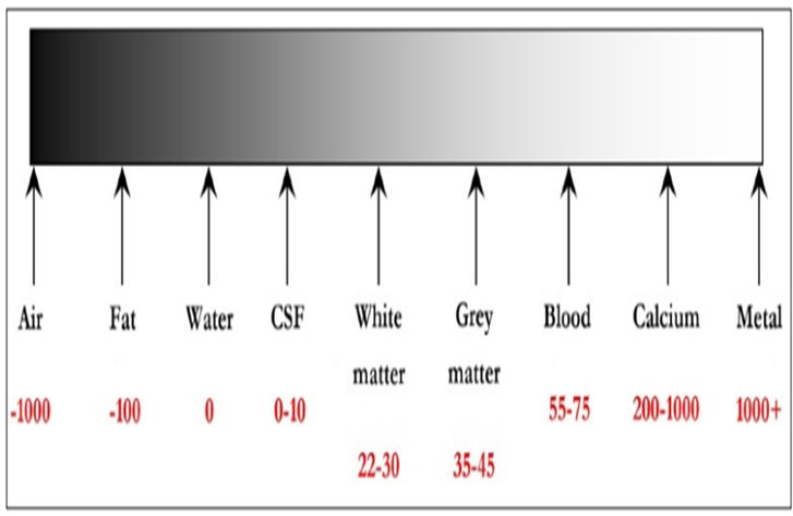
how do you interpret CT images
•CT images are displayed as varying shades of grey based on the attenuation that different tissues exhibit.
•The Hounsfield Unit (HU) is an arbitrary scale which is used to display the range of tissue densities when viewing a CT scan: the scale ranges from -1,000 to +1,000, with water by convention designated the value of 0.
The higher the HU, the brighter (or denser) the tissues displayed
label
label
label
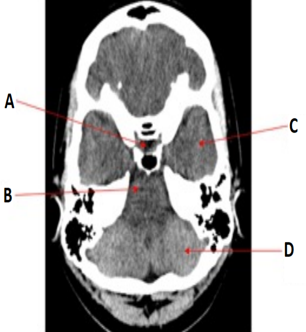
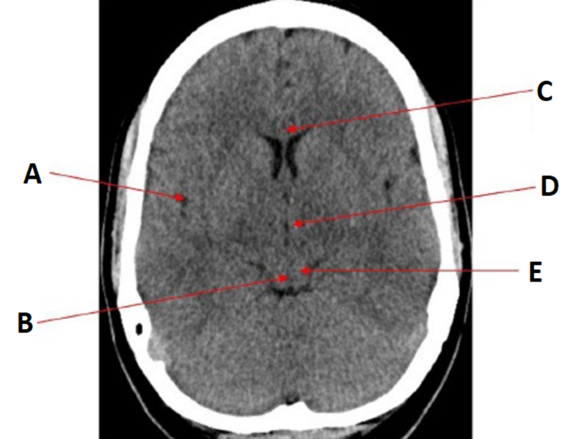
•A = Pituitary fossa
•B = Pons
•C = Temporal lobe
•D = Occipital lobe
label
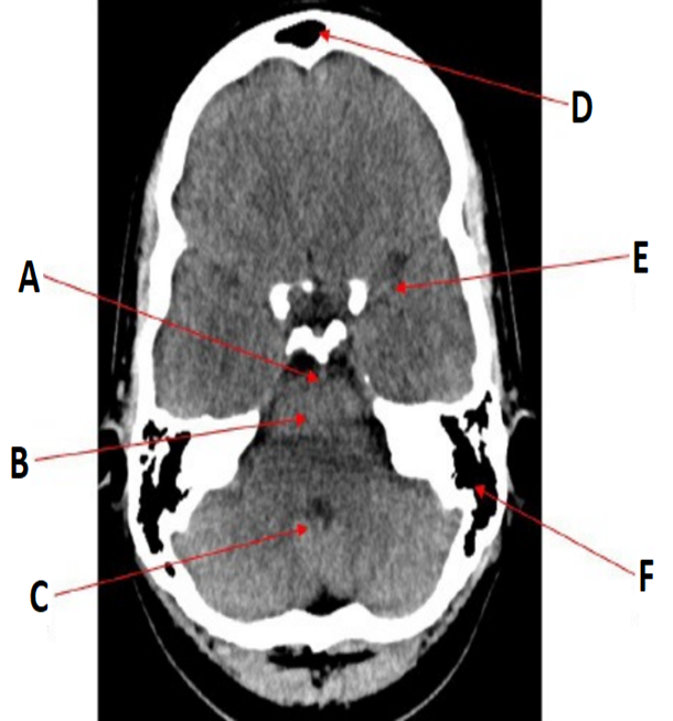
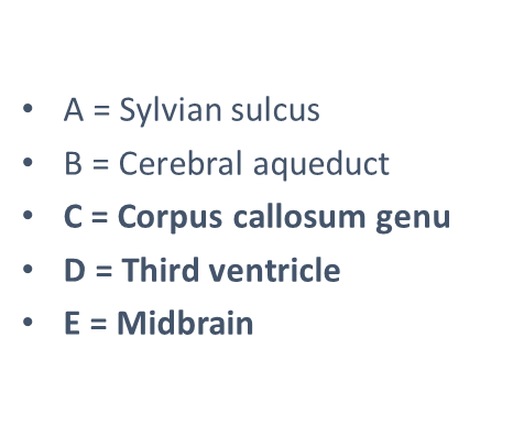
•A = Basilar artery
•B = Pons
•C = Fourth ventricle
•D = Frontal sinuses
•E = Middle cerebral artery
•F = Mastoid air cells
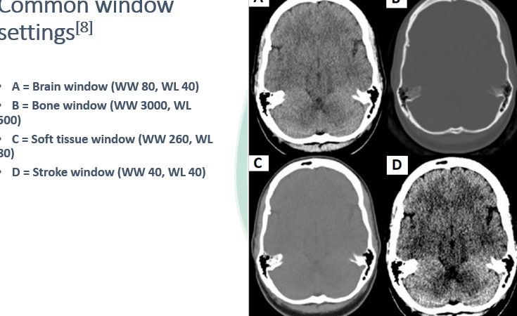
why is windowing important
•Window settings are used to aid detection of pathologies involving the brain substance (e.g. infarcts), the skull vault (e.g. fractures) or soft tissues (e.g. haematomas).
•Window settings are described in terms of window width (WW) and window level (WL) – these values are typically displayed on the computer screen.
•WW = the range of grey-scales displayed
•WL = the centre of the WW
•Altering the window settings helps reduce the range of HU displayed, which in turn helps to maximise the pickup rate of different pathologies.
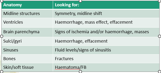
Viewing cross sectional images: a systematic approach