PSYC-2900 Chapter 6
1/60
There's no tags or description
Looks like no tags are added yet.
Name | Mastery | Learn | Test | Matching | Spaced | Call with Kai |
|---|
No analytics yet
Send a link to your students to track their progress
61 Terms
sensation
process of signal detection
relies on photoreceptors detecting different stimuli and translating them into neural signals
perception
process of interpreting neural signals
relies on organising the incoming neural signals and making sense of them to give us a conscious experience of the stimuli
top-down process that makes sense of sensation info
group stimuli together
discriminate between similar stimuli
detect when stimuli change and are constant
detecting light
photoreceptors are sensitive to certain wavelengths of light in 3 dimensions: hue, brightness, and saturation
hue
a dimension of light that defines the type of perceived colour, determined by the wavelength of light
brightness
a dimension of light determined by the intensity of light
saturation
a dimension of light that defines the intensity of colour, determined by the relative purity of light
sensory transduction
a process in which light is detected by photoreceptors (cones and rods) which convert the light into a neural signal. stimulus alters membrane potential creating a receptor potential
most photoreceptors lack axons, some of their membrane forms a synapse with dendrites
visual pathway in the eye
light → cornea → lens → retina
cornea and lens focus light onto the retina
ciliary muscle alters shape of lens to focus image
pupils regulate amount of light that enters
structures focus the image onto the retina
bones & muscles around eye
extraocular muscles attached to sclera
accommodation
the ability to focus, allowing clear vision at various distances, by the ciliary muscle changing the shape of the lens to focus the image
retina
posterior structure in the eye, consists of 3 layers: photoreceptive, bipolar, and ganglion cells
photoreceptors
specialised neurons in the retina
rods (more): detect low light
cones: detect colour and acuity
fovea only contains cones
optic disk at the back of the eye has neither
visual pathway in the retina
light signals → photoreceptors → neural signals → bipolar cells → ganglion cells → brain
horizontal & amacrine cells integrate signals between photoreceptors and pathways
photopigment molecules
molecules in the visual pathway in the retina made of opsin (protein) and retinal (lipid), that transduce light waves into a membrane potential
human rods have rhodopsin (rod opsin + retinal)
retinal synthesised from vitamin A
light → rhodopsin molecule → hyperpolarisation of membrane → neurotransmitter release (decreased glutamate)
bipolar cells
within the visual pathway of the retina, have different responses to glutamate depending on type:
light ON centre cells become hyperpolarised
light OFF centre cells become depolarised
receptive field
areas in the front of the photoreceptor layer that allows for central and peripheral vision.
location depends on the location of photoreceptor
vergence movements
a type of eye movement that helps separate environment from target
keeps both eyes fixed on same target to focus on both retinas
saccadic movements
a type of eye movement that helps separate environment from target
jerky eye movements used for scanning
pursuit movement
a type of eye movement that helps separate environment from target
slower, smoother movement by following target/environmental movement
the visual pathway
in the eye: light (travels to the back) → photoreceptors → bipolar cells → ganglion cells (signal travels forward) → axons converge into the optic nerve
lateral geniculate nucleus (LGN) of thalamus
primary visual cortex (V1, striate cortex)
visual association cortex (V2, extrastriate cortex) + additional cortical areas
retinal pathways
additional pathways use visual info in other ways:
pathways to hypothalamus involved in circadian rhythms
control eye movements: iris constriction, ciliary muscles of lens
lateral geniculate nucleus (LGN)
6 layers of neurons:
layers 1, 4, 6 receive input from contralateral eye
layers 2, 3, 5 receive input from ipsilateral (same side) eye
3 layers, each containing different types of cells that process different aspects of visual info (e.g., motion, colour, detail):
magnocellular layers (2 inner layers) → 4 Cα (striate cortex)
parvocellular layers (4 outer layers) → 4 Cβ (striate cortex)
koniocellular sublayers (under other types) → 2nd & 3rd layers of striate cortex
primary visual cortex (V1)
processes basic features
receives input directly from LGN + combines info from other areas
contains neurons that respond to specific features of stimuli (coding)
firing rate depends on where stimulus is on receptive field
more coding organises further before sending to V2
organised into modules that process info from diff visual fields
receive input from other modules → analysis → output to other modules
cytochrome oxidase (CO) blobs
a part of modules in the 2 & 3 layers of V1, surrounded by interblob regions
receives input from parvocellular & koniocellular layer (LGN) (colour info)
project to thin stripes in V2 (colour)
visual association area (V2)
combines input from V1 to build entire visual scene. contains 3 stripes:
thin stripe (dark): colour
thick (dark) & pale stripes: orientation, spatial frequency, movement, retinal disparity
and specialised regions:
receives information from “lower” regions
passes info to “higher” regions for higher processing
dorsal stream
a pathway from the V2 that processes where objects are located, and speed/direction of movement (action)
receives from magnocellular pathway → parietal lobe
ventral stream
a pathway from the V2 that processes what an object is and its colours (perception)
receives signals from all layers → temporal lobe
vertical occipital fasciculus
a white matter tract that connects the ventral and dorsal visual streams, allowing for exchange of info
perceiving light and dark
ON & OFF bipolar cells → (light/dark signals) → ganglion cells
ganglion cells
3 types:
ON cells
OFF cells
ON/OFF cells
briefly excited when light is turned on/off
trichromatic theory
a theory of colour perception that states each of the 3 types of cones are sensitive to a single hue
supported by physiology in primates: 3 types of cones that have diff peak absorption wavelengths
can’t explain colour perception on its own
opponent-colour system theory
a theory of colour perception that states colours are presented as opponents
explains why you can’t see reddish-green or blueish-yellow
trichromatic coding
changes in colour vision due to cone abnormalities
some X-linked, leading to higher rate in XY
protanopia
first-colour defect in trichromatic coding
red cones have green cone opsin
see shades of yellow and blue
red & green look yellowish
deuteranopia
second-colour defect in trichromatic coding
green cones filled with red cone opsin
confuse red and green
tritanopia
a rare genetic condition in trichromatic coding in which the individual lacks blue cones
monochromatic vision
a very rare genetic conditions in which the individual completely lacks cones
retinal ganglion cells
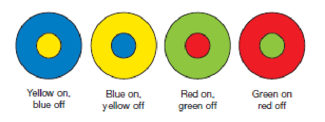
specialised cells supporting the opponent-colour theory that respond to opposing pairs of primary colours. 2 types of colour sensitive cells:
yellow-blue
red-green
when centre on, surround off, firing rate increases
when surround on, centre off, firing rate decreases
other cells are black-and-white detectors
red colour
stimulates its corresponding cone and excites red-green ganglion cells
green colour
stimulates its corresponding cone, inhibiting red-green ganglion cells
blue colour
stimulates its corresponding cone, inhibiting yellow-blue ganglion cells
yellow colour
an intermediate wavelength received by red and green cones
excites & inhibits red-green ganglion cells (no change)
excites yellow-blue ganglion cells
rebound effect
a change of firing rate shown in ganglion cells that are excited or inhibited for a long time
(opposite-coloured apple) big green stimulus inhibits red-green ganglion cells, change to neutral stimulus, cells become excited & fire faster than normal
cortical processing of colour
ganglion cells end in different layers of LGN
parvocellular layer receives wavelength info from cones & receive red and green cone info
koniocellular layer receives wavelength info from cones & info from blue cones
magnocellular layer cells colourblind, detect differences between light & dark movement
neurons from LGN → areas of V1 → areas of V2
perceiving form
V1 → V2 → ventral stream pathway
neurons in V1 sensitive to particular spatial frequency (important for perceiving size & detail)
inferior temporal cortex
has associations with visual pattern & object recognition/identification
fusiform face area (FFA)
in the V2. associated with face perception & other areas of expertise
expansion associated with age & performance
pattern of activity altered in ASD and William’s syndrome
extrastriate body area (EBA)
overlaps with the fusiform face in the V2, active when perceiving silhouettes, stick figures, body parts (not faces or objects)
parahippocampal place area (PPA)
in the V2, active during perception of scenes and backgrounds
visual agnosias
a condition that results from damage to part of V2, leading to deficits in visual recognition, and discrimination in areas of expertise & details
prosopagnosia
the inability to perceive faces
can occur at birth
can be associated with differences in nearby structures (EBA) or connectivity
monocular vision
vision requiring 1 eye, allows for location perception using perspective & relative retinal size,
loss of detail through effects of atmospheric haze & appearance of movements
binocular vision
vision requiring both eyes, allows for location perception using vivid depth perception (stereopsis)
consists of most V1 neurons, project to posterior parietal cortex (dorsal stream)
respond to retinal disparity, difference in retinal image that reveals change in depth
flat vision
impaired depth perception caused by damage to the parieto-occipital areas involved in processing retinal disparity
parietal lobe
receives auditory, somatosensory, and vestibular info
involved in spatial & somatosensory perception
damage can impair perception, memory of location, & influence movements of eyes & limbs
intraparietal sulcus (IPS)
a groove on the parietal lobe containing 5 important dorsal stream areas:
LIP & VIP: control saccadic eye movements
VIP & MIP: visual control of reaching & pointing
AIP: visual control of grasping & manipulation
CIP: depth perception
perceiving orientation & movement
feature detectors → higher visual areas perceiving movement (medial temporal (MT) (← superior colliculus (visual reflexes)) & V5)
processing motion is faster than form & colour
feature detectors
neurons in the V1 that fire at most rapid rates to certain orientations of stimuli
medial superior temporal area
receives input from V5, responds to pattern of movement
dorsolateral region involved in processing optic flow (how retinal image changes as it moves)
centre of expansion
the process by which the centre of field changes size instead of changing position
akinetopsia
damage to the bilateral V5, impairing perception of motion
form from motion
perception of movement that helps perceive 3D objects
does not involve V5
suggested association with right superior temporal gyrus