Musculoskeletal System
5.0(1)
Studied by 37 people Call Kai
 Learn
Learn Practice Test
 Spaced Repetition
 Match
Match Flashcards
 Knowt Play
Card Sorting
1/68

Earn XP
Description and Tags
Last updated 4:19 PM on 12/6/22
Name | Mastery | Learn | Test | Matching | Spaced | Call with Kai |
|---|
No analytics yet
Send a link to your students to track their progress
69 Terms
1
New cards
two primary functions of the musculoskeletal system
1) movement and stability
2) storage of minerals
2) storage of minerals
2
New cards
What is the name of the connection btwn the nervous system and the skeletal system?
neuromuscular junction
3
New cards
name the three types of muscle tissue, whether they are under voluntary or involuntary control, and which nervous systems control each type
1) skeletal muscle: voluntary, somatic
2) cardiac: involuntary, autonomic (heart beating)
3) smooth muscle: involuntary, autonomic (digestion)
2) cardiac: involuntary, autonomic (heart beating)
3) smooth muscle: involuntary, autonomic (digestion)
4
New cards
functions of the skeletal system
1) support and protect the body
2) aid in movement
3) store nutrients
4) produce blood cells
2) aid in movement
3) store nutrients
4) produce blood cells
5
New cards
functions of the muscular system
1) bodily movements
2) stability
3) internal movement of substances within the body
2) stability
3) internal movement of substances within the body
6
New cards
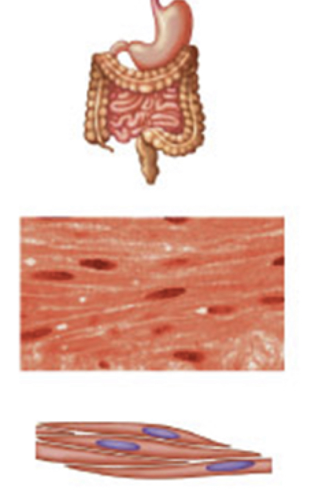
smooth muscle
- contains spindle-shaped, NON-STRIATED, UNI-NUCLEATED fibers
- occurs in walls of internal organs
- INVOLUNTARY
- occurs in walls of internal organs
- INVOLUNTARY
7
New cards
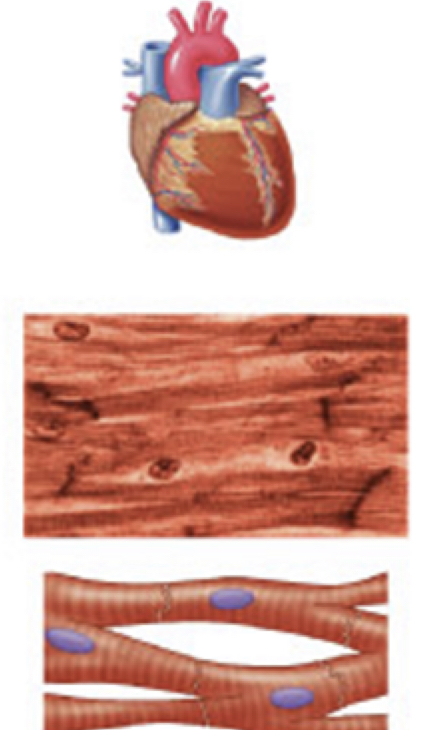
cardiac muscle
- has STRIATED, branched, UNI-NUCLEATED fibers
- occurs in walls of the heart
- INVOLUNTARY
- occurs in walls of the heart
- INVOLUNTARY
8
New cards
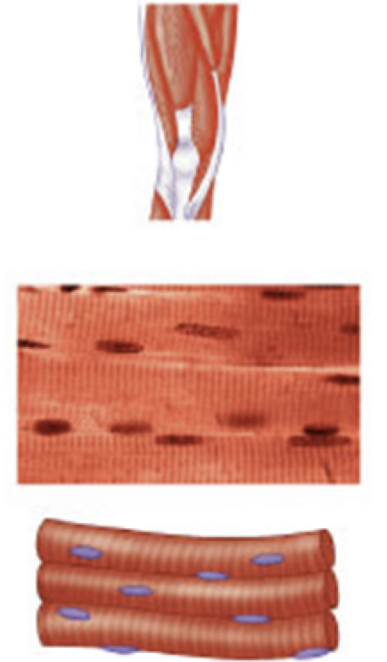
skeletal muscle
- has STRIATED, tubular, MULTI-NUCLEATED fibers
- usually attached to the skeleton
- VOLUNTARY
- usually attached to the skeleton
- VOLUNTARY
9
New cards
connective tissue connecting the musculoskeletal system
1) ligament: short band of tough, flexible, fibrous connective tissue that connects TWO BONES or cartilage, holding together a joint
2) tendon: connective tissue attaching MUSCLE TO BONE; when a muscle contracts to move a joint, it is the tendon that pulls on the bone
3) cartilage: soft cushioning substance that covers the ends of bones; acts as shock absorber and prevents friction btwn bones
2) tendon: connective tissue attaching MUSCLE TO BONE; when a muscle contracts to move a joint, it is the tendon that pulls on the bone
3) cartilage: soft cushioning substance that covers the ends of bones; acts as shock absorber and prevents friction btwn bones
10
New cards
skeletal muscle movement
1) when a muscle contracts it shortens (usually)
2) tension is placed on tendons connecting muscle to bone
3) the tension moves the bone at a joint
4) movement of the bone is toward the point of tendon insertion
2) tension is placed on tendons connecting muscle to bone
3) the tension moves the bone at a joint
4) movement of the bone is toward the point of tendon insertion
11
New cards
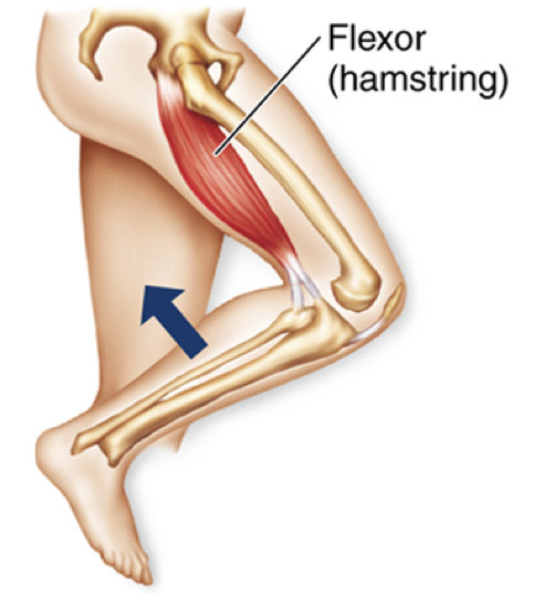
flexor
muscles DECREASE the angle btwn two bones at a joint
12
New cards
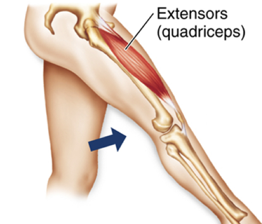
extensor
muscles INCREASE the angle btwn two bones at a joint
13
New cards
antagonist vs agonist muscles
- antagonist: flexors and extensors that act on the same joint to produce opposite actions; ex. triceps in a bicep curl
- agonist: muscle is the prime mover of any skeletal movement; ex. quadriceps when straightening the leg
- agonist: muscle is the prime mover of any skeletal movement; ex. quadriceps when straightening the leg
14
New cards
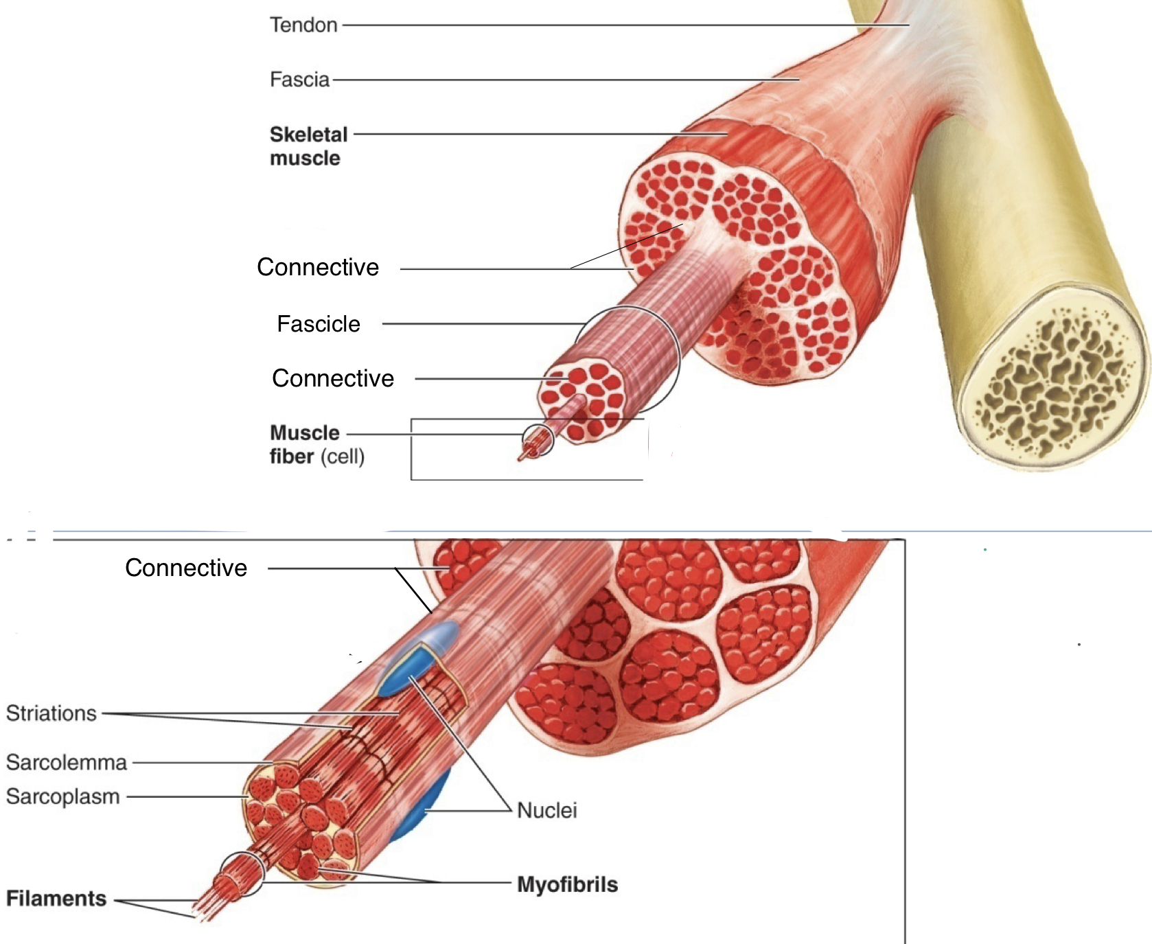
"layers" of muscle tissue from superficial to deep
muscle --> fascicle --> muscle fiber/myofiber/myocyte/muscle cell, --> myofibril --> myofilament
15
New cards
sarcolemma
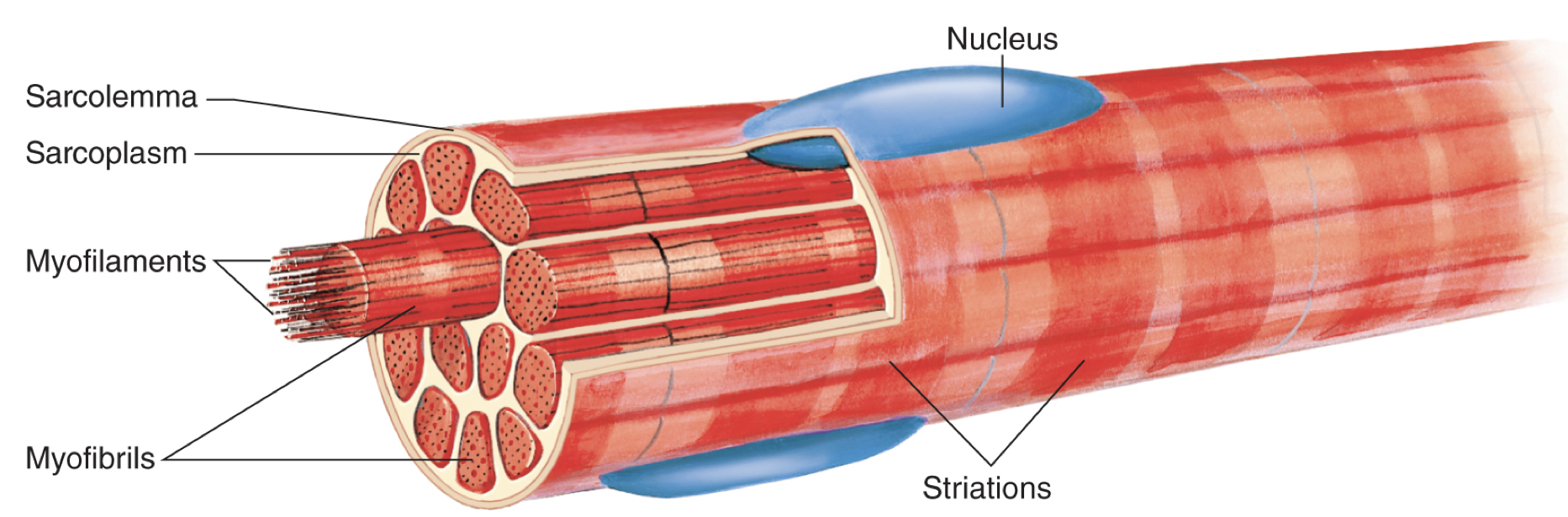
specialized plasma membrane that surrounds ONE MUSCLE CELL
16
New cards
muscle cell/fiber/myofiber/myocyte
- each fiber has densely packed subunits called MYOFIBRILS that run the entire length of the muscle fiber/cell as well as the entire muscle
- myofibrils are composed of thick and thin MYOFILAMENTS called actin and myosin (proteins that pull on each other to cause contractions)
- nuclei shoved to the side bc myofibrils fill up most of the cell space
- myofibrils are composed of thick and thin MYOFILAMENTS called actin and myosin (proteins that pull on each other to cause contractions)
- nuclei shoved to the side bc myofibrils fill up most of the cell space
17
New cards
sarcomere
unit of contraction
z-line to z-line
many sarcomeres repeated over the length of the muscle fiber=> myofibril
z-line to z-line
many sarcomeres repeated over the length of the muscle fiber=> myofibril
18
New cards
"parts" of sarcomeres
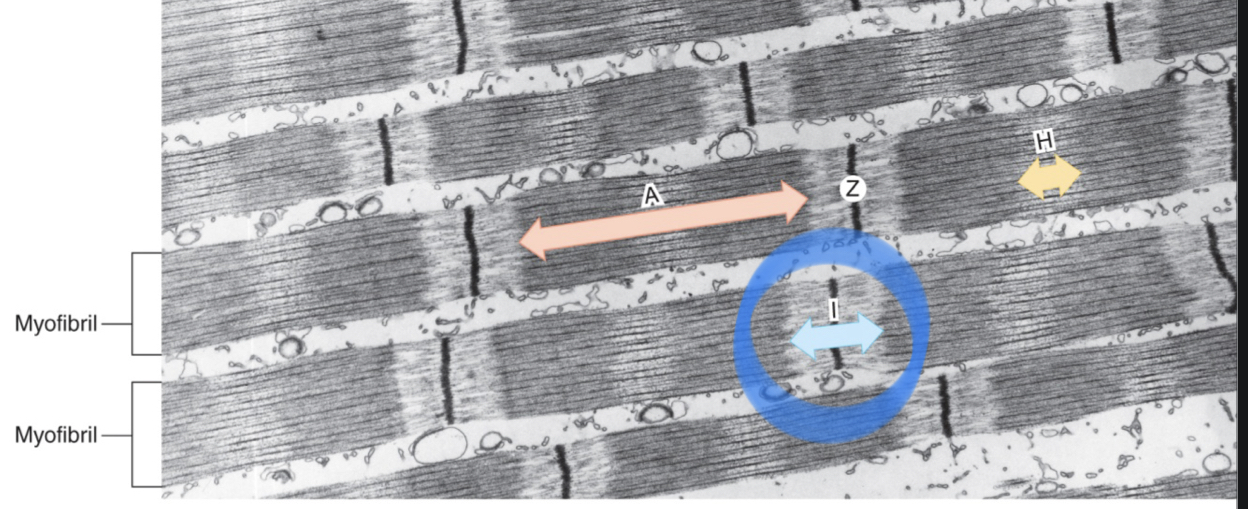
I bands, A bands, H zones, and z-lines/discs
19
New cards
I band
- LIGHT in color
- contain only THIN filaments; primarily of the protein ACTIN
- contain only THIN filaments; primarily of the protein ACTIN
20
New cards
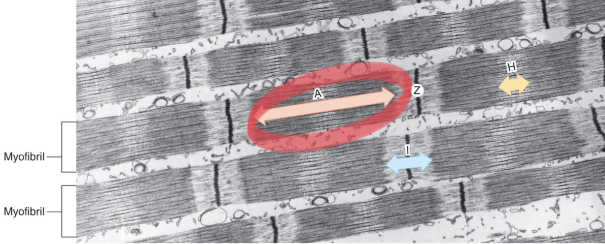
A band
- DARK in color
- contain all of the THICK filament composed of protein called MYOSIN with some thin overlap
- contain all of the THICK filament composed of protein called MYOSIN with some thin overlap
21
New cards
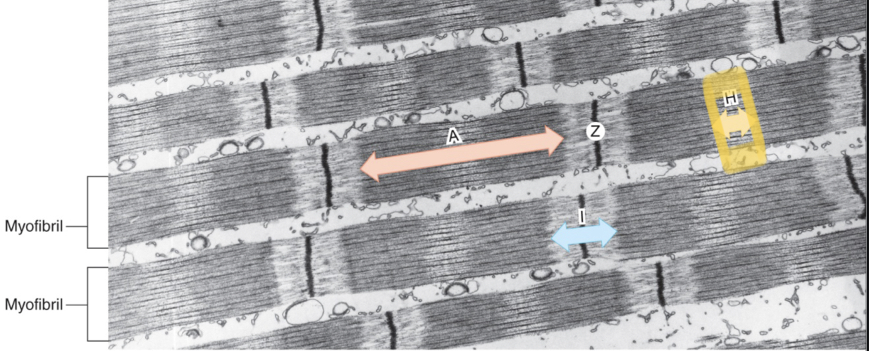
H zones
- center of the A band with NO THIN filament overlap
22
New cards
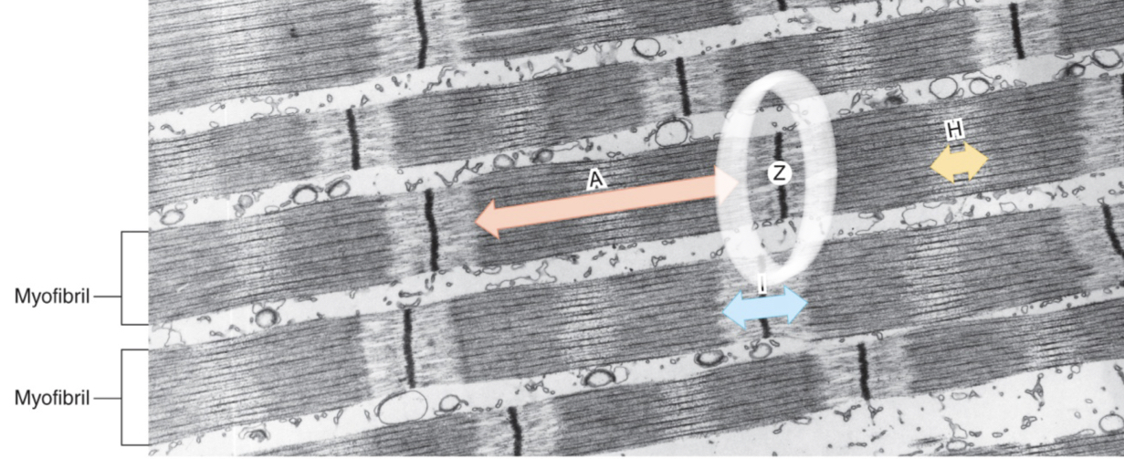
Z disc/lines
dark lines found in the center of each I band; end/beginning of sarcomere
23
New cards
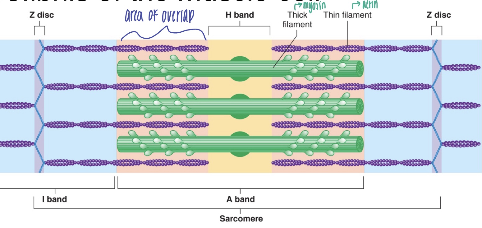
darkest area of sarcomere
where there is overlap of both myosin (thick filaments) and actin (thin filaments)
24
New cards
M lines
- found in the center of each A BAND
- help hold down thick filaments
- help hold down thick filaments
25
New cards
titan
- elastic filament protein that runs from Z disc to M line and allows ELASTIC RECOIL
- holds thick filaments in place; aid in sliding filament theory
- holds thick filaments in place; aid in sliding filament theory
26
New cards
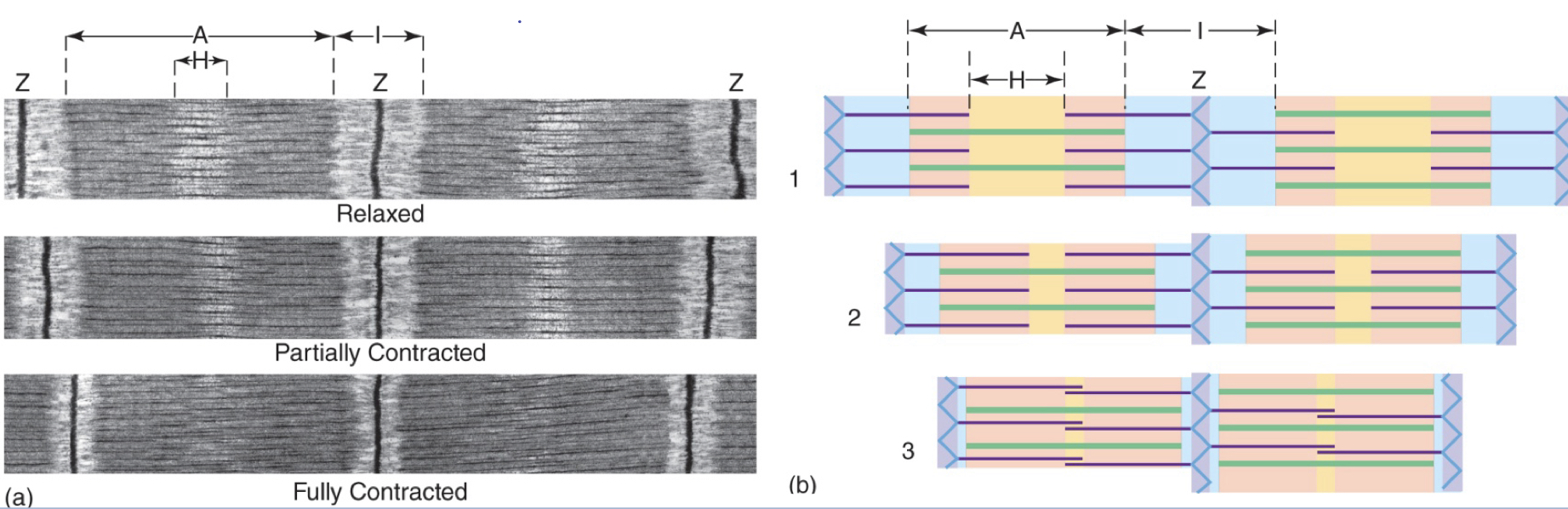
sliding filament theory
- when a muscle contracts, SARCOMERES SHORTEN; proteins overlap, they do NOT get shorter
- A bands move CLOSER together, but NO CHANGE in length of the A band
- I bands SHORTEN, and "move closer" to H band; the region of overlap increases
- H band shortens or disappears
- Z lines/discs are moving in closer together
- ONLY ACTIN AND Z-LINES MOVING IN
- A bands move CLOSER together, but NO CHANGE in length of the A band
- I bands SHORTEN, and "move closer" to H band; the region of overlap increases
- H band shortens or disappears
- Z lines/discs are moving in closer together
- ONLY ACTIN AND Z-LINES MOVING IN
27
New cards
how do muscles contract? (summary)
1) actin filaments will slide past myosin filaments
2) myosin proteins form cross-bridges with the actin filaments, linking the two chains together (and HOLDING your muscle in a CONTRACTED state)
2) myosin proteins form cross-bridges with the actin filaments, linking the two chains together (and HOLDING your muscle in a CONTRACTED state)
28
New cards
tropomyosin
protein that blocks the myosin binding sites on actin to prevent unwanted contractions
29
New cards
troponin
protein that binds actin to tropomyosin and calcium; moves tropomyosin out of the way to allow binding
30
New cards
role of calcium
1) when muscle is relaxed, tropomyosin blocks myosin binding sites
2) when muscle cells are stimulated by a signal from neurons, calcium is released inside the muscle fiber cytoplasm
3) calcium attaches to troponin, causing a shape change in troponin and tropomyosin; tropomyosin is pushed up and no longer blocking the binding sites on actin
4) myosin is allowed access to actin and forms CROSS BRIDGES
2) when muscle cells are stimulated by a signal from neurons, calcium is released inside the muscle fiber cytoplasm
3) calcium attaches to troponin, causing a shape change in troponin and tropomyosin; tropomyosin is pushed up and no longer blocking the binding sites on actin
4) myosin is allowed access to actin and forms CROSS BRIDGES
31
New cards
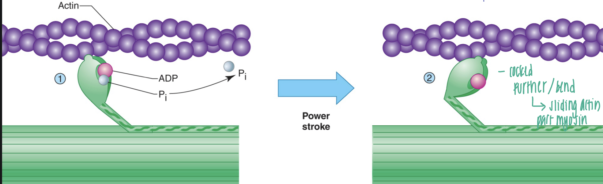
power stroke
the release of phosphate upon binding cocks the myosin head, producing a power stroke that pulls the THIN filament; keep pulling actin in one direction
32
New cards
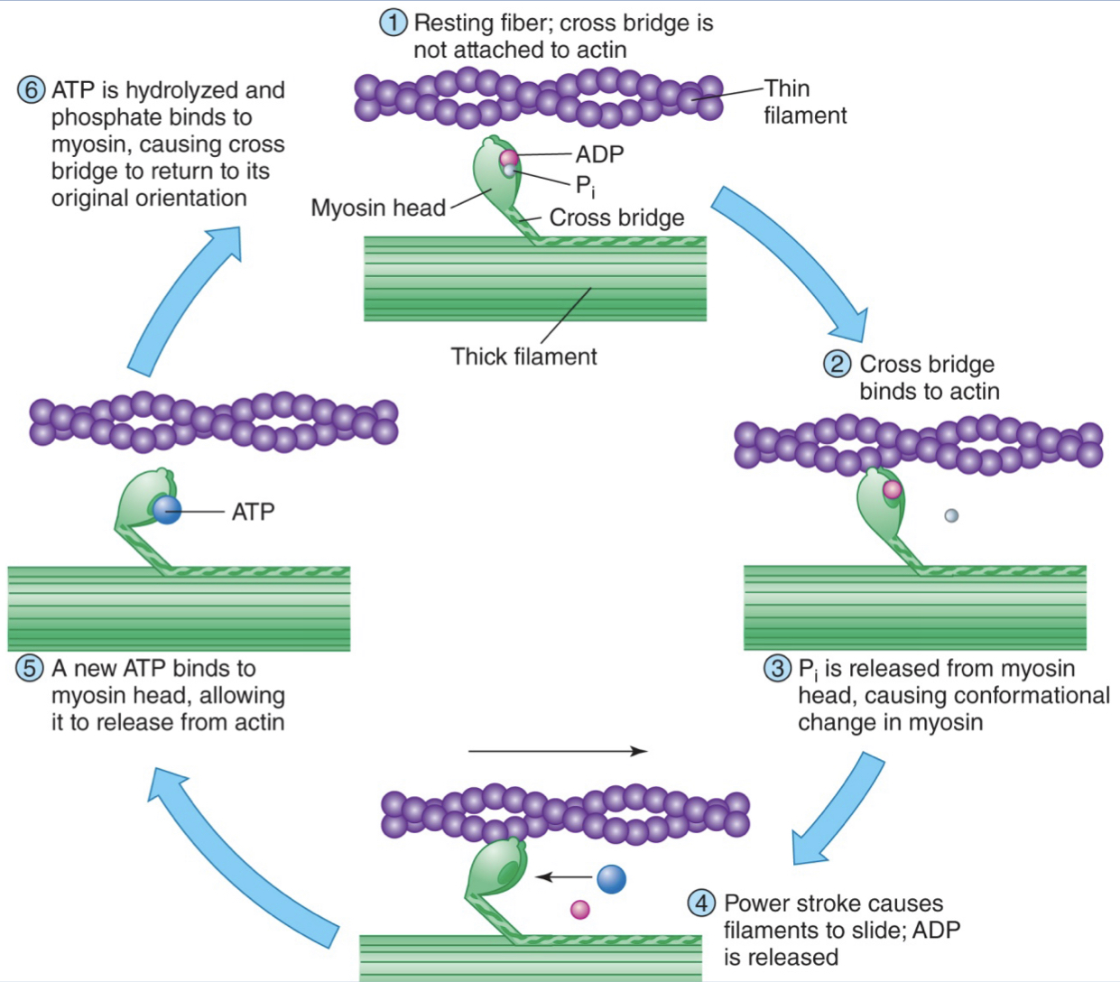
cross bridge cycling
1) resting fiber; cross bridge is not attached to actin; tropomyosin blocking binding sites
2) cross bridge binds to actin
3) Phosphate intermediate is released from myosin head, causing a shape change in myosin
4) power stroke causes filaments to slide; ADP is released; myosin head is still bound to actin
5) a new ATP bind to myosin head, allowing it to release from actin
6) ATP is hydrolyzed snd phosphate binds to myosin causing cross bridge to return to its original orientation
* process only happens if calcium is present to move tropomyosin from blocking binding sites*
2) cross bridge binds to actin
3) Phosphate intermediate is released from myosin head, causing a shape change in myosin
4) power stroke causes filaments to slide; ADP is released; myosin head is still bound to actin
5) a new ATP bind to myosin head, allowing it to release from actin
6) ATP is hydrolyzed snd phosphate binds to myosin causing cross bridge to return to its original orientation
* process only happens if calcium is present to move tropomyosin from blocking binding sites*
33
New cards
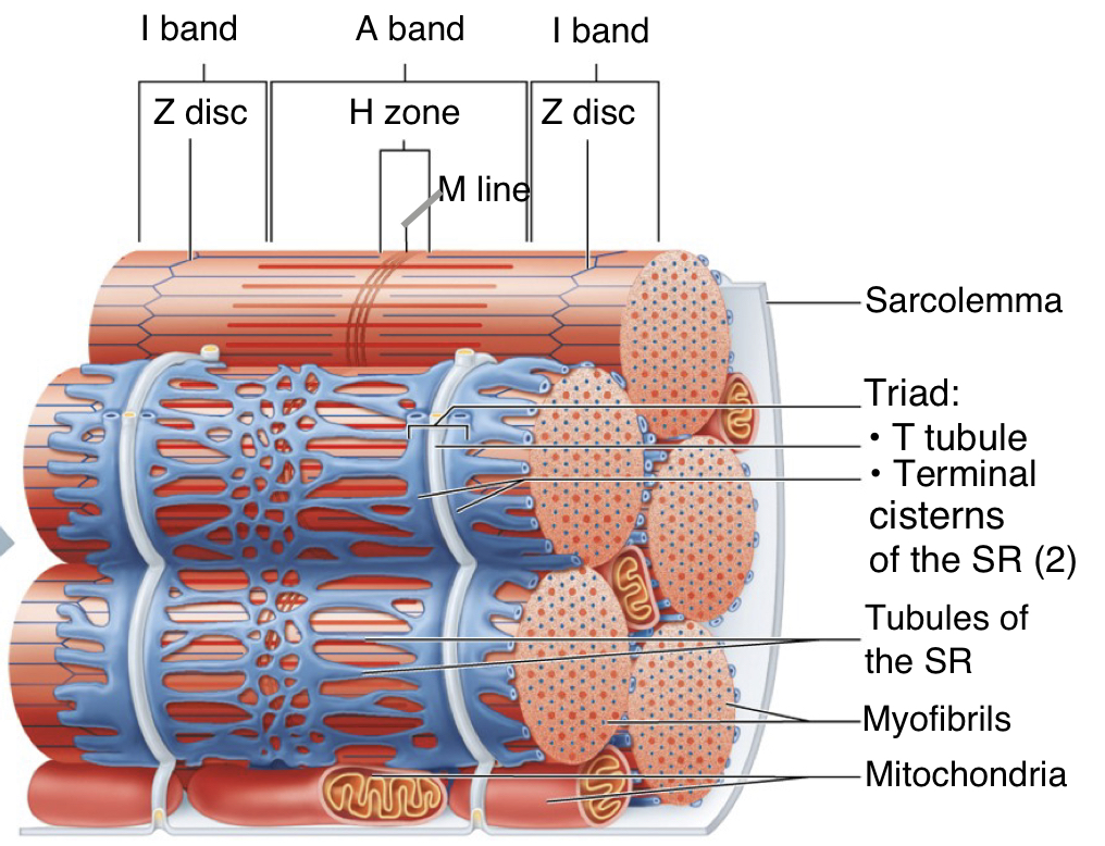
function of sarcoplasmic reticulum (SR)
calcium storage and control
34
New cards
SR calcium control
- stores calcium when muscle is at rest
- when muscle fiber is stimulated, calcium diffuses out of calcium release channels
- at the end of a contraction, calcium is actively pumped back into the SR
- when muscle fiber is stimulated, calcium diffuses out of calcium release channels
- at the end of a contraction, calcium is actively pumped back into the SR
35
New cards
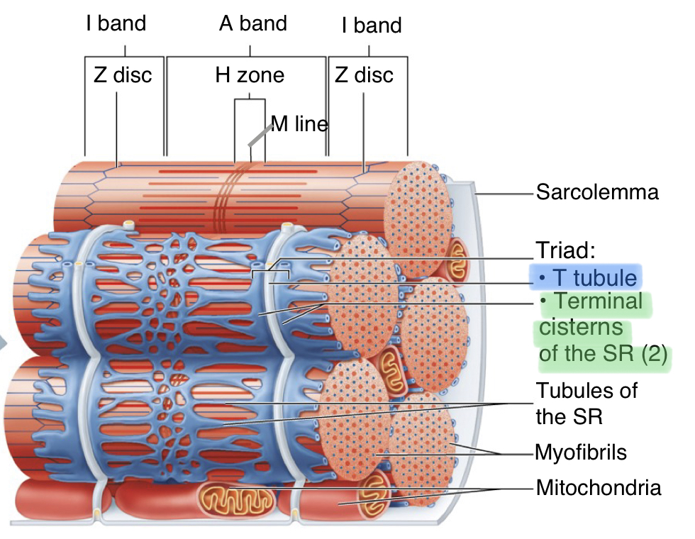
transverse tubules
- narrow membranous tunnels formed from the sarcolemma
- open to the extracellular environment
- ABLE TO CONDUCT ACTION POTENTIALS
- closely situated next to terminal cisternae of SR --> faster stimulation of SR from sarcolemma
- open to the extracellular environment
- ABLE TO CONDUCT ACTION POTENTIALS
- closely situated next to terminal cisternae of SR --> faster stimulation of SR from sarcolemma
36
New cards
stimulating a muscle fiber
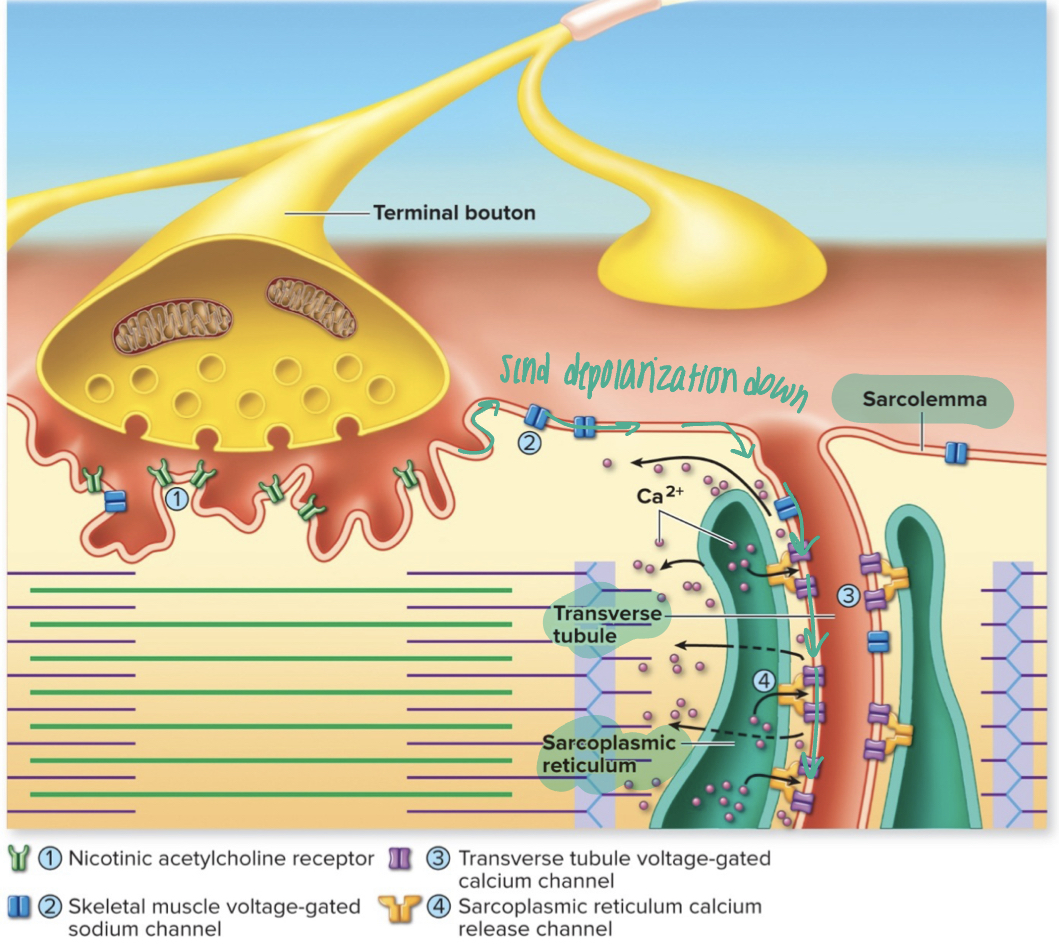
1) acetylcholine (Ach) is released from the motor neuron, binds to Ach receptors in sarcolemma, Na+ enters muscle cell through channels
2) causes local membrane depolarization of sarcolemma (end plate potential), that stimulates voltage-gated channels
3) end plate potentials are generated and conducted along transverse tubules
4) voltage-gated calcium channels (VGCaC) in transverse tubules change shape and mechanically cause calcium channels in SR to open
5) calcium is released into the cytoplasm and can bind to troponin to push tropomyosin out of the way and stimulate a muscle contraction
2) causes local membrane depolarization of sarcolemma (end plate potential), that stimulates voltage-gated channels
3) end plate potentials are generated and conducted along transverse tubules
4) voltage-gated calcium channels (VGCaC) in transverse tubules change shape and mechanically cause calcium channels in SR to open
5) calcium is released into the cytoplasm and can bind to troponin to push tropomyosin out of the way and stimulate a muscle contraction
37
New cards
relaxing a muscle fiber
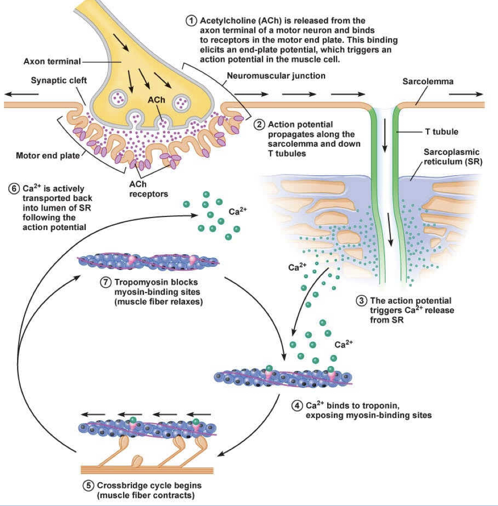
1) end plate/action potentials cease
2) calcium release channels close
3) calcium-ATPase pumps move calcium back into SR through active transport
4) no more calcium is available to bind to troponin
5) tropomyosin moves to block the myosin heads from binding to actin
2) calcium release channels close
3) calcium-ATPase pumps move calcium back into SR through active transport
4) no more calcium is available to bind to troponin
5) tropomyosin moves to block the myosin heads from binding to actin
38
New cards
why does rigor mortis occur?
- no longer making ATP, therefore the body is no longer pumping calcium into SR, allowing to stay bound to troponin to move tropomyosin out if the way for myosin to bind to actin --> contractions
- no more ATP to release myosin from actin, so the body is stuck in a contracted state
- no more ATP to release myosin from actin, so the body is stuck in a contracted state
39
New cards
myasthenia gravis
- autoimmune disorder where the body makes antibodies that improperly recognize and block Ach receptors in the sarcolemma
- result: severe muscle weakness
- treatment: therapies with reversible inhibitors of acetylcholinesterase (AChE)- enzyme that breaks down Ach
- result: severe muscle weakness
- treatment: therapies with reversible inhibitors of acetylcholinesterase (AChE)- enzyme that breaks down Ach
40
New cards
latrotoxin
- causes increased release of Ach vesicles --> increased muscle contraction
41
New cards
motor unit
- single motor neuron and all the muscle fibers it innervates
- all the muscle fibers in a motor unit contract AT ONCE
- the more innervated the motor unit, the stronger the contraction (different muscles require different amount of power- ex. eyelid vs quad)
- finer muscle control requires many smaller motor units (ex. eye lid, lots of control-neuron:muscle fiber ratio 1:4)
- larger, stronger muscles may have motor units with 1: several hundred ratio (ex. quad- many more muscle fibers contracting from one neuron- less control)
- all the muscle fibers in a motor unit contract AT ONCE
- the more innervated the motor unit, the stronger the contraction (different muscles require different amount of power- ex. eyelid vs quad)
- finer muscle control requires many smaller motor units (ex. eye lid, lots of control-neuron:muscle fiber ratio 1:4)
- larger, stronger muscles may have motor units with 1: several hundred ratio (ex. quad- many more muscle fibers contracting from one neuron- less control)
42
New cards
graded contractions
- varied contraction strength of overall muscle due to DIFFERENT numbers of motor units being stimulated
- add up based on load
- add up based on load
43
New cards
neuromuscular junction
- site where a motor neuron stimulates a muscle fiber
- one motor unit has many neuromuscular junctions
- one motor unit has many neuromuscular junctions
44
New cards
motor end plate
- area of the muscle fiber sarcolemma where a motor neuron stimulates it using the NT Ach
45
New cards
further contracting strength comes from...
1) increased FREQUENCY of stimulation: increases contraction strength of a motor unit (fire more rapidly)
2) motor unit recruitment: if a stronger contraction is needed, MORE motor units will fire to cause more cells to contract
2) motor unit recruitment: if a stronger contraction is needed, MORE motor units will fire to cause more cells to contract
46
New cards
twitch
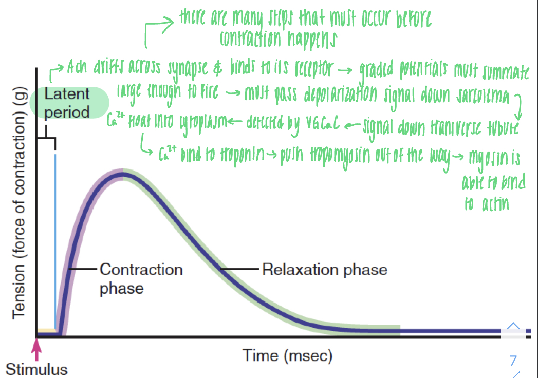
- a stimulation of a muscle fiber/cell
- small, single stimulation
- ONE muscle fiber is a part of only ONE motor unit
- has a latent period after stimulus and before contraction phase
- small, single stimulation
- ONE muscle fiber is a part of only ONE motor unit
- has a latent period after stimulus and before contraction phase
47
New cards
latent phase
- time between action potential and contraction
- occurs bc there are many steps that must occur before contraction happens
- action potential always releases same amount of calcium, therefore there is the same amount of contraction or strength
- occurs bc there are many steps that must occur before contraction happens
- action potential always releases same amount of calcium, therefore there is the same amount of contraction or strength
48
New cards
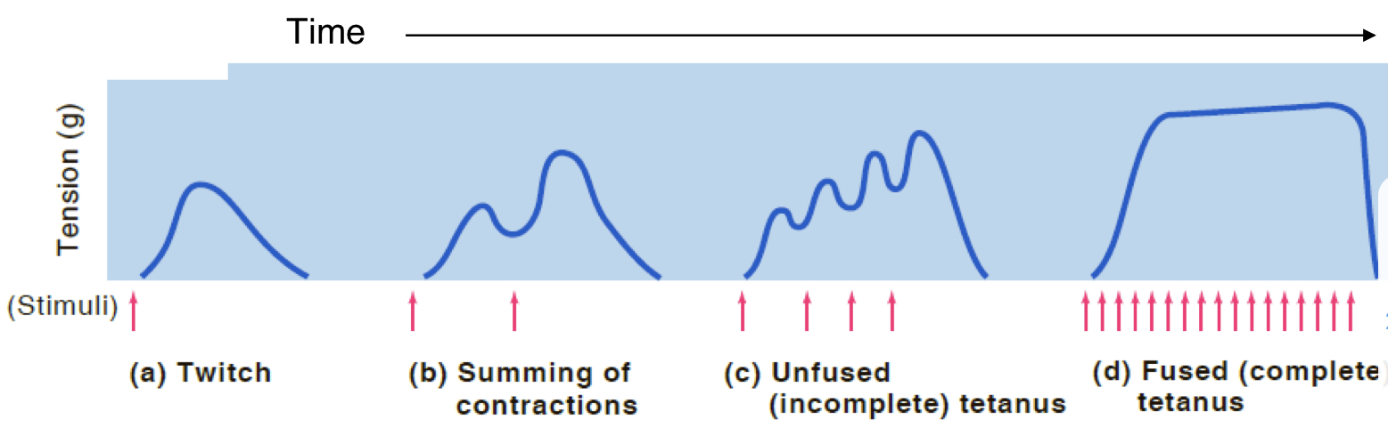
muscle contractions
1) twitch: small, single stimulation
2) summation: twitch + twitch (before relaxation)
3) unfused (incomplete) tetanus: summation of many twitches
4) fused (complete) tetanus: stimulation with no relaxation- max amount of force your muscles can provide; all the muscle fibers are contracted to their full length (myosin and actin have slid their max)
2) summation: twitch + twitch (before relaxation)
3) unfused (incomplete) tetanus: summation of many twitches
4) fused (complete) tetanus: stimulation with no relaxation- max amount of force your muscles can provide; all the muscle fibers are contracted to their full length (myosin and actin have slid their max)
49
New cards
motor unit recruitment
- asynchronous activation of motor units: some motor units START to twitch when others RELAX
- produces continuous contraction of the whole muscle
recruitment of additional motor units makes muscle contractions stronger
- certain motor units are preferred (small forces = small motor units; large forces = large motor units)
- produces continuous contraction of the whole muscle
recruitment of additional motor units makes muscle contractions stronger
- certain motor units are preferred (small forces = small motor units; large forces = large motor units)
50
New cards
are both temporal and spatial summation possible with skeletal muscle cells?
- we see temporal (being close to temporal) but do not really see spatial
- to achieve similar outcome to spatial in skeletal muscle cells is motor unit recruitment
- to achieve similar outcome to spatial in skeletal muscle cells is motor unit recruitment
51
New cards
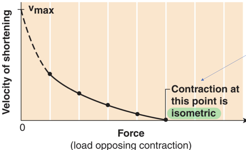
velocity and force of muscle contractions
- for muscle to contract, they must generate force that is GREATER than the opposing forces
- the greater the FORCE, the SLOWER the contraction
- the greater the FORCE, the SLOWER the contraction
52
New cards
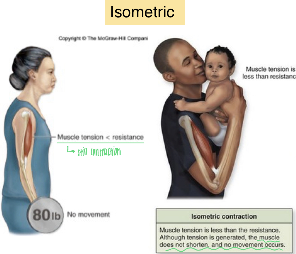
isometric contraction
-muscle is working, but holding a pose (planks or yoga)
- muscle does NOT change length
- muscle tension is LESS than resistance
- muscle can't shorten because the load is too great
- still contracting but no movement occurs
- muscle does NOT change length
- muscle tension is LESS than resistance
- muscle can't shorten because the load is too great
- still contracting but no movement occurs
53
New cards
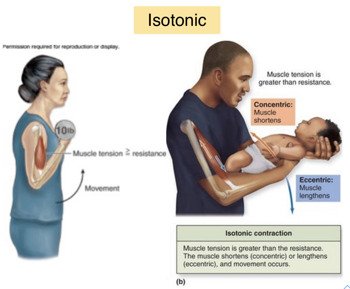
isotonic contraction
- muscle is working and movement occurs
- muscle changes length (shortens or lengthens)
- amount of power stays the same
- muscle tension is greater than resistance
- muscle fibers SHORTEN when the TENSION produced is just GREATER than the load
- muscle changes length (shortens or lengthens)
- amount of power stays the same
- muscle tension is greater than resistance
- muscle fibers SHORTEN when the TENSION produced is just GREATER than the load
54
New cards
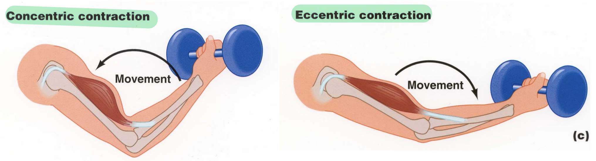
types of isotonic contractions
- concentric contraction: muscle fiber SHORTENS when force is GREATER than load
- eccentric contraction: muscle may actually LENGTHEN, despite contraction, if the load is too great ( allow you to lower a weight gently after full contraction)
- eccentric contraction: muscle may actually LENGTHEN, despite contraction, if the load is too great ( allow you to lower a weight gently after full contraction)
55
New cards
factors that increase the force of skeletal muscle contraction
1) high frequency of stimulation (temporal summation and tetanus)
2) large number of muscle fibers recruited
3) large muscle fibers: if have larger fibers, you have more (contractile) myofilaments, and therefore have more sliding filaments
4) muscle and sarcomere stretched to slightly over 100% of resting length (length-tension relationship)
2) large number of muscle fibers recruited
3) large muscle fibers: if have larger fibers, you have more (contractile) myofilaments, and therefore have more sliding filaments
4) muscle and sarcomere stretched to slightly over 100% of resting length (length-tension relationship)
56
New cards
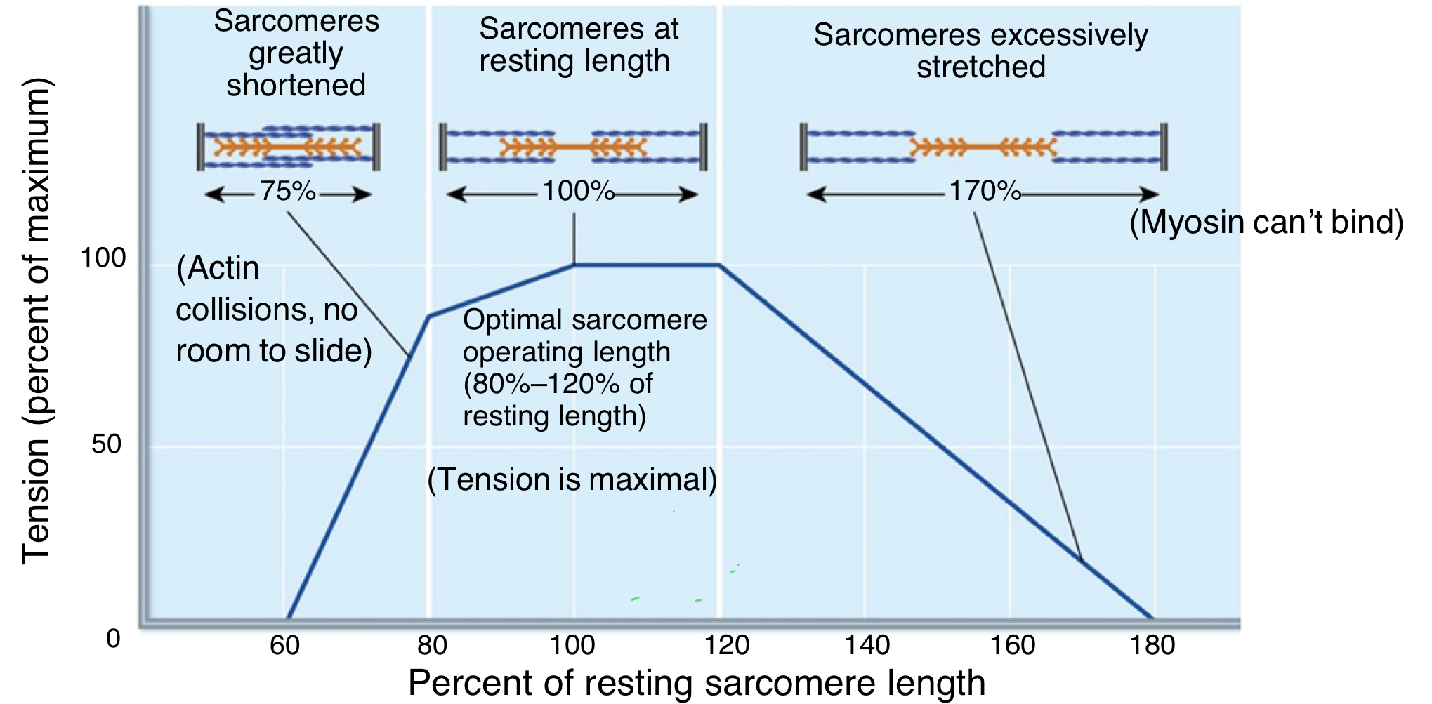
length-tension relationship
- there is an ideal length of the sarcomere, where the length is just long enough to support the correct amount of muscle tension for contraction to occur
- sarcomeres at resting length: optimal sarcomere operating length is 80-120% of resting length; tension is maximal (amount of actin and myosin overlap that is desired- give a little more contraction and generate more tension)
- sarcomeres greatly shortened: actin collisions, no room to slide (already so close to z-lines)
- sarcomeres excessively stretched: myosin can't bind to actin bc too far apart
- sarcomeres at resting length: optimal sarcomere operating length is 80-120% of resting length; tension is maximal (amount of actin and myosin overlap that is desired- give a little more contraction and generate more tension)
- sarcomeres greatly shortened: actin collisions, no room to slide (already so close to z-lines)
- sarcomeres excessively stretched: myosin can't bind to actin bc too far apart
57
New cards
what affects how fast muscles can contract and how long they can continue to contract until failure?
muscle type, load, and recruitment
58
New cards
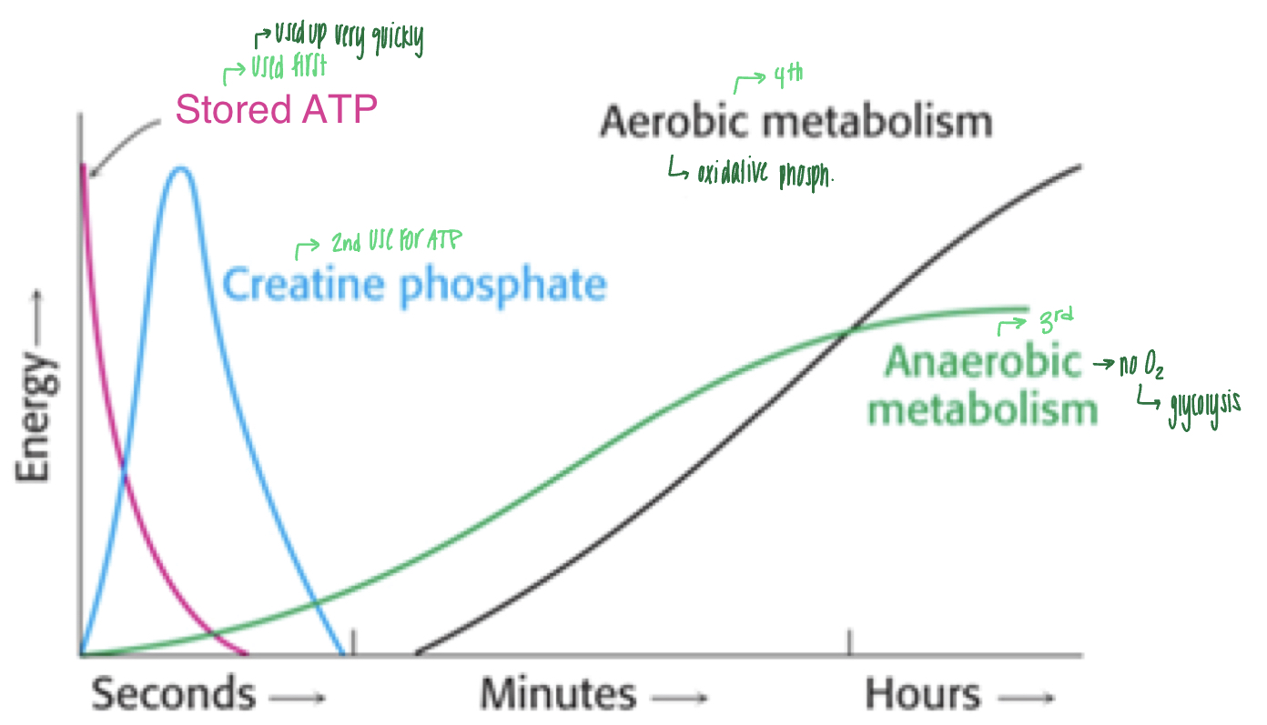
metabolism and muscle contractions
- muscle cells use energy from various sources over time
1) stored ATP: used up very quickly (within seconds)
2) creatine phosphate ( within seconds)
3) anaerobic metabolism: doesn't require oxygen and uses glycolysis for ATP production (within minutes)
4) aerobic metabolism: oxidative phosphorylation (within minutes or hours)
1) stored ATP: used up very quickly (within seconds)
2) creatine phosphate ( within seconds)
3) anaerobic metabolism: doesn't require oxygen and uses glycolysis for ATP production (within minutes)
4) aerobic metabolism: oxidative phosphorylation (within minutes or hours)
59
New cards
types of skeletal muscle fibers
slow twitch (1) and fast twitch (2)
60
New cards
slow twitch fiber
- slow oxidative fibers
- slowest velocity of contraction; long distance runner
- go through krebs cycle and ETC to create ATP, therefore takes longer
- slow bc need ATP for every myosin to release from actin
- ex. soleus muscle
- slowest velocity of contraction; long distance runner
- go through krebs cycle and ETC to create ATP, therefore takes longer
- slow bc need ATP for every myosin to release from actin
- ex. soleus muscle
61
New cards
fast twitch fibers
1) fast oxidative fibers: middle ground, mixed oxidative and glycolytic production, middle distance runner
2) fast glycolytic fibers: fastest velocity of contraction, use of glycolysis for ATP production, ex. eyelid, sprinter
2) fast glycolytic fibers: fastest velocity of contraction, use of glycolysis for ATP production, ex. eyelid, sprinter
62
New cards
what determines the cross bridge cycle rate?
- the rate of ATP production: if can make ATP faster, can go through cross bridge cycle faster
63
New cards
glycolytic vs oxidative muscle fibers
glycolytic: ATP production through glycolysis, few mitochondria, large fiber diameter, small capillary supply (not as reliant on oxygen), few myoglobin (anaerobic), fast fatigue rate (lactic acid build up)
oxidative: ATP production through oxidative phosphorylation in mitochondria, many mitochondria, small fiber diameter, large capillary supply (relies on presence of oxygen), many myoglobin (aerobic), slow fatigue rate (lactic acid build up)
oxidative: ATP production through oxidative phosphorylation in mitochondria, many mitochondria, small fiber diameter, large capillary supply (relies on presence of oxygen), many myoglobin (aerobic), slow fatigue rate (lactic acid build up)
64
New cards
inactivation of which protein(s) in a myofiber would result in an increased muscle contraction?
acetylcholinesterase, tropomyosin, calcium- ATPase pump
65
New cards
which protein provides elasticity to allow a sarcomere to recover from a contraction?
titin
66
New cards
Which part(s) of a sarcomere would you expect to shrink or disappear during a contraction?
H bands and I bands
67
New cards
Which step of the cross-bridge cycle allows for the filaments to slide past one another during a contraction?
myosin releasing Pi
68
New cards
Which of these events take place during stimulation of both neurons and muscle cells?
-opening of VGKC
- opening of VGNaC
- opening of VGCaC
- release of Ach from synaptic vesicles
- opening of VGNaC
- opening of VGCaC
- release of Ach from synaptic vesicles
69
New cards
true or false: The calcium that binds troponin and the calcium that stimulates synaptic vesicle release come from the same intracellular source.
false