Upper Respiratory Passageways: Comparative Anat. and Structure/Function Considerations (Week 1, Mod 9)

0.0(0)
studied byStudied by 0 people
0.0(0)
full-widthCall Kai
learnLearn
examPractice Test
spaced repetitionSpaced Repetition
heart puzzleMatch
flashcardsFlashcards
GameKnowt Play
Card Sorting

1/13

encourage image

There's no tags or description

Looks like no tags are added yet.

Study Analytics
Name
Mastery
Learn
Test
Matching
Spaced

No study sessions yet.

14 Terms

1
New cards

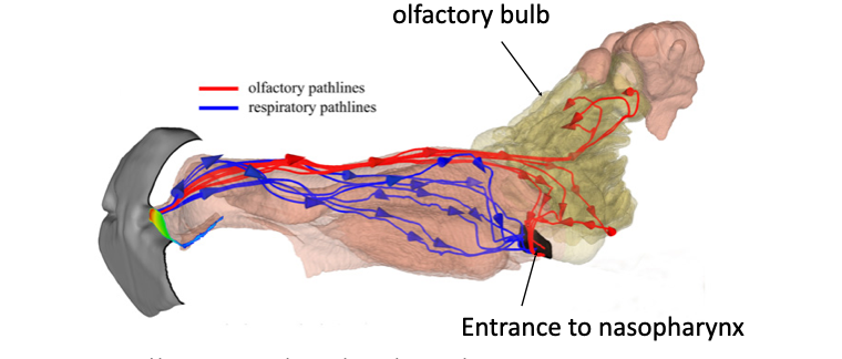
There are 2 nasal passageways… how do they modify and regulate the volume of inspired air? (hint: one does most of this, the other is just for smell)

Respiratory pathway (blue) - respiratory epithelium

  • Warms the air via rich blood supply of nasal lining

    • Protects lower respiratory tracts

  • Moistens the air- glandular secretions → mucus

    • Goblet cells and glands produce mucus that adds moisture to inspired air

  • Cleanses the air- mucus and cilia

    • Mucus traps dust and particles

    • Cilia move the mucus toward the pharynx to be swallowed or removed

  • Regulates airflow- blood vessel congestion or constriction

    • Blood vessels can swell (CONGESTION) or constrict, altering airflow

      • EX: constrict during exercise to increase air flow

Olfactory pathway (red) - olfactory epithelium

  • Runs alongside the respiratory pathway and is responsible for detecting smells

<p><strong><span>Respiratory pathway (blue)&nbsp;- respiratory epithelium </span></strong></p><ul><li><p><span>Warms the air via rich blood supply of nasal lining</span></p><ul><li><p>Protects lower respiratory tracts</p></li></ul></li><li><p><span>Moistens the air- glandular secretions → mucus</span></p><ul><li><p>Goblet cells and glands produce mucus that adds moisture to inspired air</p></li></ul></li><li><p><span>Cleanses the air- mucus and cilia</span></p><ul><li><p>Mucus traps dust and particles</p></li><li><p>Cilia move the mucus toward the pharynx to be swallowed or removed</p></li></ul></li><li><p><span>Regulates airflow- blood vessel congestion or constriction</span></p><ul><li><p>Blood vessels can swell (CONGESTION) or constrict, altering airflow</p><ul><li><p>EX: constrict during exercise to increase air flow</p></li></ul></li></ul></li></ul><p></p><p><strong>Olfactory pathway (red) - olfactory epithelium </strong></p><ul><li><p>Runs alongside the respiratory pathway and is responsible for detecting smells</p></li></ul><p></p>
2
New cards

Where in the respiratory passageways is it NOT lined by respiratory epithelium?

Ethmoidal and middle conchae

Vomeronasal gland

3
New cards

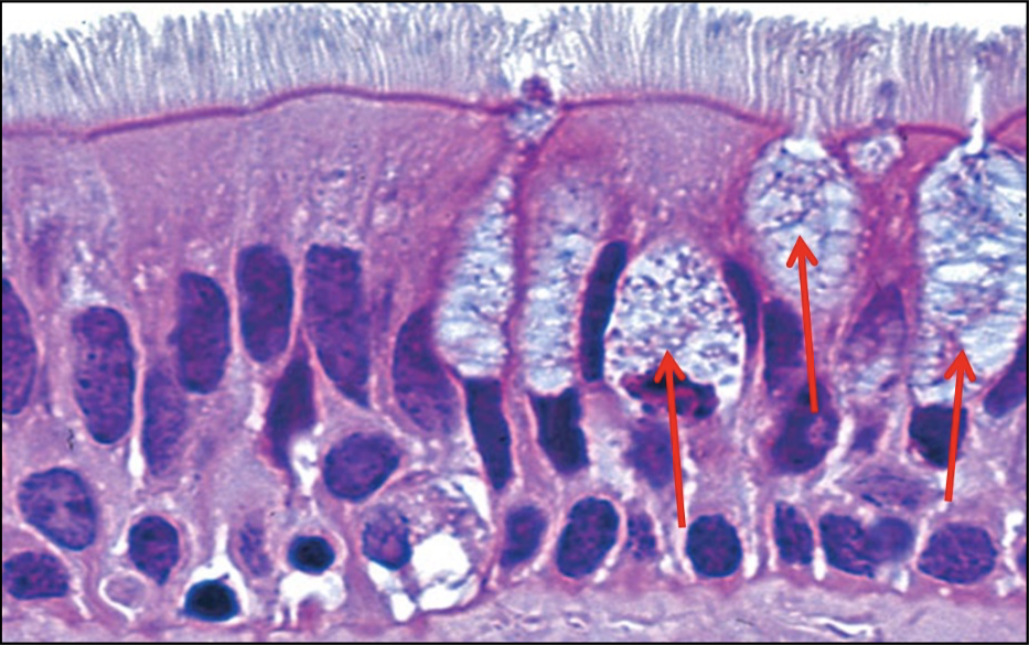
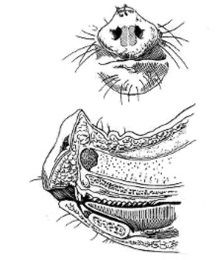
What is the histological classification for “respiratory” epithelium, and how does its structure relate to its function?

Histological classification: Pseudostratified columnar ciliated epithelium with goblet cells

  • 5:1 ratio of epithelial cells to goblet cells

Function: “air conditioning”

  • Mucus sheet 

    • Produced by goblet cells and sub-mucosal glands

    • Moistens air 

    • Traps particulate matter

  • Cilla

    • Motile- beat in concert

    • Mucus and trapped particulate is wafted towards pharynx  where it is swallowed or expectorated

  • Submucosa is also highly vascular (bvs)

    • Warms the incoming air

    • Acts like an erectile tissue

      • Congestion- disrupts airflow

      • Vasoconstriction- allows increased airflow during exercise

<p>Histological classification: <strong><em><span>Pseudostratified columnar ciliated epithelium with goblet cells</span></em></strong></p><ul><li><p>5:1 ratio of epithelial cells to goblet cells</p></li></ul><p></p><p><strong>Function: “</strong>air conditioning”</p><ul><li><p class="p2"><strong><span>Mucus sheet</span></strong><span>&nbsp;</span></p><ul><li><p><span>Produced by goblet cells and sub-mucosal glands</span></p></li><li><p><span>Moistens air&nbsp;</span></p></li><li><p><strong><span>Traps particulate matter</span></strong></p></li></ul></li><li><p class="p2"><strong><span>Cilla</span></strong></p><ul><li><p><strong><span>Motile- beat in concert</span></strong></p></li><li><p><span>Mucus and trapped particulate is wafted towards pharynx&nbsp; where it is swallowed or expectorated</span></p></li></ul></li><li><p><strong><span>Submucosa is also highly vascular (bvs)</span></strong></p><ul><li><p><span>Warms the incoming air</span></p></li><li><p><span>Acts like an erectile&nbsp;tissue</span></p><ul><li><p><span>Congestion- disrupts airflow</span></p></li><li><p><span>Vasoconstriction- allows increased airflow during exercise</span></p></li></ul></li></ul></li></ul><p></p>
4
New cards

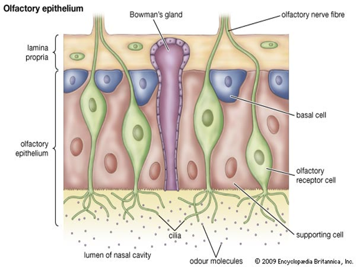
What are the 4 main cell types found in olfactory epithelium? Describe their structure and function.

Olfactory receptor cells (green)

  • Bipolar neurons - apical pole carries a non motile cilia with specific odorant binding receptors

  • Unite to form an olfactory  nerve which passes through the cribriform plate to terminate in olfactory bulb 

Bowman’s gland (purple)

  • secretion solubilizes incoming odorants and washes away excess

Supporting cells (red)

  • Provide metabolic and physical support to olfactory cells

Basal cells (blue)

  • stem cells- can differentiate and replace olfactory receptor cells

<p><strong><span>Olfactory receptor cells (green)</span></strong></p><ul><li><p><span>Bipolar neurons - apical pole carries a non motile cilia with specific odorant binding receptors</span></p></li><li><p><span>Unite to form an olfactory&nbsp; nerve which passes through the cribriform plate to terminate in olfactory bulb&nbsp;</span></p></li></ul><p class="p1"><strong><span>Bowman’s gland (purple)</span></strong></p><ul><li><p><span>secretion solubilizes incoming odorants and washes away excess</span></p></li></ul><p class="p1"><strong><span>Supporting cells (red)</span></strong></p><ul><li><p><span>Provide metabolic and physical support to olfactory cells</span></p></li></ul><p class="p1"><strong><span>Basal cells (blue)</span></strong></p><ul><li><p><span>stem cells- can differentiate and replace olfactory receptor cells</span></p></li></ul><p></p>
5
New cards

How many olfactory receptors do canines have in their nose? How does the structure of their nose overall enhance their smell?

Dog’s sense of smell is their PRIMARY sense

  • Have 300 million olfactory receptors

  • Olfactory bulb also 40 times larger than a human; means dogs can distinguish and remember more smells than we can

Structure for smell:

  • Have a wet nose, that helps them determine the direction of the air current containing the smell. 

  • Have slits in their noses that allow them to exhale air separately from the inhaled air, unlike humans who use the same passageways for both. 

    • This means that dogs can keep the scent molecules in their noses longer and avoid washing them out with the exhaled air.

6
New cards

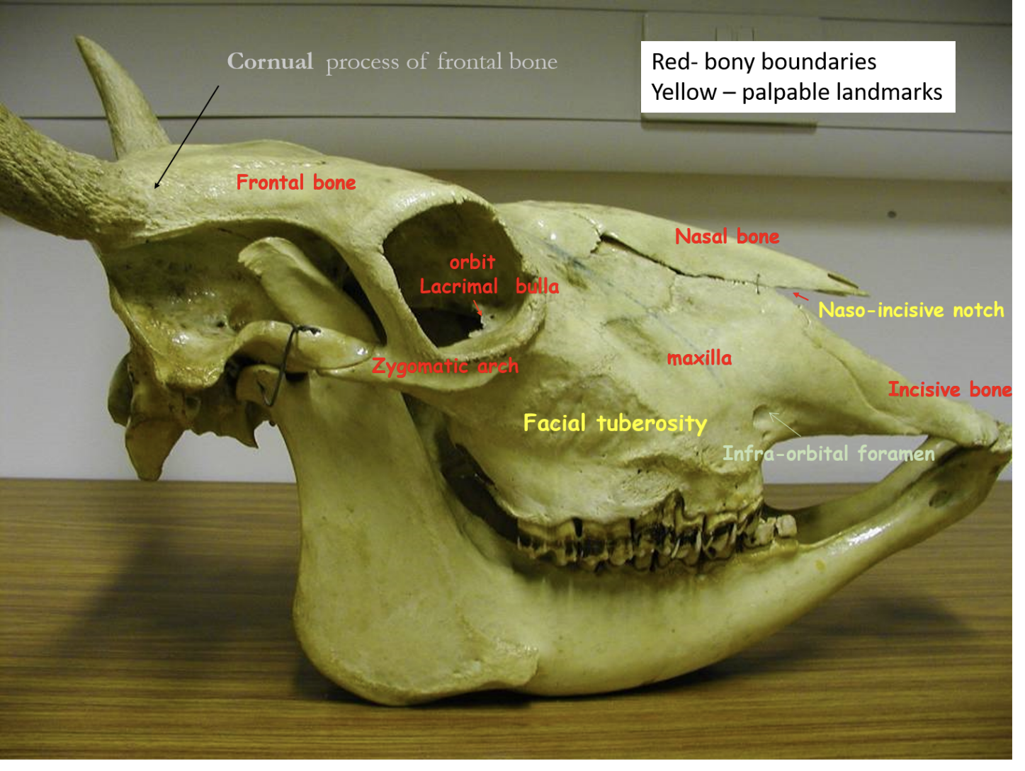
What are the palpable bony landmarks on the bovine skull?

Naso-incisive notch

Facial tuberosity

Cornual process of frontal bone (right by horn)

<p>Naso-incisive notch</p><p>Facial tuberosity </p><p>Cornual process of frontal bone (right by horn)</p>
7
New cards

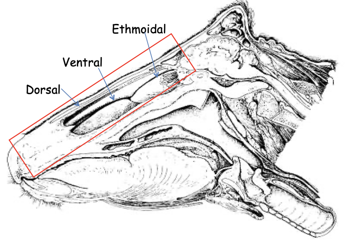
How are the nasal cavities of the ruminant different from that of the carnivore?

Conchae are LESS COMPLEX compared to the carnivore, less scrolled (doesn’t look like a wrinkled prune)

  • Has dorsal, ventral, and ethmoidal conchae… middle conchae is VERY small, not significant

  • Dorsal and ventral conchae are both scrolled ROSTRALLY, but form a sinus CAUDALLY

<p>Conchae are LESS COMPLEX compared to the carnivore, less scrolled (doesn’t look like a wrinkled prune) </p><ul><li><p>Has dorsal, ventral, and ethmoidal conchae… middle conchae is VERY small, not significant </p></li><li><p>Dorsal and ventral conchae are both scrolled ROSTRALLY, but form a sinus CAUDALLY</p></li></ul><p></p>
8
New cards

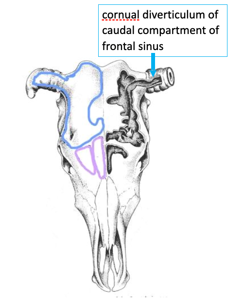
How are the frontal sinuses of the LARGE ruminant different from that of the carnivore? Structurally and functionally?

  • Frontal sinuses are extensive in adult

    • Give rise to adult head conformation 

    • Each frontal sinus consists of Rostral & Caudal compartments

    • Invade the cornual process @6 months (has implications for dehorning & fracture)

      • The cornual diverticulum is the part of the CAUDAL COMPARTMENT of the FRONTAL SINUS that extends into the horn (see image)

      • This is why disbudding is recommended instead; performed BEFORE the frontal sinus invades cornual process

FUNCTION:

  • Drainage : 

    • Poor; drain directly into nasal cavity via Ethmoidal meatuses, worse when mucosa inflamed

  • Protection:

    • Protects the cranial cavity… is a target for euthanasia in cattle

<ul><li><p><strong><span>Frontal sinuses </span></strong><span>are extensive in adult</span></p><ul><li><p><span>Give rise to adult head conformation&nbsp;</span></p></li><li><p><span>Each frontal sinus consists of Rostral &amp; Caudal compartments</span></p></li><li><p><span>Invade the </span><strong><span>cornual process </span></strong><span>@6 months (has implications for dehorning &amp; fracture)</span></p><ul><li><p>The <strong>cornual diverticulum </strong>is the part of the CAUDAL COMPARTMENT of the FRONTAL SINUS that extends into the horn (see image)</p></li><li><p>This is why disbudding is recommended instead; performed BEFORE the frontal sinus invades cornual process</p></li></ul></li></ul></li></ul><p></p><p><strong>FUNCTION:</strong></p><ul><li><p><strong><span>Drainage :&nbsp;</span></strong></p><ul><li><p><span>Poor; drain directly into nasal cavity via Ethmoidal meatuses, worse when mucosa inflamed</span></p></li></ul></li><li><p><strong>Protection:</strong></p><ul><li><p>Protects the cranial cavity… is a target for euthanasia in cattle </p></li></ul></li></ul><p></p>
9
New cards

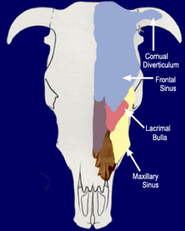
Describe the maxillary sinus in ruminants… why is it so important compared to the maxillary recess of the carnivore? What are 2 factors of clinical significance of the maxillary sinus?

Is a large sinus of major clinical importance

  • Extends medially to form the palatine sinus and caudally to form the lacrimal bulla, which protrudes into the orbit

  • Communicates with the nasal cavity via the nasomaxillary opening*, positioned dorsal to the main sinus compartment

  • Clinical significance: sinus infections are difficult to drain because the drainage opening* is high (dorsal) relative to the sinus floor (1)

    • Roots of the upper cheek teeth project into the ventral floor (Sinus enlarges with age, increasing dental association)… so the cheek teeth are WITHIN the maxillary sinus (2)

      • Dental disease is the the MOST FREQUENT cause of secondary sinusitis

So basically, maxillary sinus can become infected by dental disease, and the infection won’t be able to drain due to the position of the nasomaxillary opening

<p><span>Is a large sinus of major </span><strong><span>clinical</span></strong><span> importance</span></p><ul><li><p><span>Extends medially to form the </span><strong><span>palatine sinus</span></strong><span> and caudally to form the</span><strong><span> lacrimal bulla</span></strong><span>, which protrudes into the orbit</span></p></li><li><p><span>Communicates with the nasal cavity via the </span><strong><span>nasomaxillary opening*</span></strong><span>, positioned dorsal to the main sinus compartment</span></p></li><li><p><strong><span>Clinical significance:</span></strong><span> sinus infections are difficult to drain because the drainage opening* is high (dorsal) relative to the sinus floor (1)</span></p><ul><li><p><span>Roots of the upper cheek teeth project into the ventral floor (Sinus enlarges with age, increasing dental association)… so the cheek teeth are WITHIN the maxillary sinus (2)</span></p><ul><li><p>Dental disease is the the MOST FREQUENT cause of secondary sinusitis</p></li></ul></li></ul></li></ul><p></p><p>So basically, maxillary sinus can become infected by dental disease, and the infection won’t be able to drain due to the position of the nasomaxillary opening </p><p></p>
10
New cards

Describe the nasopharynx of the ruminant… what are 2 features of the nasopharynx that are unique to ruminants?

Contains auditory openings and the pharyngeal tonsil like carnivores, but ALSO has:

  • Pharyngeal septum - partially divides nasopharynx; is where the pharyngeal tonsil is located

  • Tubal tonsil - found close to the entrance of both auditory tubes

11
New cards

What is Oestrus ovis, and how does it effect the frontal sinus of small ruminants?

Oestrus ovis → Adult Sheep Nasal Fly / Sheep Gad Fly

  • Cause NOSE BOT in their larval stage

    • Deposited by adult fly in or around the nostrils of the host. 

      • Larvae migrate into the nasal cavity and the frontal and maxillary sinuses, where they feed on the mucous membrane and grow for several weeks or months

      • When mature, the larvae are expelled by sneezing and pupate in the soil.

12
New cards

How is the pig nose different from other species? What do they have that others don’t?

Thickened skin over rostral plate

  • Strengthens the snout region

  • Allows for rooting behavior

Also has rostral bones UNDERNEATH the plate, strengthening it

<p>Thickened skin over rostral plate </p><ul><li><p>Strengthens the snout region</p></li><li><p>Allows for rooting behavior</p></li></ul><p></p><p>Also has <strong>rostral bones </strong>UNDERNEATH the plate, strengthening it </p><p></p>
13
New cards

Describe the frontal sinuses of the porcine…

Frontal sinuses

  • Excavates entire dorsal surface of the skull

  • Cranial cavity 5cm below skull

  • Difficult to stun reliably and humanely by mechanical bolt

  • Maxillary sinus also extensive

14
New cards

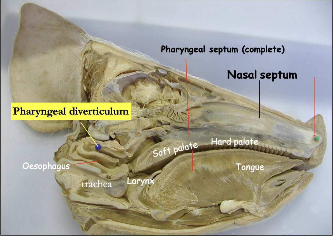
What is the pharyngeal diverticulum, and what species is it ONLY found in?

ONLY FOUND IN PIGS

  • Is a pouch-like structure that extends from the roof of the nasopharynx

    • Most prominent in piglets

Functions:

  • Supporting the nasopharyngeal tonsil (immunity for resp. system)

  • Helps to create negative pressure to allow auditory tubes to open

CAN BE AN ISSUE WHEN TUBING PIGLETS

  • Tube goes there instead of esophagus… leads to meds/nutrients not getting to piglet AND aspiration

  • Can become ulcerated / damaged, leading to secondary infections of the resp. system

<p>ONLY FOUND IN PIGS</p><ul><li><p>Is a pouch-like structure that extends from the roof of the nasopharynx</p><ul><li><p>Most prominent in piglets</p></li></ul></li></ul><p></p><p>Functions: </p><ul><li><p>Supporting the nasopharyngeal tonsil (immunity for resp. system)</p></li><li><p>Helps to create negative pressure to allow auditory tubes to open</p></li></ul><p></p><p>CAN BE AN ISSUE WHEN TUBING PIGLETS </p><ul><li><p>Tube goes there instead of esophagus… leads to meds/nutrients not getting to piglet AND aspiration </p></li><li><p>Can become ulcerated / damaged, leading to secondary infections of the resp. system</p></li></ul><p></p>