Special Senses
1/47
There's no tags or description
Looks like no tags are added yet.
Name | Mastery | Learn | Test | Matching | Spaced | Call with Kai |
|---|
No analytics yet
Send a link to your students to track their progress
48 Terms
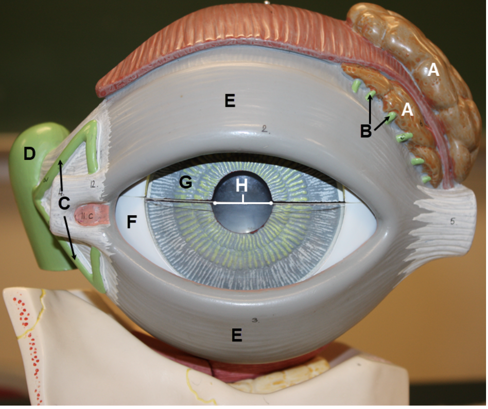
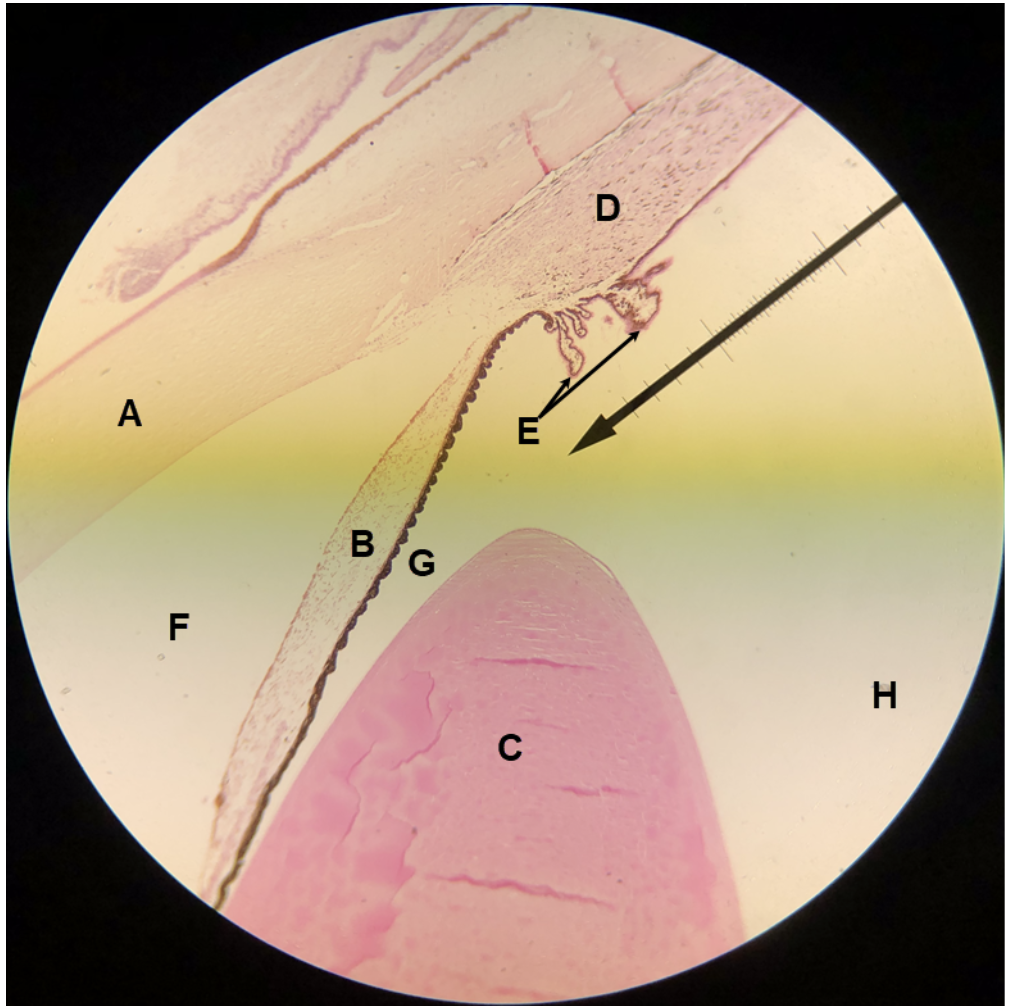
In the image above, identify structures A-G, and opening H.
A. lacrimal gland
B. lacrimal gland ducts
C. lacrimal canaliculi
D. lacrimal sac
E. eyelids (palpebrae)
F. sclera
G. iris
H. pupil
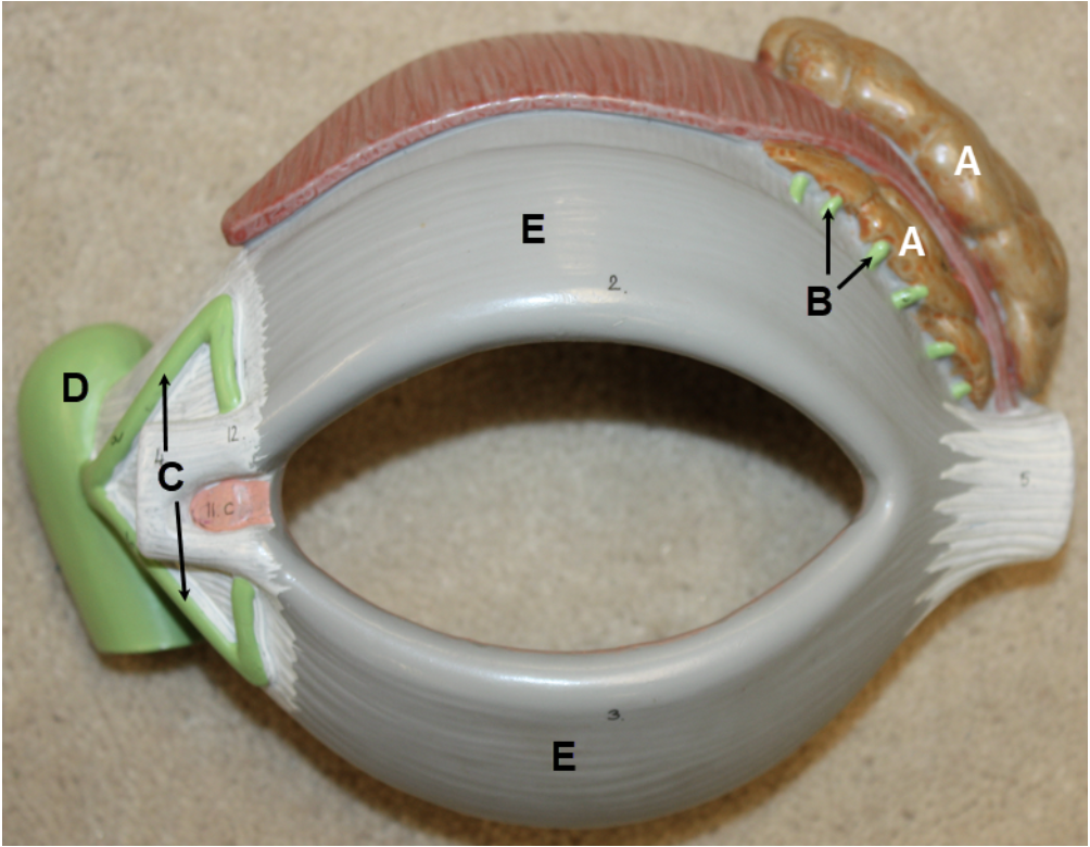
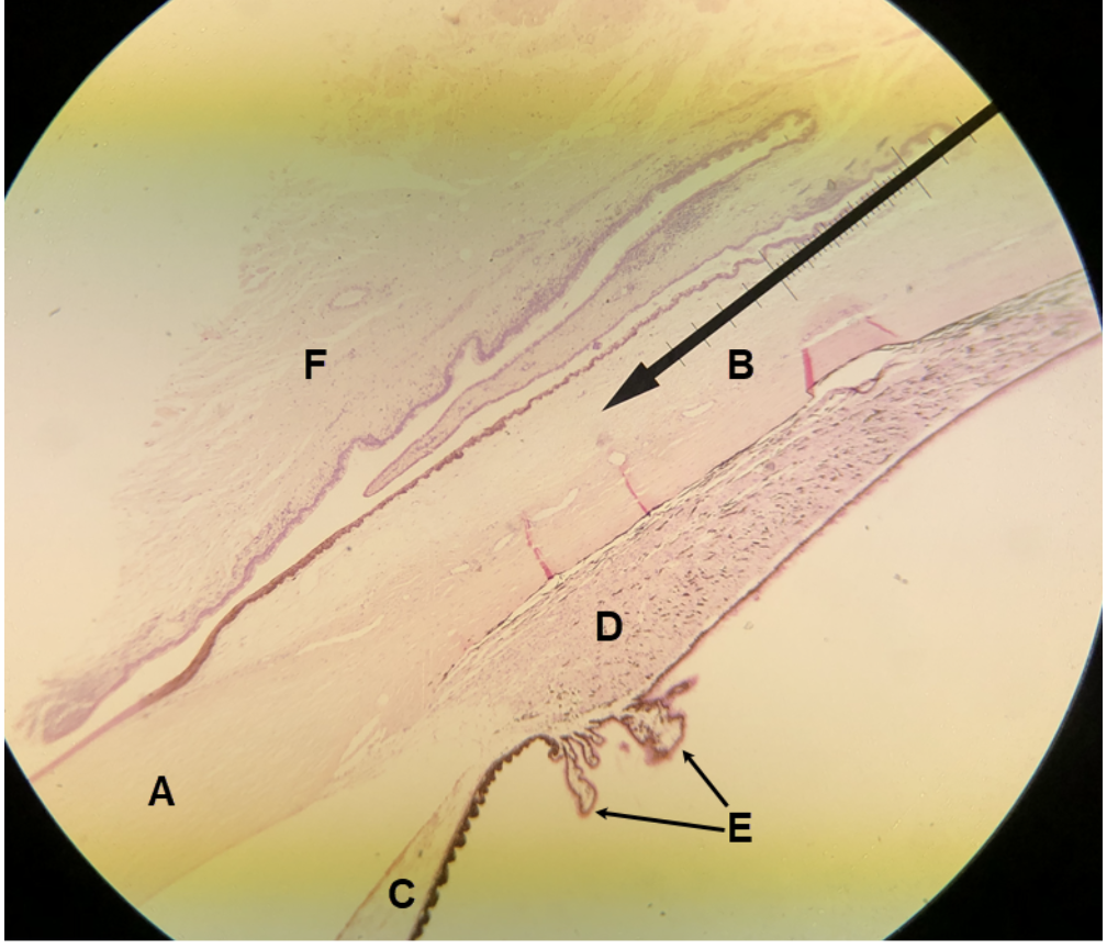
In the image above, identify structures A-E.
A. lacrimal gland
B. lacrimal gland ducts
C. lacrimal canaliculi
D. lacrimal sac
E. eyelids (palpebrae)
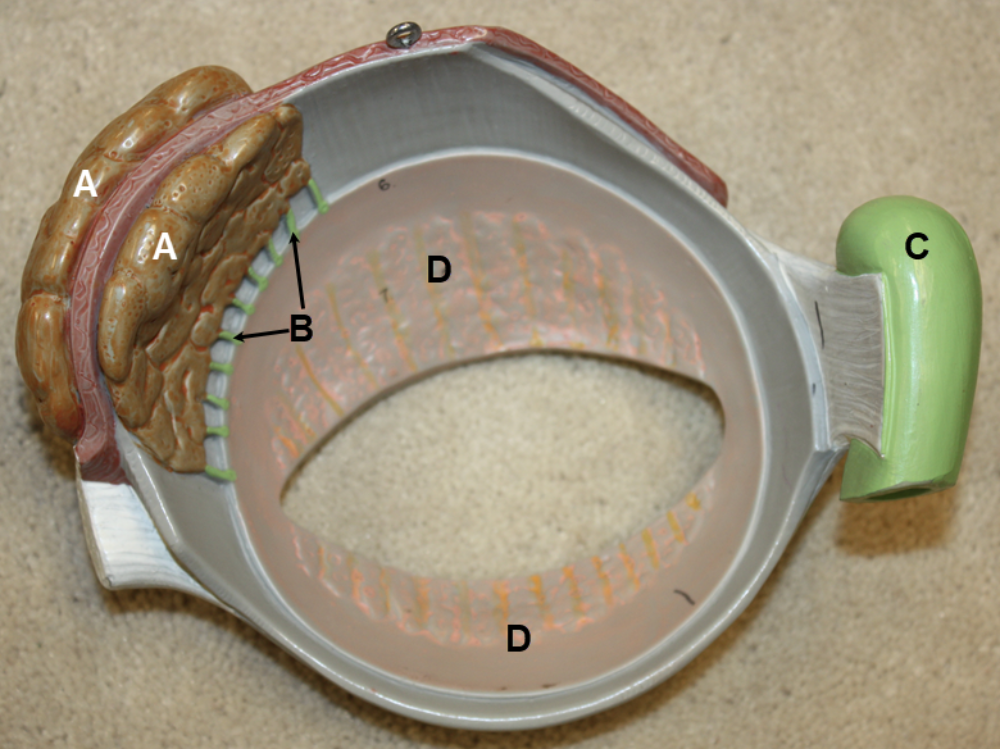
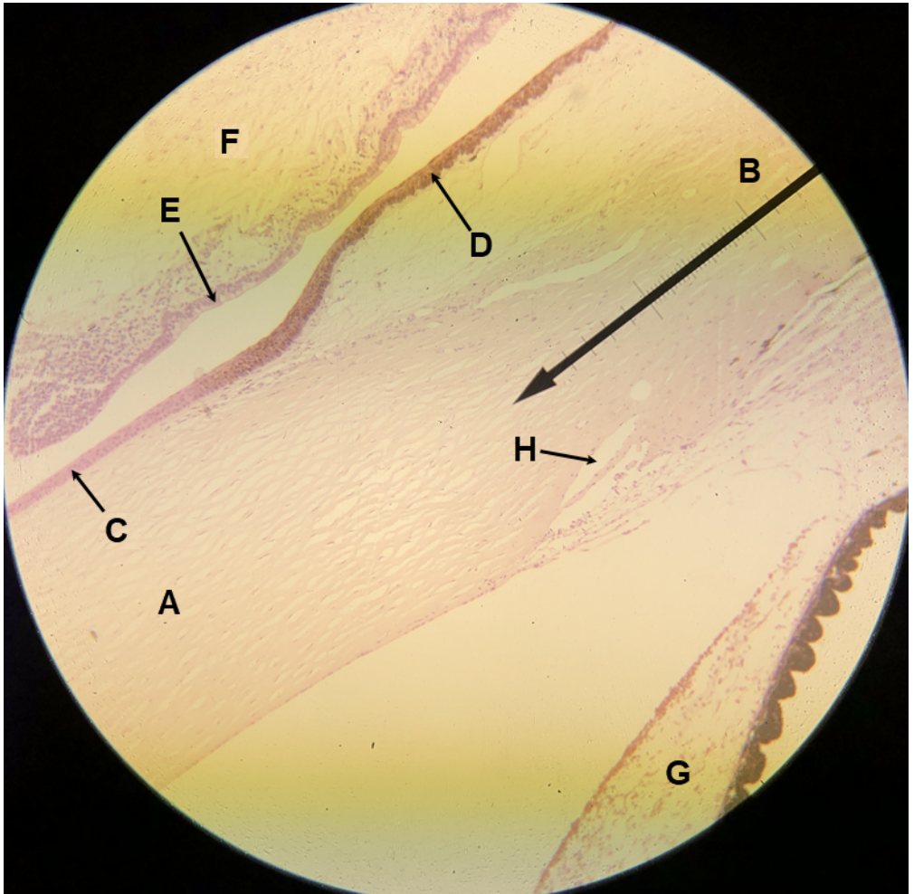
In the image above, identify structures A-D.
A. lacrimal gland
B. lacrimal gland ducts
C. lacrimal sac
D. palpebral conjunctiva
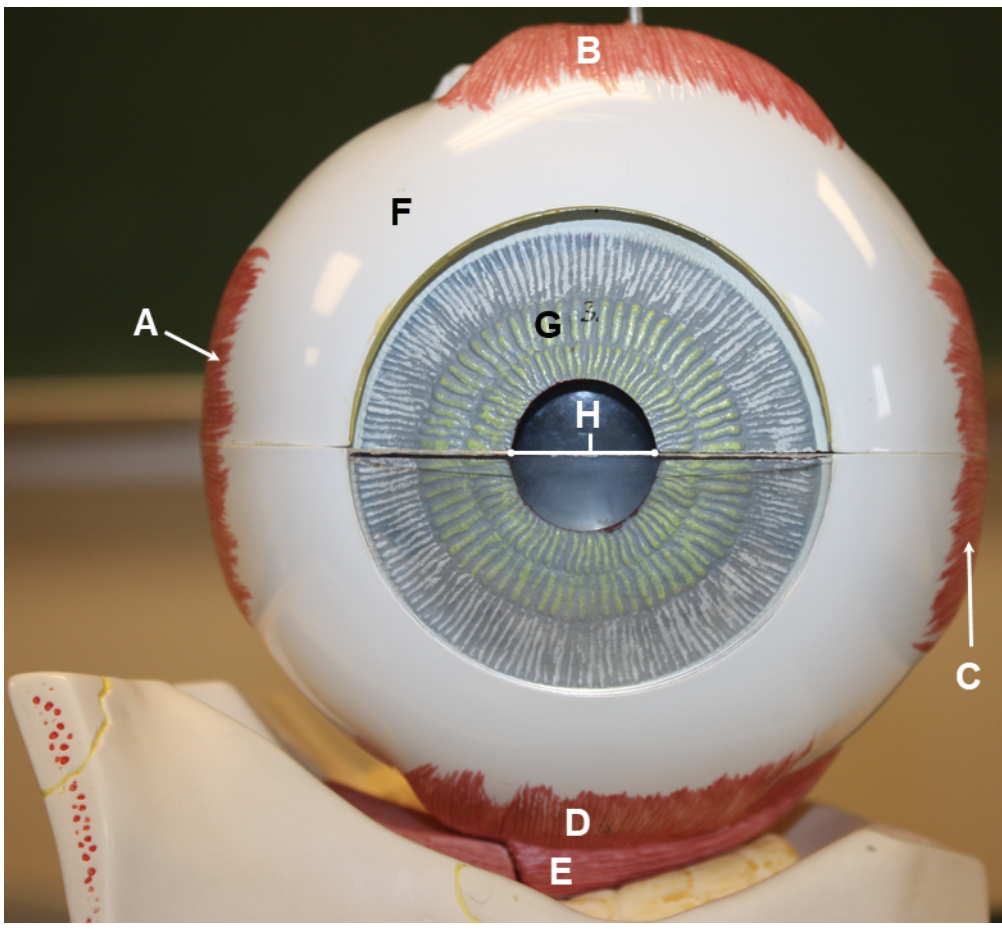
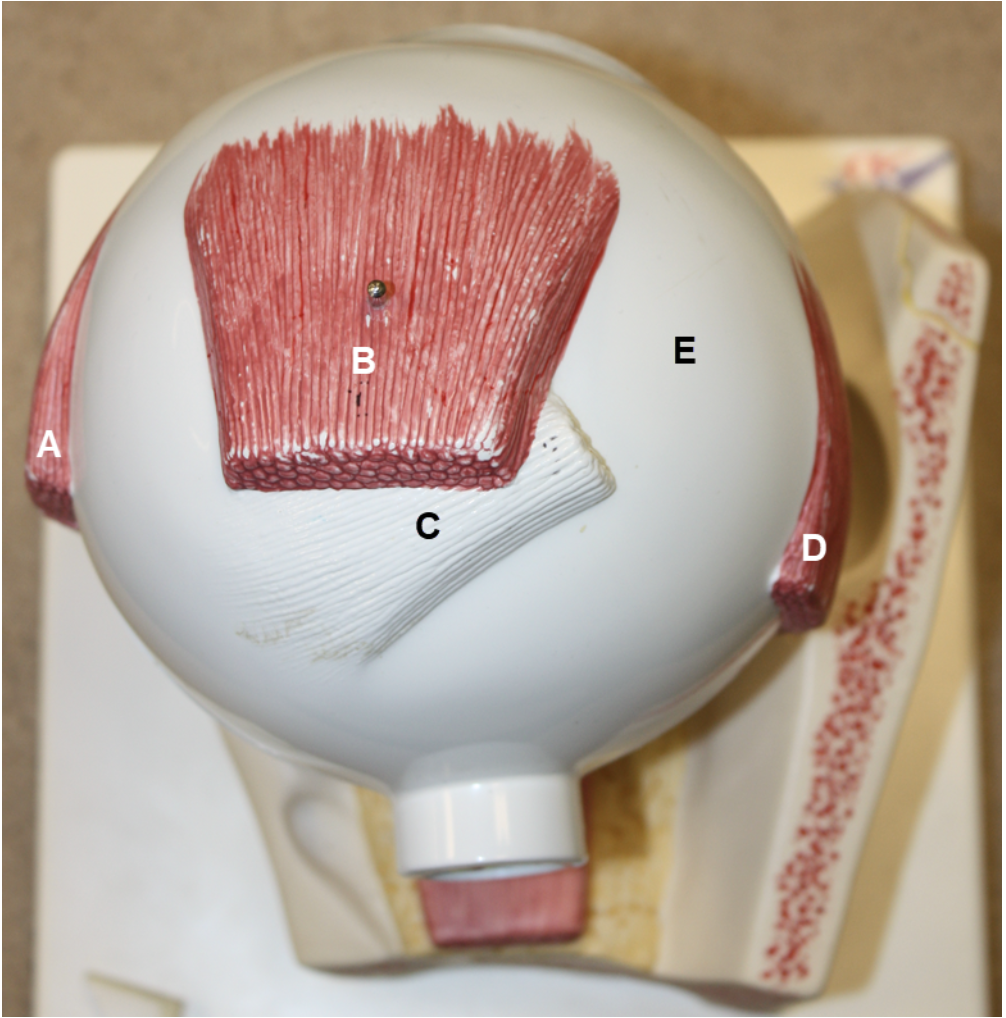
In the image above, identify structures A-G, and opening H.
A. medial rectus
B. superior rectus
C. lateral rectus
D. inferior rectus
E. inferior oblique
F. sclera
G. iris
H. pupil
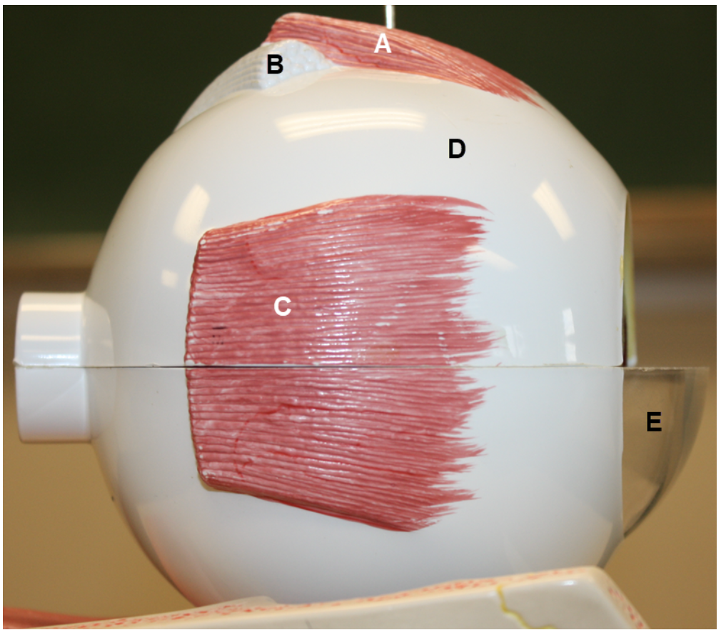
In the image above, identify structures A-E.
A. lateral rectus
B. superior rectus
C. (tendon of) superior oblique
D. medial rectus
E. sclera
In the image above, identify structures A-E.
A. superior rectus
B. (tendon of) superior oblique
C. medial rectus
D. sclera
E. cornea
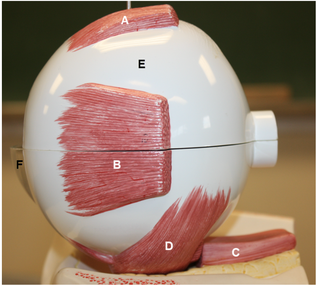
In the image above, identify structures A-F.
A. superior rectus
B. lateral rectus
C. inferior rectus
D. inferior oblique
E. sclera
F. cornea
In the image above, identify structure A, and structures/parts of the eye wall B and C.
A. cornea
B. sclera
C. choroid
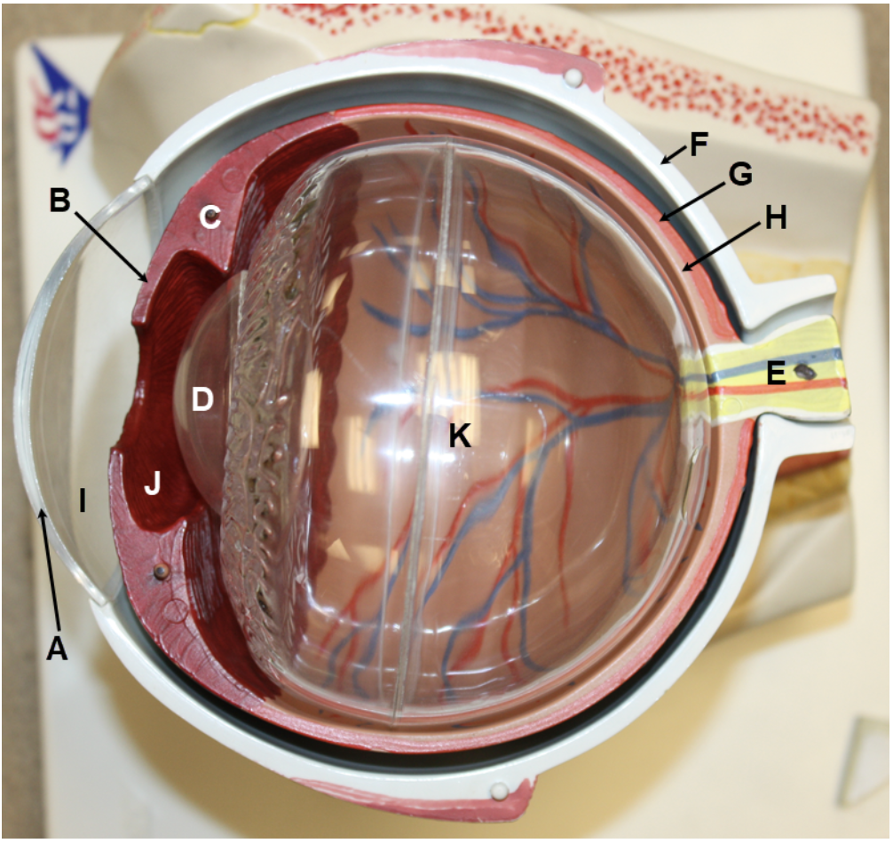
In the image above, identify structures A-E, structures/parts of the eye wall F-H, and spaces I-K. For L, name the space that consists of I and J together.
A. cornea
B. iris
C. ciliary muscle (of ciliary body)
D. lens
E. optic nerve
F. sclera
G. choroid
H. retina
I. anterior chamber
J. posterior chamber
K. posterior cavity (vitreous chamber)
L. anterior cavity
In the image above, identify structures/parts of the eye wall A and B, and structures C-E.
A. choroid
B. retina
C. fovea centralis
D. optic disc (blind spot)
E. optic nerve
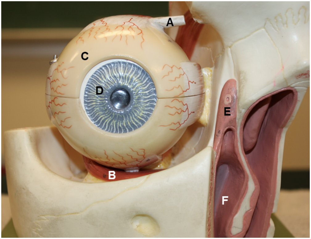
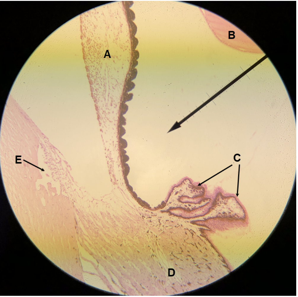
In the image above, identify structures A-H, and opening I.
A. lacrimal gland
B. lacrimal gland ducts
C. lacrimal canaliculi
D. lacrimal sac
E. nasolacrimal duct
F. eyelids (palpebrae)
G. sclera
H. iris
I. pupil
In the image above, identify structures A-E.
A. superior rectus
B. lateral rectus
C. lacrimal gland
D. eyelids (palpebrae)
E. sclera
In the image above, identify structures A-F.
A. (tendon of) superior oblique
B. inferior oblique
C. sclera
D. iris
E. lacrimal sac
F. nasolacrimal duct
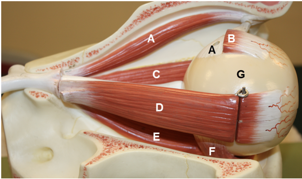
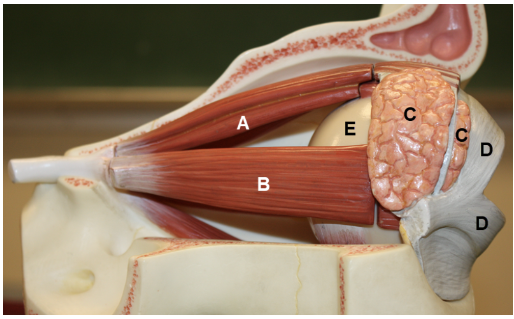
In the image above, identify structures A-G.
A. superior oblique (muscle belly and tendon)
B. superior rectus
C. medial rectus
D. lateral rectus
E. inferior rectus
F. inferior oblique
G. sclera
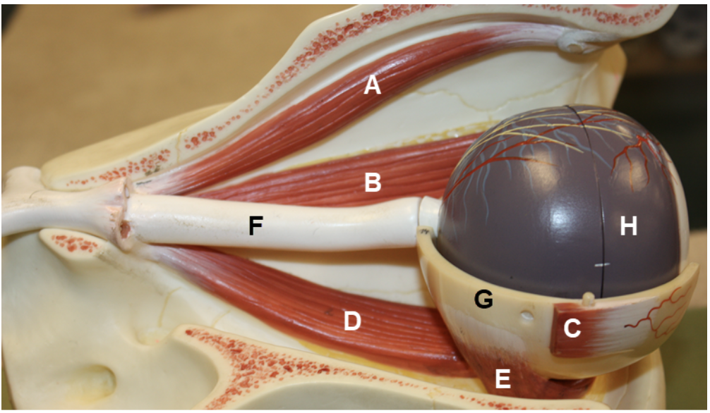
In the image above, identify structures A-F, and structures/parts of the eye wall G and H.
A. superior oblique
B. medial rectus
C. lateral rectus
D. inferior rectus
E. inferior oblique
F. optic nerve
G. sclera
H. choroid
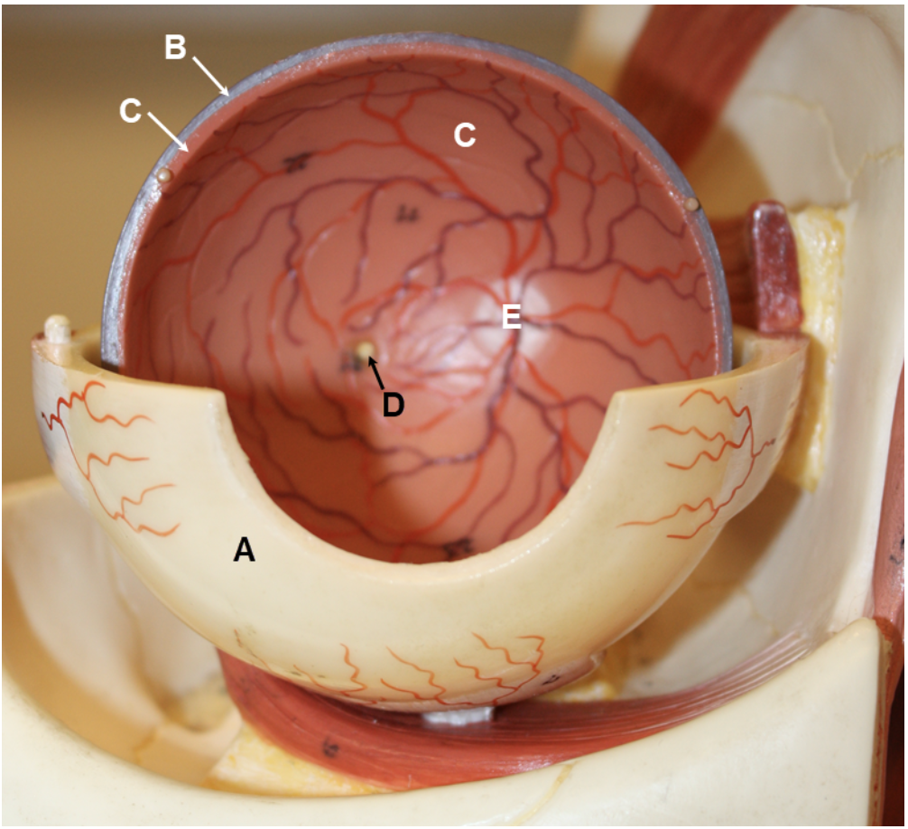
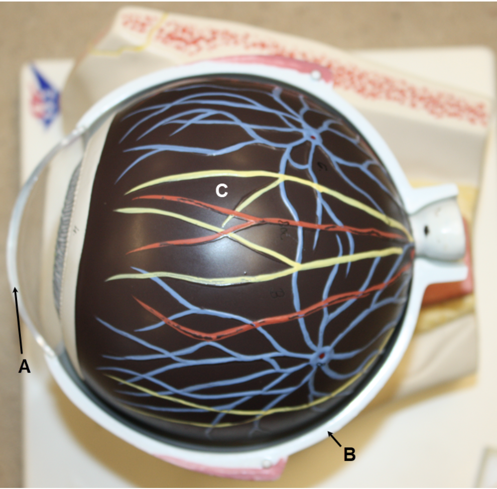
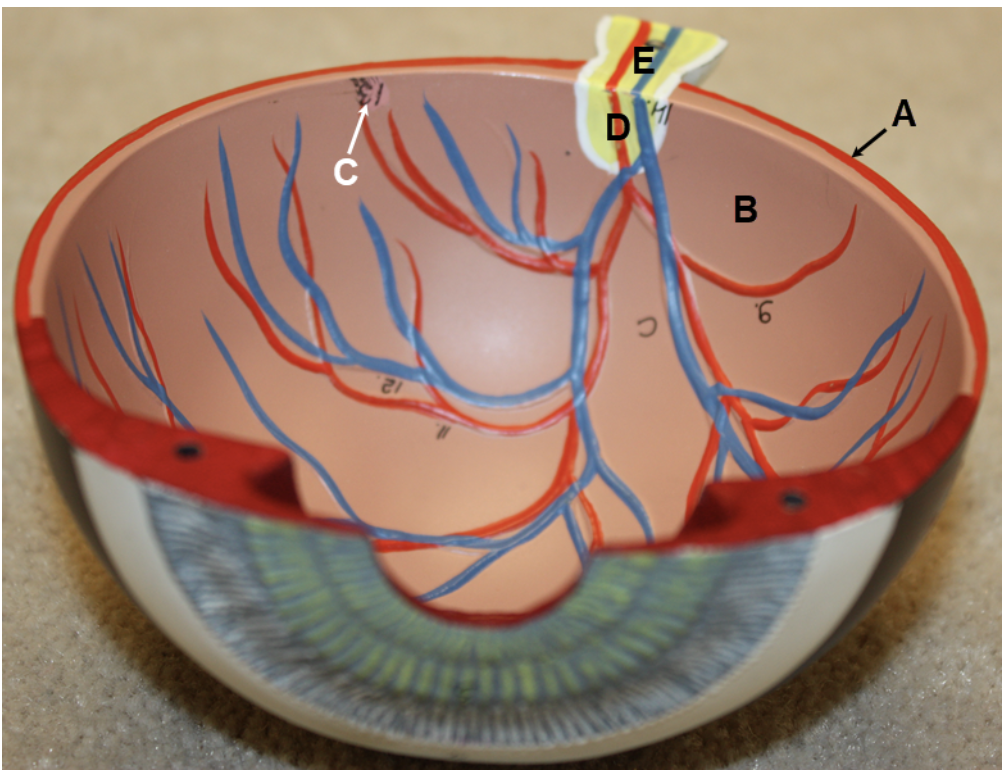
In the image above, identify structures/parts of the eye wall A-C, and structures D and E.
A. sclera
B. choroid
C. retina
D. fovea centralis
E. optic disc (blind spot)
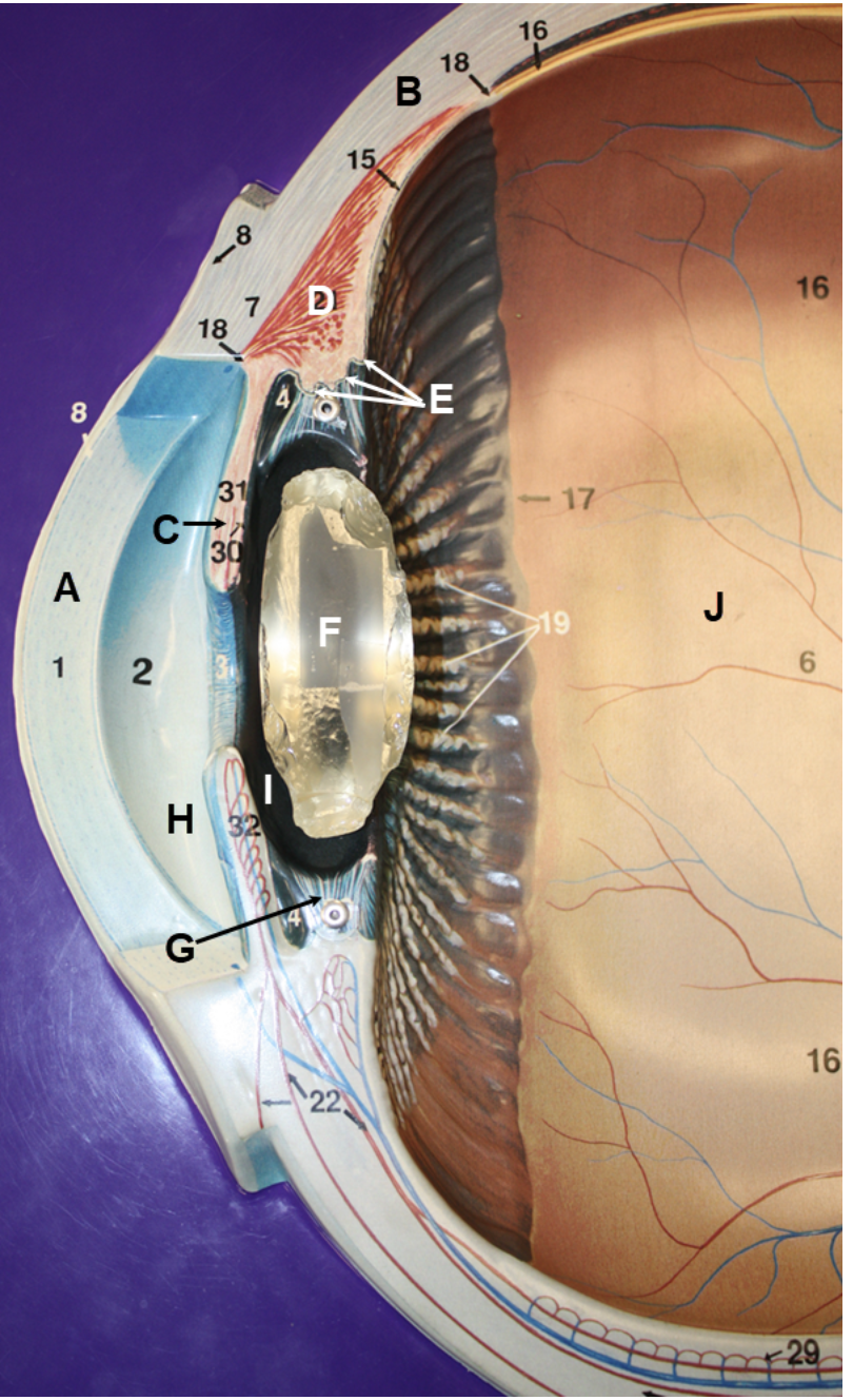
In the image above, identify structures/parts of the eye wall A and B, structures C-G, and spaces H-J. For K, name the structure that consists of D and E together. For L, name the space that consists of H and I together.
A. cornea
B. sclera
C. iris
D. ciliary muscle
E. ciliary processes
F. lens
G. ciliary zonule (suspensory ligament)
H. anterior chamber
I. posterior chamber
J. posterior cavity (vitreous chamber)
K. ciliary body
L. anterior cavity
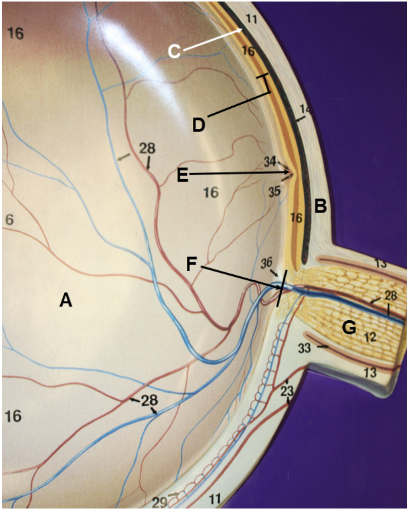
In the image above, identify space A, structures/parts of the eye wall B-D, and structures E-G.
A. posterior cavity (vitreous chamber)
B. sclera
C. choroid
D. retina
E. fovea centralis
F. optic disc (blind spot)
G. optic nerve
In the image above, identify structure/part of the eye wall A, structure/tissue B, structures C and D, and spaces E and F. For G, name the space that consists of E and F together.
A. cornea
B. corneal epithelium
C. iris
D. lens
E. anterior chamber
F. posterior chamber
G. anterior cavity
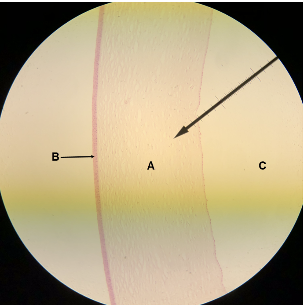
In the image above, identify structure/part of the eye wall A, structure/tissue B, and space C.
A. cornea
B. corneal epithelium
C. anterior chamber
In the image above, identify structure/part of the eye wall A, structures B-E, and spaces F-H. For I, name the structure that consists of D and E together. For J, name the space that consists of F and G together.
A. cornea
B. iris
C. lens
D. ciliary muscle
E. ciliary processes
F. anterior chamber
G. posterior chamber
H. posterior cavity (vitreous chamber)
I. ciliary body
J. anterior cavity
In the image above, identify structures/parts of the eye wall A and B, and structures C-F. For G, name the structure that consists of D and E together.
A. cornea
B. sclera
C. iris
D. ciliary muscle
E. ciliary processes
F. part of an eyelid (palpebra)
G. ciliary body
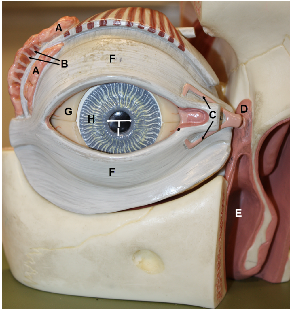
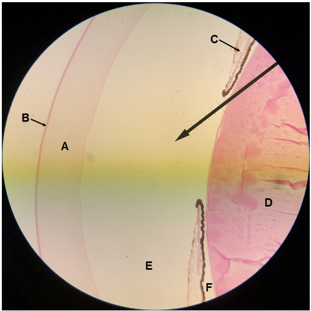
In the image above, identify structures/parts of the eye wall A and B, structures/tissues C-E, and structures F-H.
A. cornea
B. sclera
C. corneal epithelium
D. bulbar (ocular) conjunctiva
E. palpebral conjunctiva
F. part of an eyelid (palpebra)
G. iris
H. scleral venous sinus (canal of Schlemm)
In the image above, identify structures A-E. For F, name the structure that consists of C and D together.
A. iris
B. lens
C. ciliary processes
D. ciliary muscle
E. scleral venous sinus (canal of Schlemm)
F. ciliary body
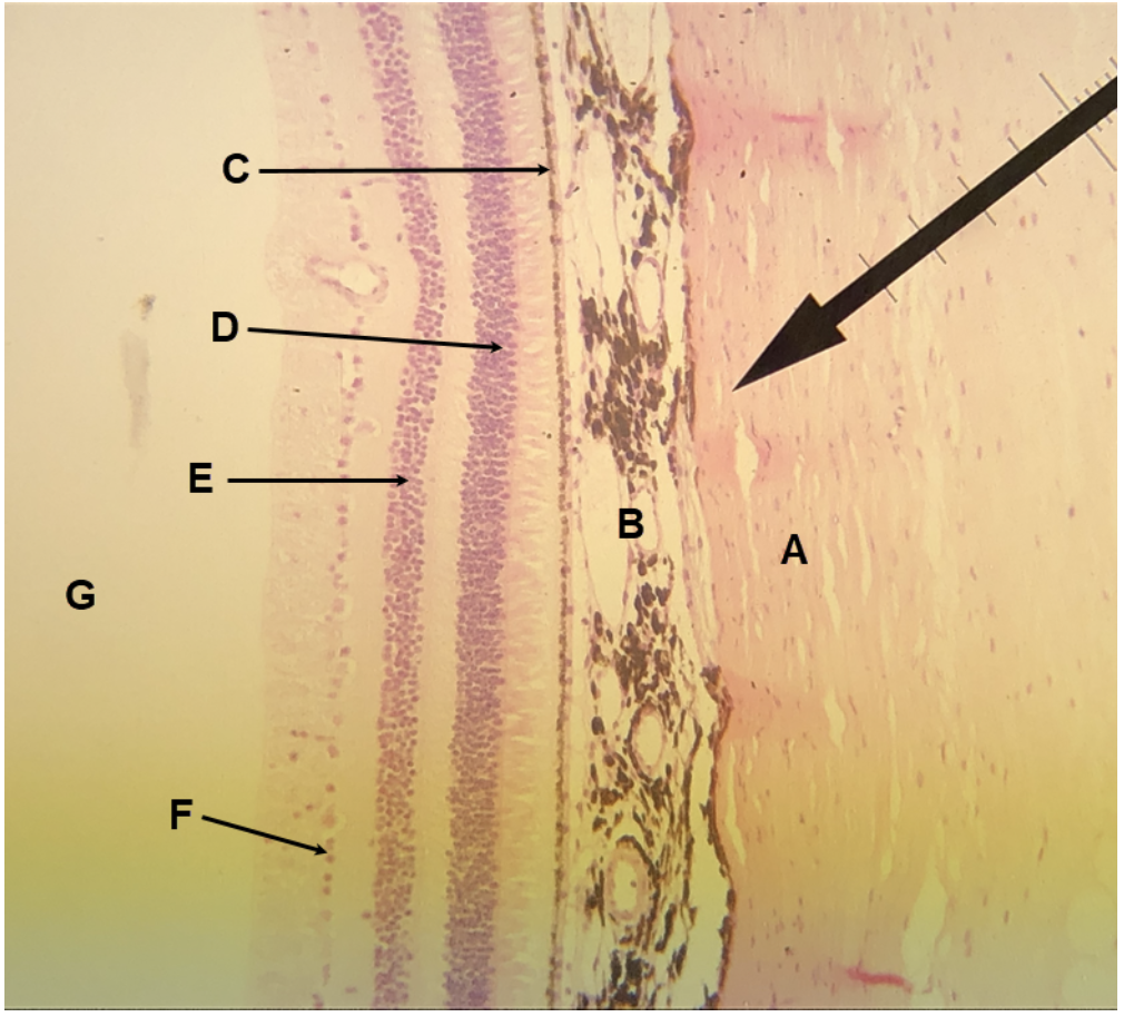
In the image above, identify structures/parts of the eye wall A and B, layers/sublayers C-F, and space G. For H, name the structure/part of the eye wall that consists of C-F together.
A. sclera
B. choroid
C. pigmented layer
D. photoreceptor (rods and cones) layer
E. bipolar cell layer
F. ganglion cell layer
G. posterior cavity (vitreous chamber)
H. retina
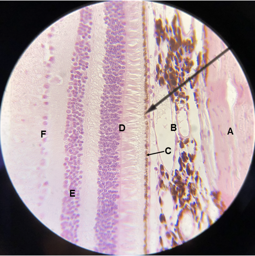
In the image above, identify structures/parts of the eye wall A and B, and layers/sublayers C-F. For G, name the structure/part of the eye wall that consists of C-F together.
A. sclera
B. choroid
C. pigmented layer
D. photoreceptor (rods and cones) layer
E. bipolar cell layer
F. ganglion cell layer
G. retina
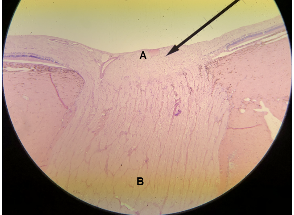
In the image above, identify structures A and B.
A. optic disc (blind spot)
B. optic nerve
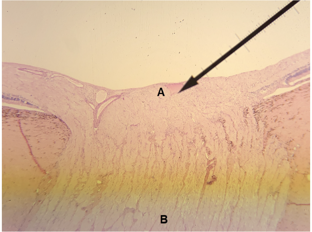
In the image above, identify structures A and B.
A. optic disc (blind spot)
B. optic nerve
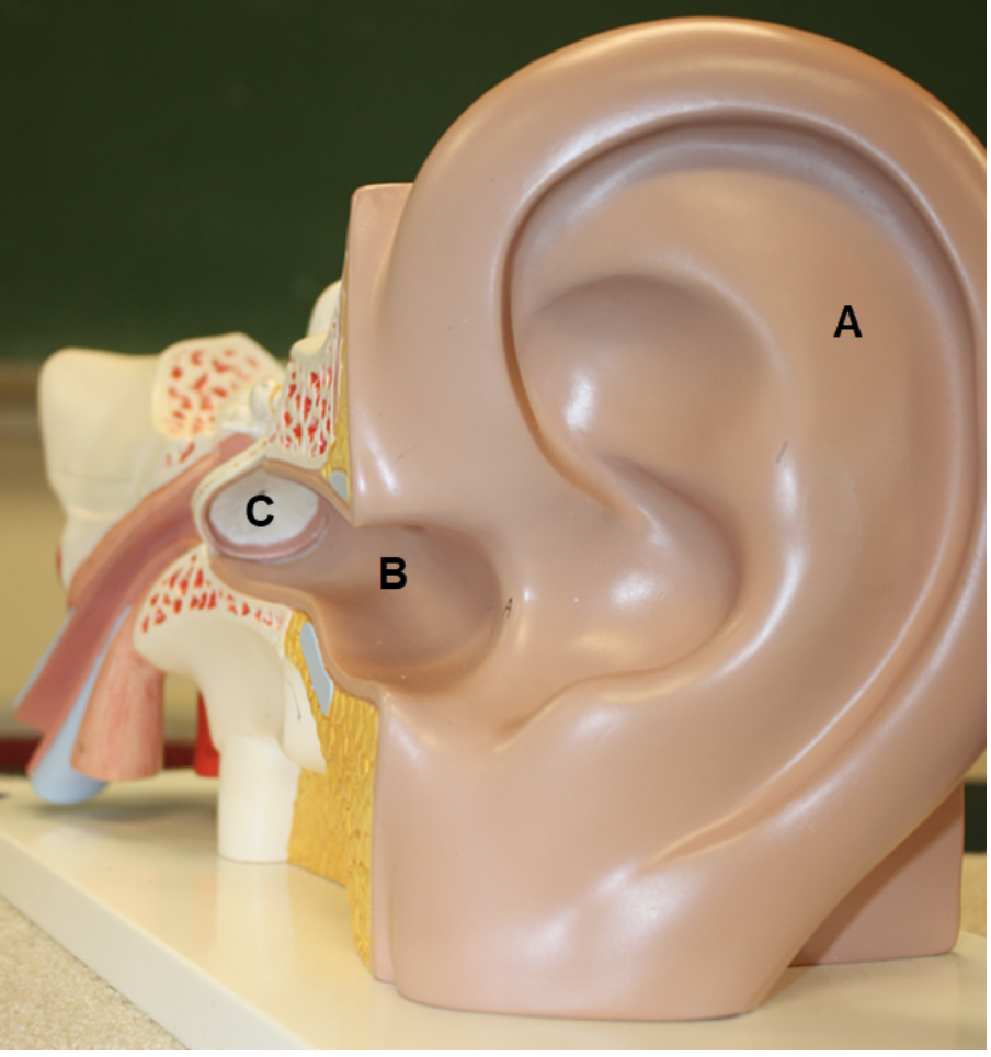
In the image above, identify structures A-C.
A. auricle (pinna)
B. external acoustic meatus (external auditory canal)
C. tympanic membrane
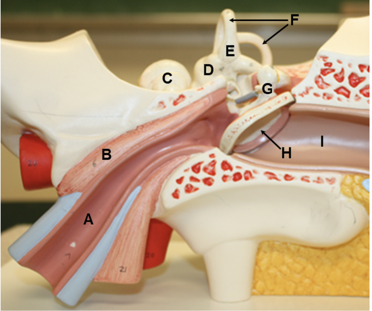
In the image above, identify structures A-I.
A. auditory tube (Eustachian tube or pharyngotympanic tube)
B. tensor tympani
C. cochlea
D. vestibule
E. ampulla
F. semicircular canals
G. malleus
H. tympanic membrane
I. external acoustic meatus (external auditory canal)
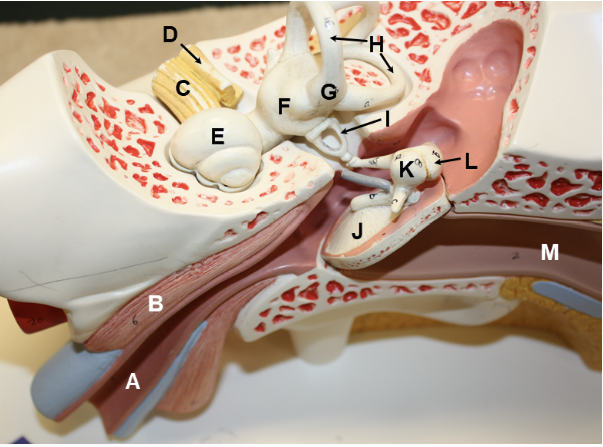
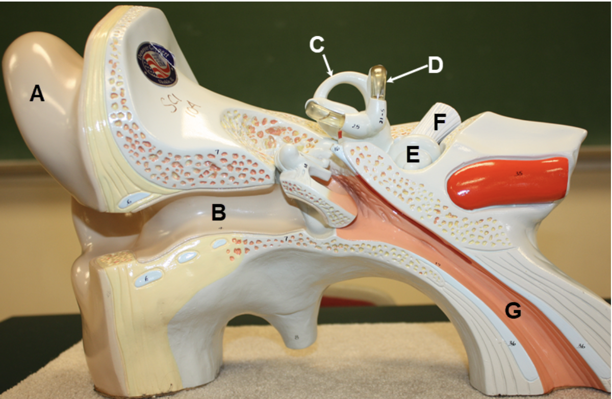
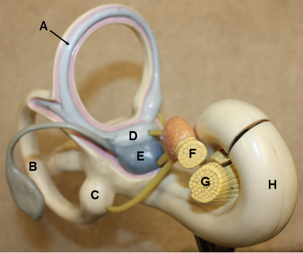
In the image above, identify structures A-M. For N, name the structure that consists of C and D together.
A. auditory tube (Eustachian tube or pharyngotympanic tube)
B. tensor tympani
C. cochlear nerve (cochlear branch of CN VIII)
D. vestibular nerve (vestibular branch of CN VIII)
E. cochlea
F. vestibule
G. ampulla
H. semicircular canals
I. stapes
J. tympanic membrane
K. malleus
L. incus
M. external acoustic meatus (external auditory canal)
N. vestibulocochlear nerve (CN VIII)
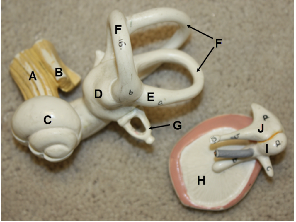
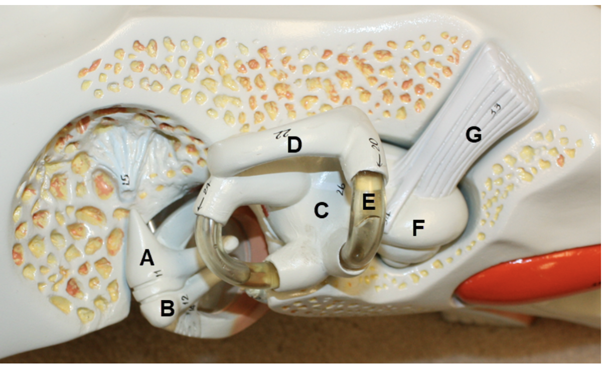
In the image above, identify structures A-J. For K, name the structure that consists of A and B together.
A. cochlear nerve (cochlear branch of CN VIII)
B. vestibular nerve (vestibular branch of CN VIII)
C. cochlea
D. vestibule
E. ampulla
F. semicircular canals
G. stapes
H. tympanic membrane
I. malleus
J. incus
K. vestibulocochlear nerve (CN VIII)
In the image above, identify structures A-G.
A. auricle (pinna)
B. external acoustic meatus (external auditory canal)
C. semicircular canal
D. semicircular duct
E. cochlea
F. cochlear nerve (cochlear branch of CN VIII)
G. auditory tube (Eustachian tube or pharyngotympanic tube)
In the image above, identify structures A-G.
A. incus
B. malleus
C. vestibule
D. semicircular canal
E. semicircular duct
F. cochlea
G. cochlear nerve (cochlear branch of CN VIII)
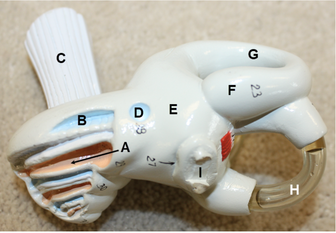
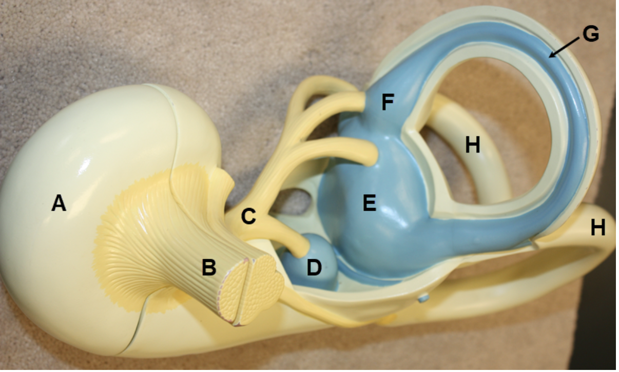
In the image above, identify structures A-G.
A. scala vestibuli (vestibular duct)
B. scala tympani (tympanic duct)
C. cochlear nerve (cochlear branch of CN VIII)
D. round window
E. vestibule
F. ampulla
G. semicircular canal
H. semicircular duct
I. stapes
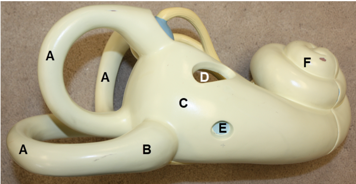
In the image above, identify structures A-F.
A. cochlea
B. vestibule
C. oval window
D. ampulla
E. semicircular duct
F. semicircular canals
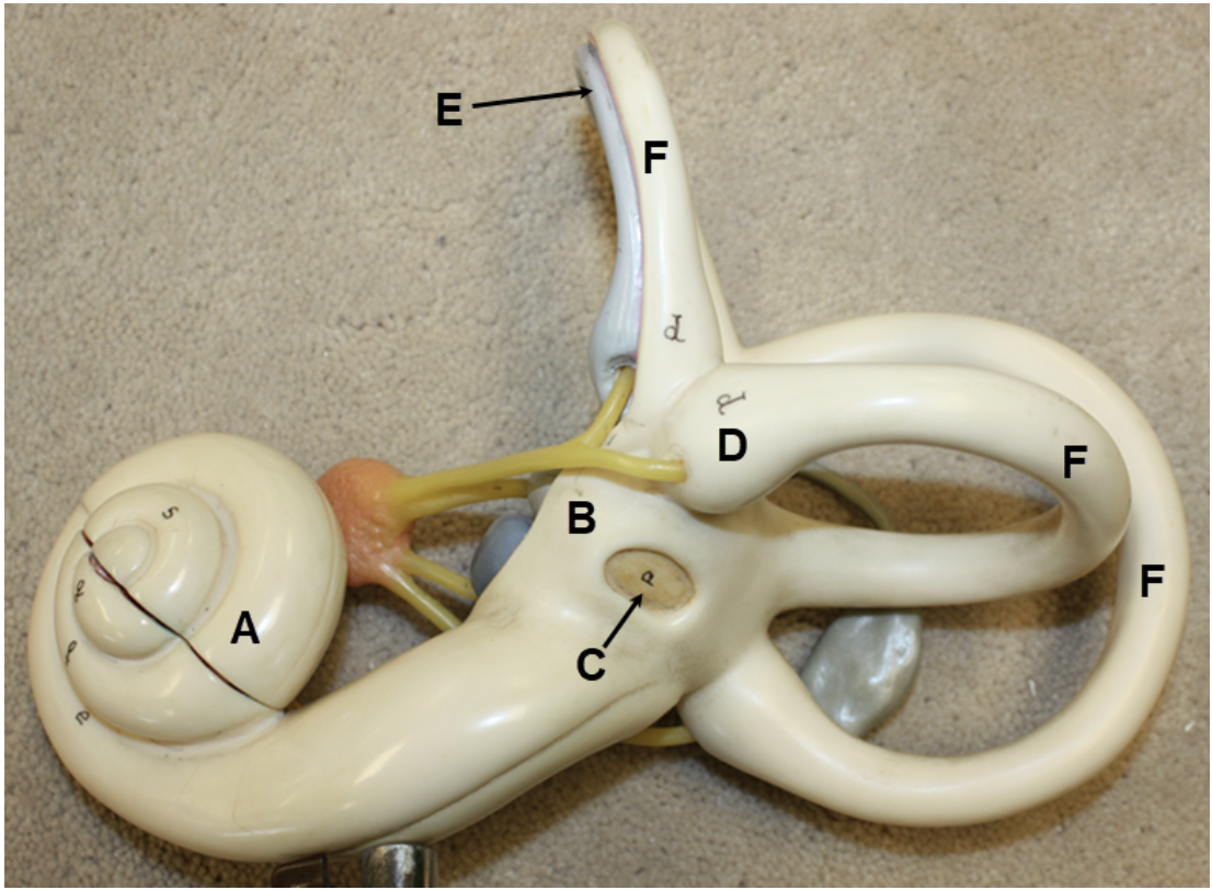
In the image above, identify structures A-H. For I, name the structure that consists of F and G together.
A. semicircular duct
B. semicircular canal
C. ampulla
D. utricle
E. saccule
F. vestibular nerve (vestibular branch of CN VIII)
G. cochlear nerve (cochlear branch of CN VIII)
H. cochlea
I. vestibulocochlear nerve (CN VIII)
In the image above, identify spaces A-C, and structures D and E. For F, name the structure that consists of D and E together.
A. scala vestibuli (vestibular duct)
B. cochlear duct (scala media)
C. scala tympani (tympanic duct)
D. cochlear nerve (cochlear branch of CN VIII)
E. vestibular nerve (vestibular branch of CN VIII)
F. vestibulocochlear nerve (CN VIII)
In the image above, identify structures A-F.
A. semicircular canals
B. ampulla
C. vestibule
D. oval window
E. round window
F. cochlea
In the image above, identify structures A-H. For I, name the structure that consists of B and C together.
A. cochlea
B. cochlear nerve (cochlear branch of CN VIII)
C. vestibular nerve (vestibular branch of CN VIII)
D. saccule
E. utricle
F. ampulla
G. semicircular duct
H. semicircular canals
I. vestibulocochlear nerve (CN VIII)
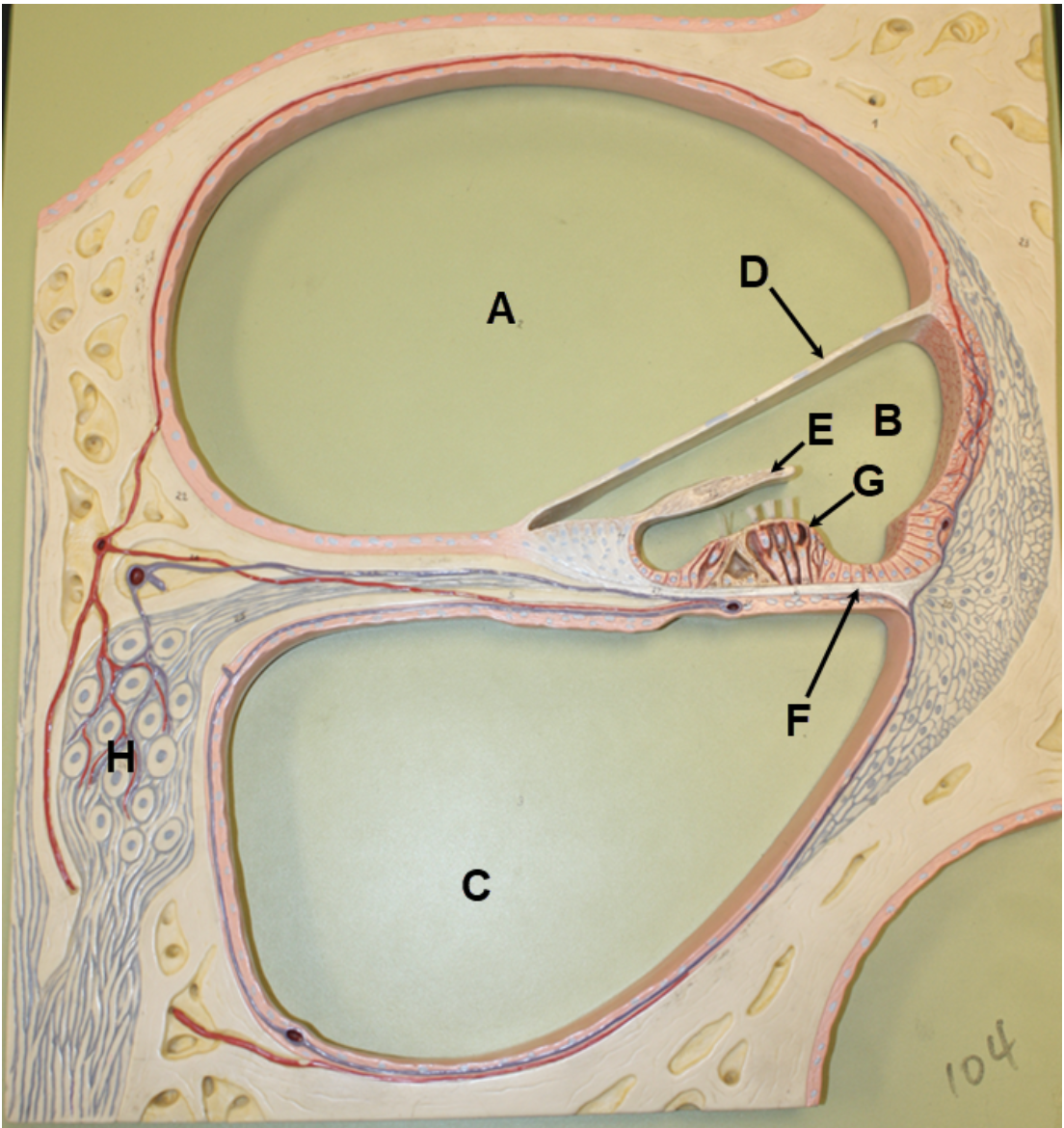
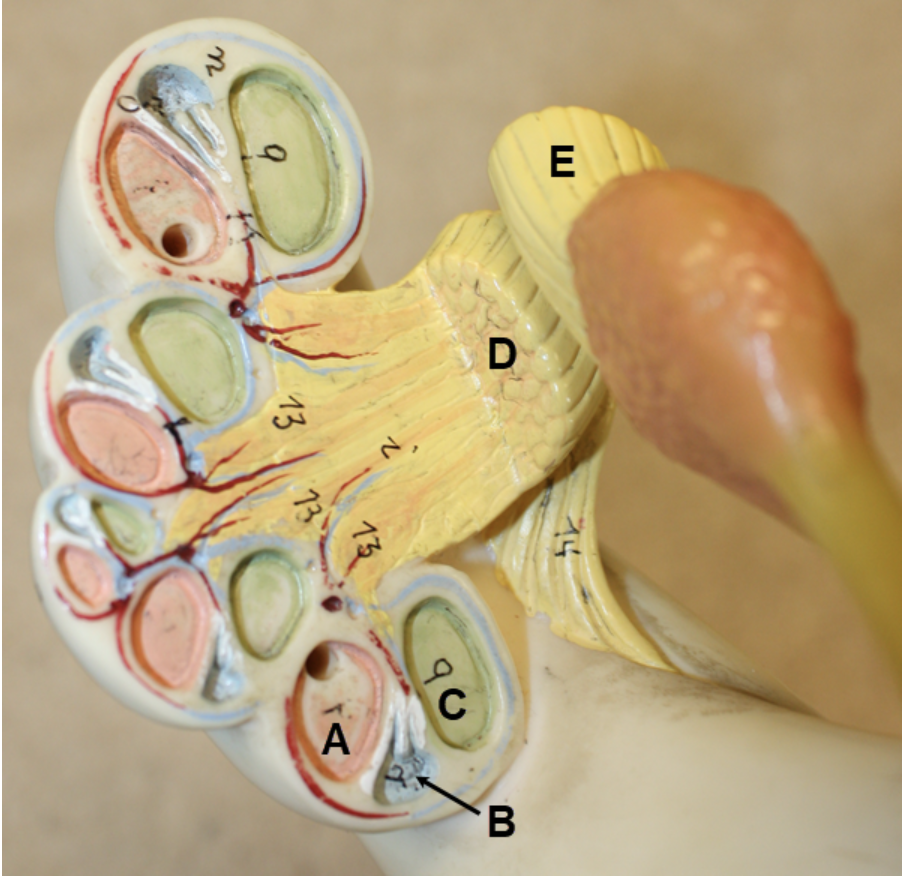
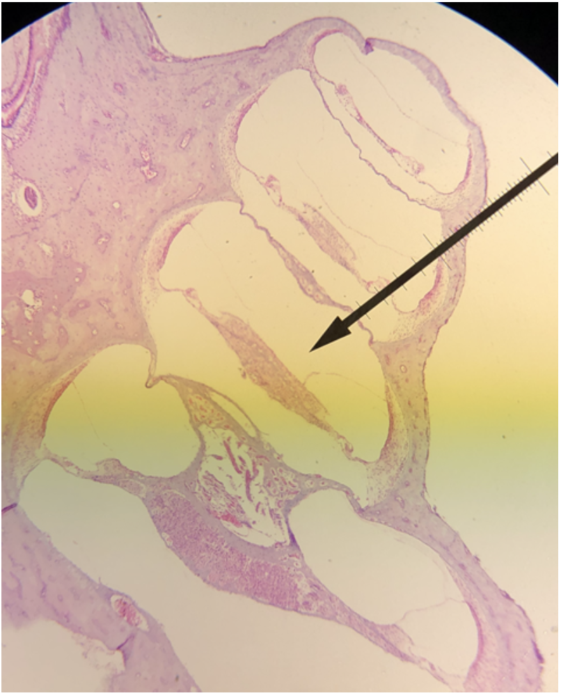
In the image above, identify spaces A-C, and structures D-H.
A. scala vestibuli (vestibular duct)
B. cochlear duct (scala media)
C. scala tympani (tympanic duct)
D. vestibular membrane
E. tectorial membrane
F. basilar membrane
G. spiral organ (organ of Corti)
H. spiral ganglion
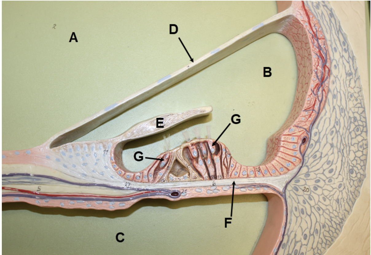
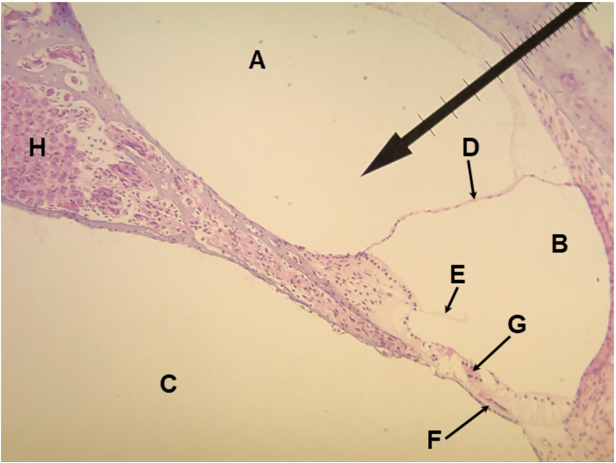
In the image above, identify spaces A-C, structures D-F, and cells G. For H, name the structure that cells G are a part of.
A. scala vestibuli (vestibular duct)
B. cochlear duct (scala media)
C. scala tympani (tympanic duct)
D. vestibular membrane
E. tectorial membrane
F. basilar membrane
G. hair cells
H. spiral organ (organ of Corti)
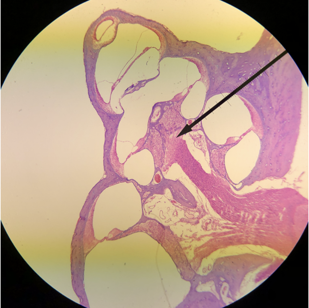
In the image above, identify the main structure seen in the field of view.
cochlea
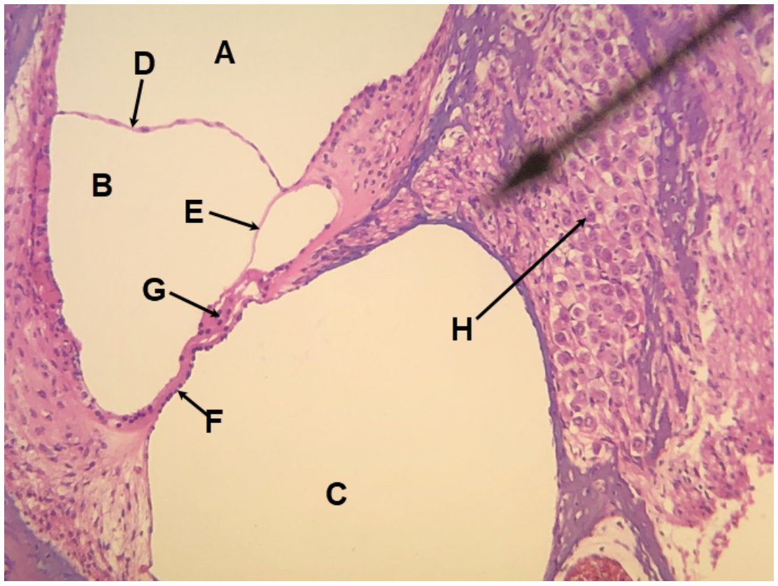
In the image above, identify spaces A-C, and structures D-H.
A. scala vestibuli (vestibular duct)
B. cochlear duct (scala media)
C. scala tympani (tympanic duct)
D. vestibular membrane
E. tectorial membrane
F. basilar membrane
G. spiral organ (organ of Corti)
H. spiral ganglion
In the image above, identify the main structure seen in the field of view.
cochlea
In the image above, identify spaces A-C, and structures D-H.
A. scala vestibuli (vestibular duct)
B. cochlear duct (scala media)
C. scala tympani (tympanic duct)
D. vestibular membrane
E. tectorial membrane
F. basilar membrane
G. spiral organ (organ of Corti)
H. spiral ganglion
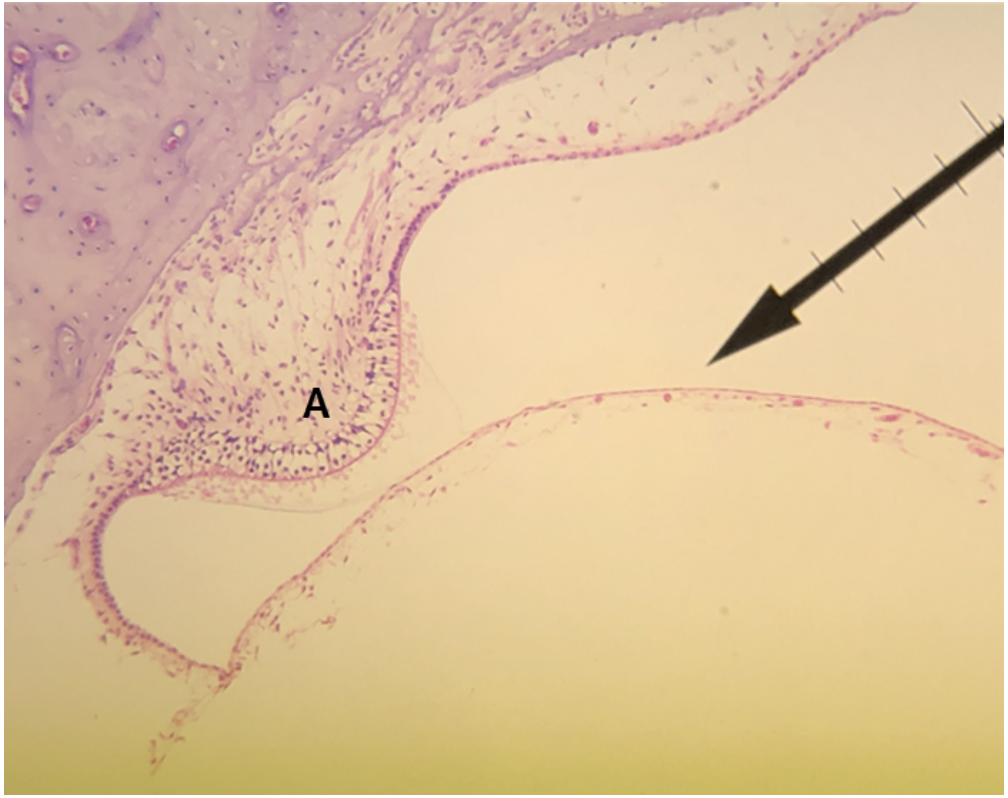
In the image above, identify structure A.
A. ampullary crest (crista ampullaris)
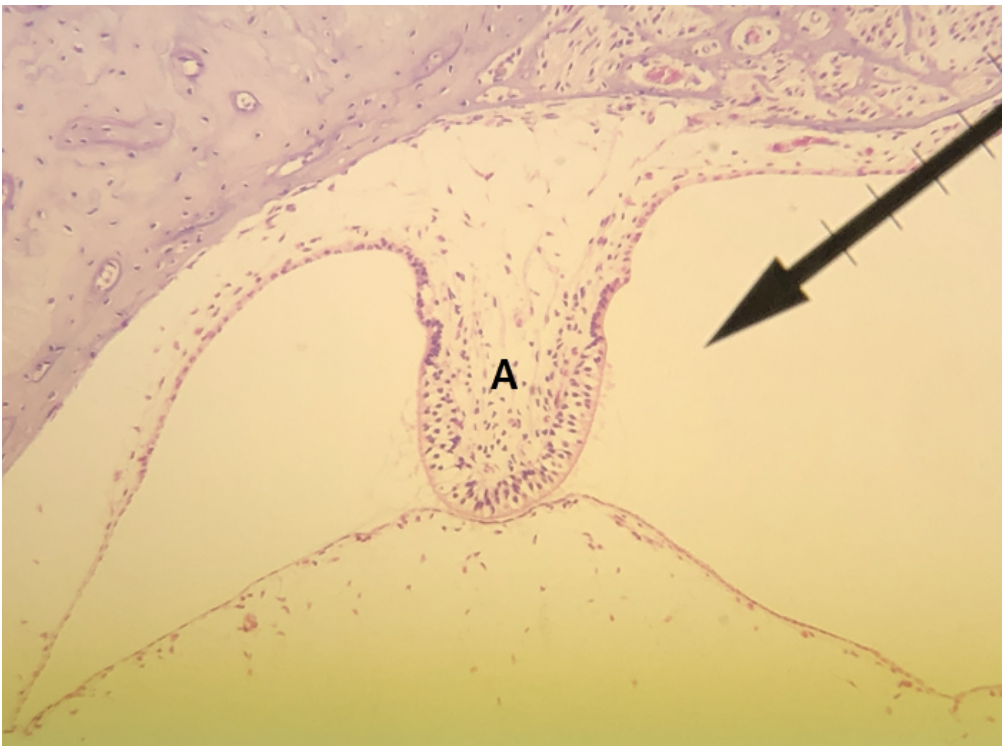
In the image above, identify structure A.
A. ampullary crest (crista ampullaris)