Introduction to histology | Quizlet
1/22
Earn XP
Description and Tags
➢ List the steps in histological preparation of tissues and understand how this determines microscopic appearance. ➢ Describe the four basic tissue types and how they contribute to organ structure & function. ➢ Relate epithelium and connective tissue to the structure of the common integument.
Name | Mastery | Learn | Test | Matching | Spaced | Call with Kai |
|---|
No analytics yet
Send a link to your students to track their progress
23 Terms
What is histology?
histology = microanatomy
what is histopathology?
study of changes in the microscopic anatomy, caused by disease e.g. in biopsies, post-mortem samples
typical steps of histology prep
1. Take a sample
• Handle sample with care (avoid crushing) • If > 1 cm thick, incise to allow fixative to penetrate
• Can mark areas with ink or suture material
2. Fixation (e.g., in 10 % formalin)
• Hardens and preserves tissue (basically just conserving the tissue)
• Some tissue shrinkage
• Aim for a 1:10 ratio of sample to solution
3. Selection and trimming
• Choose your orientation and cut surface
4. Processing and embedding
• Replace water with solid medium (e.g. paraffin wax, resin)
5. Sectioning thin slices
6. Staining to make tissue structures visible
in the case of harder substances like bone - you need to have additional steps like decalcification to soften it and dissolve in acids before doing the next steps
what is formalin
it is water based solution of formaldehyde (formaldehyde is acc just a strong gas, but putting it in liquid makes the formalin)
taking a sample
should have all the constituent cells that make that sample e.g. if you take liver piece don't exclude cells that make up the liver -make sure you have the whole structure like epithelium, core etc
histology stains
colour diff things by diff amounts
• "Standard" Haematoxylin and Eosin (H&E) • Haematoxylin (purple/blue) can be considered a basic dye. It binds to acids e.g. nucleic acids (so stains nuclei blue) • Eosin (pink/red) is an acidic dye. It binds to bases, e.g. most proteins in the cytoplasm are basic
haematoxylin (purple/blue)
basic dye
binds to acids e.g. nucleic acids (so stains nuclei as blue)
eosin (pink/red)
acidic dye - binds to bases-most proteins in the cytoplasm are basic
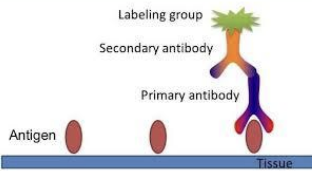
immunohistochemical stains
use antibodies to bind to tissue-specific substance, and a second labelled antibody to bind to the first
label can be coloured dye or fluorescent marker
get the actual pic
basic tissues - building blocks
epithelial
connective
nervous
muscle
organ
fully differentiated structural and functional unit in an animal that is specialised for particular function

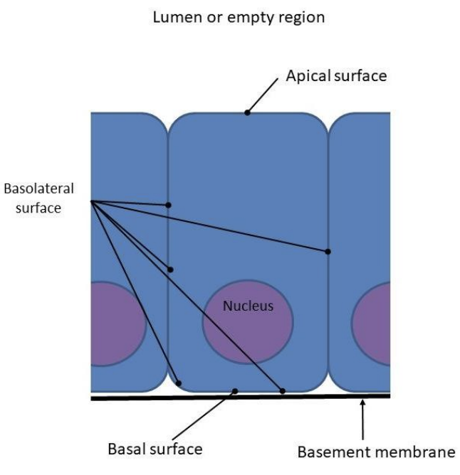
what is epithelium?
continuous sheets, minimal ECM
functions: protection, secretion, absorption
Polarity: apical, basal
Avascular, supported by connective tissue (no blood supply-depends on exchange of nutrients in the surrounding tissue)
replace all pics with slides
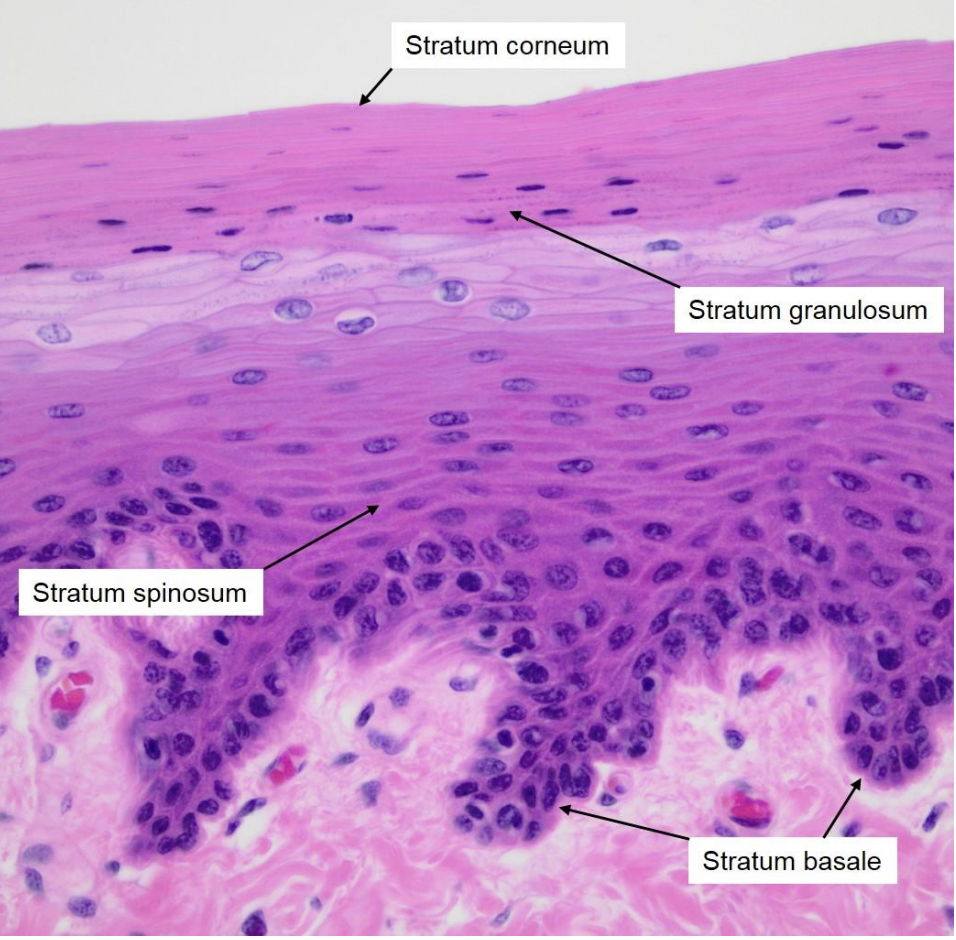
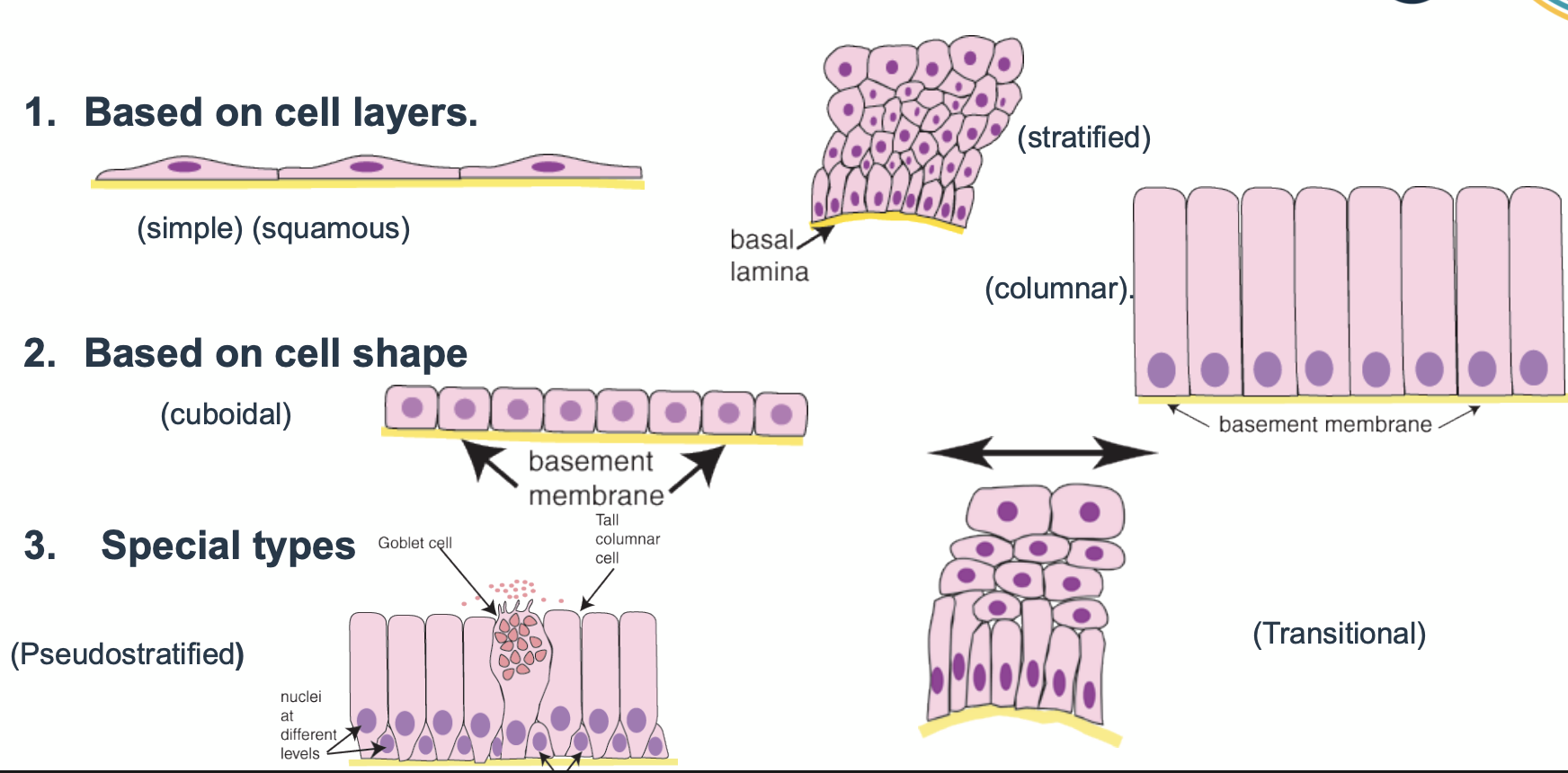
classification of epithelium
1. based on cell layers e.g. simple - single layer of cells, stratified
2. based on cell shape e.g. cuboidal, columnar, squamous (flat)
3. special types e.g. pseudostratified (cells of two different shapes), transitional
the base would be called basement membrane or basal laminar
cells closer to the base = basal cells
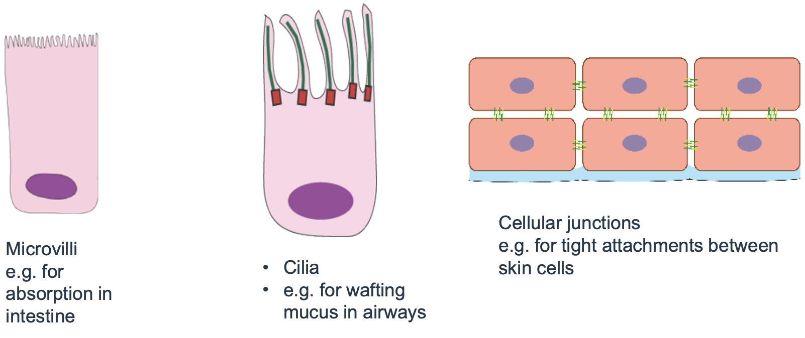
extra modifications of epithelium
can have extra features
like microvilli for absorption in intestine
cilia e.g. for wafting mucus in airways
cellular junctions e.g. tight attachments between skin cells
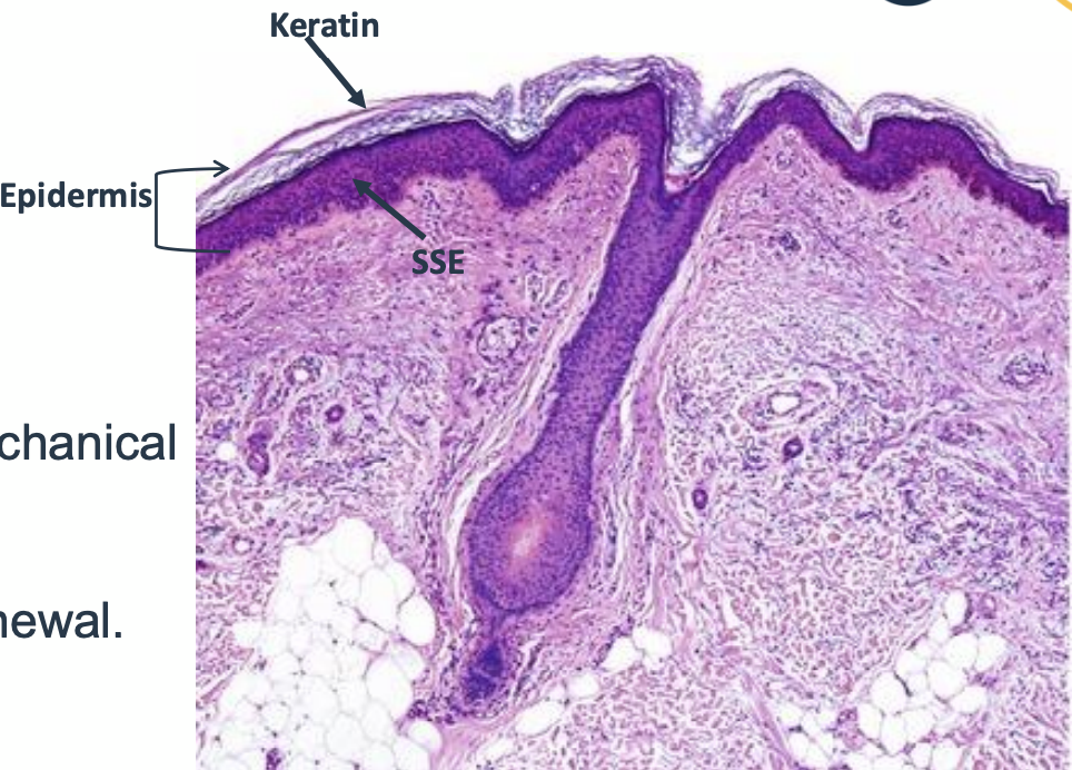
epithelium in the integument
its epidermis is made of stratified squamous keratinized epithelium
provides barrier, protection from mechanical and microbial injury
special features: keratinization, renewal

what is the integument
the outer covering of an organism, such as the skin, hair, nails, and glands
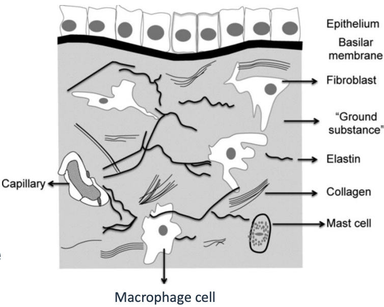
what is connective tissue (CT)
derived from mesoderm (During embryonic development, the mesoderm gives rise to various tissues, including connective tissues, muscle, bone, and specific epithelial linings) and forms a matrix beneath the epithelium
unlike epithelium, it is relatively few cells
instead, mostly made of extracelullar matrix: consisting of protein fibres and ground substance
functions of epithelium tissue
provide structural support to other tissues
mediate the exchange of nutrients and waste
CT - types of fibres
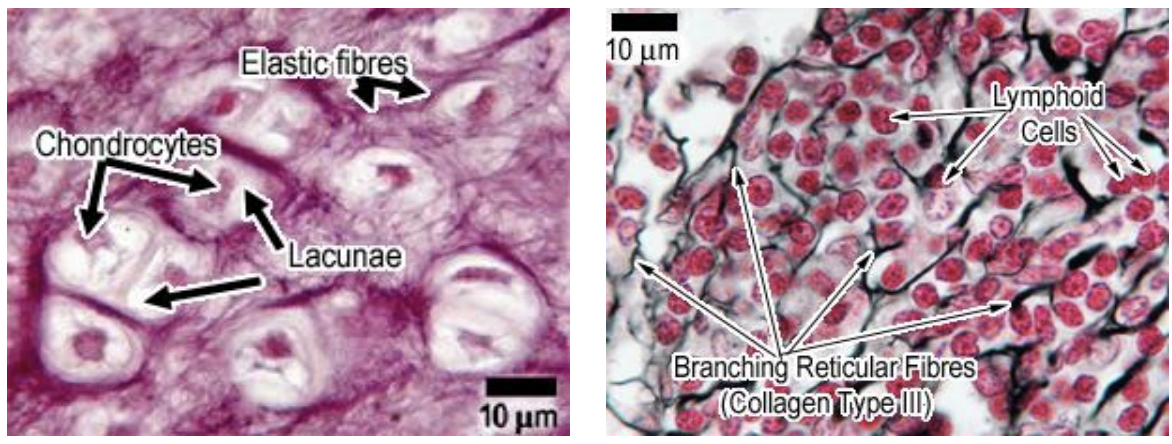
collagen (thick and strong)
• Composed of the protein collagen • Great tensile strength • Five sub-types (I-V), found in different areas of the body
elastic (heart has it)
• Composed of the protein elastin • Ability to stretch and bounce back
reticular (like lymph nodes, spleen)
• Modified type III collagen filaments • Secreted by reticular cells
what is ground substance?
found in connective tissue
it is gel-like material
transparent
fills spaces between cells and fibres
what is ground substance made up of?
made of up large molecules called glycosaminoglycans (GAGs)
to describe them they are…
very hydrophilic (because negatively charged polysaccharides)
inflexible
they can link up into even large proteoglycans
they are good at binding to water (90% of extracellular matrix is water)
high water content and inflexible molecules mean very good at resisting compressive force
watery ‘space’ faciliates movement of cells and diffusion of molecules

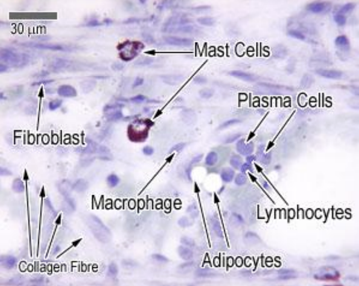
what cells are found in connective tissue>
• Fixed Vs Migratory Cells
• Fibroblasts - Least specialised cells, secrete the collagen and ground substance
• Adipocytes (fat cells)
• Immune cells
Macrophages
Mast cells
Plasma cells
• Other specialised cells (depend on the type of connective tissue) e.g.
Osteocytes and osteoblasts (osteo = bone)
Chondrocytes and chondroblasts (chondro = cartilage)
uno blood, bone, cartilage are specialised forms of connective tissue
CT in the integument
• Dermis = dense irregular connective tissue.
• Contains collagen, elastic fibers, fibroblasts.
• Provides tensile strength, elasticity, support for epidermis.