FULL Practical 2 COMBINED
1/122
There's no tags or description
Looks like no tags are added yet.
Name | Mastery | Learn | Test | Matching | Spaced | Call with Kai |
|---|
No analytics yet
Send a link to your students to track their progress
123 Terms
Which un-paired artery off the abdominal aorta supplies the stomach?
Celiac trunk
Left Gastric Artery
Parietal Peritoneum
Splenic Artery
Common Hepatic Artery
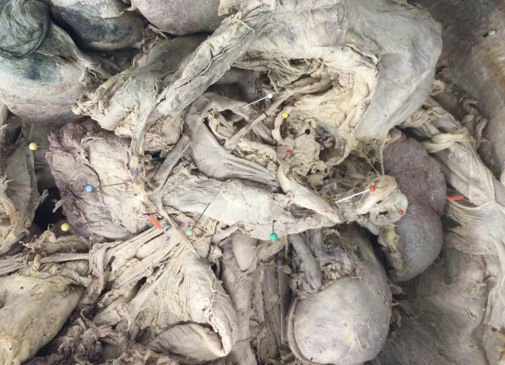
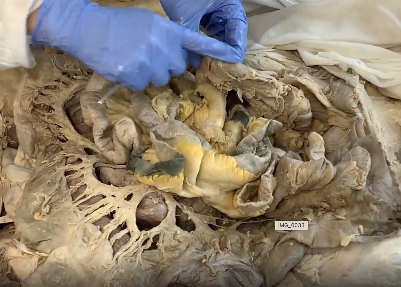
black - celiac trunk
yellow - left gastric a.
red - splenic a.
white - common hepatic a
gold - duodenum
not on structure list
red starbust - spleen
pancreas uncinate/head/neck/body - orange, lt blue, lt green, dark green
What plane travels through the pylorus of the stomach and at what vertebral level?
Transpyloric plane at L1
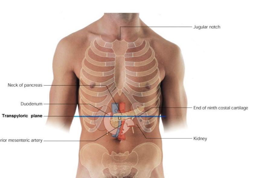

List the structures located at the transpyloric plane.
Pyloric orifice, fundus of gallbladder, hila of kidneys, origin of SMA, hepatic portal v., colic and duodenojejunal flexures, 1st part of duodenum, body of pancreas, spleen, cisterna chyli, and conus medullaris
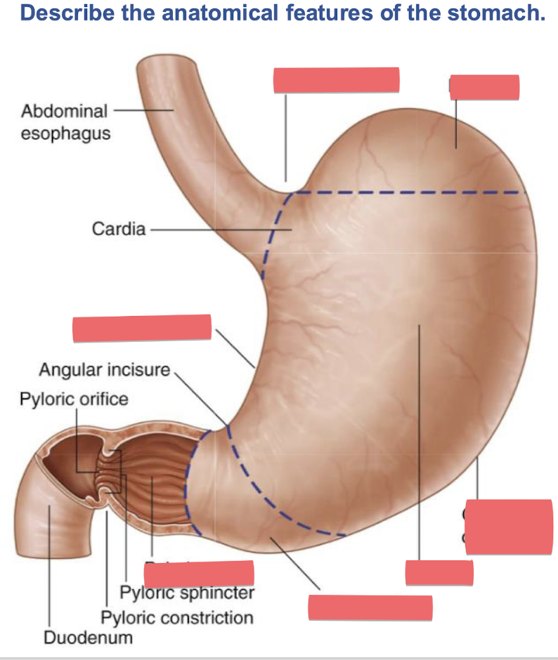
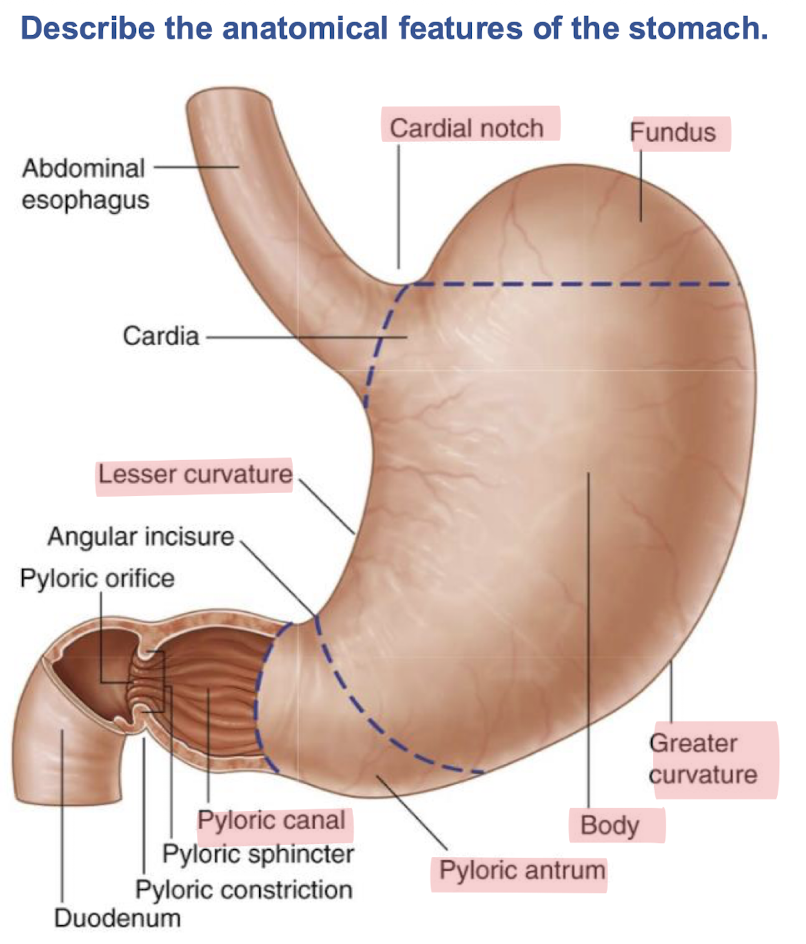
pink cardiac region
red fundus
blue greater curvature
orange lesser curvature
black pylorus (aka pyloric antrum/canal)
yellow parietal peritoneum
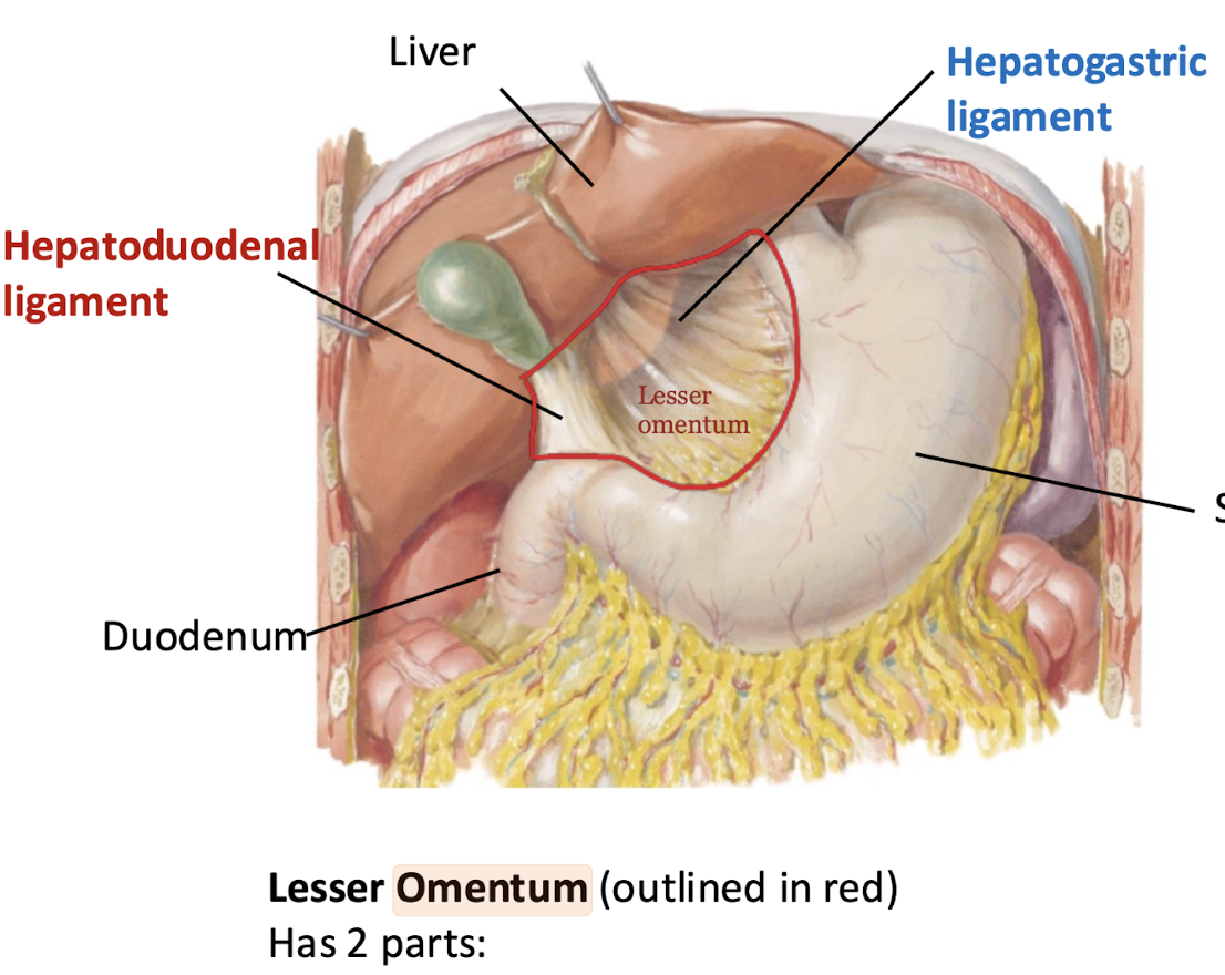
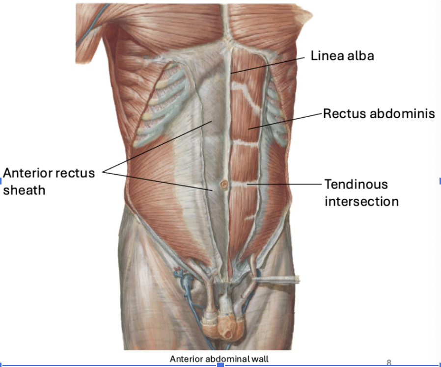
Falciform Ligament
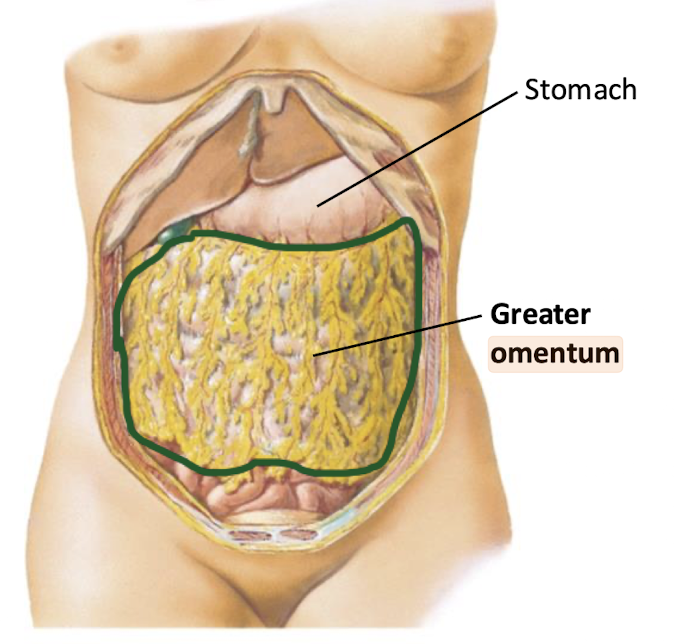
Which omentum connects to the greater curvature of the stomach?
Greater omentum
(omenta are double-layered, fatty peritoneum folds connecting the stomach to adjacent organs)
Which omentum connects to the lesser curvature of the stomach?
Lesser omentum
At what costal cartilage and vertebral level does the pyloric part of the stomach lie?
9th costal cartilage and L1 vertebra (left of the midline)
On which side of the abdomen is the pylorus usually located?
Right side
Between what vertebral levels is the pylorus usually located?
Can vary from L2-L4
What specific part of the stomach is continuous with the superior (1st part) of the duodenum?
Pylorus
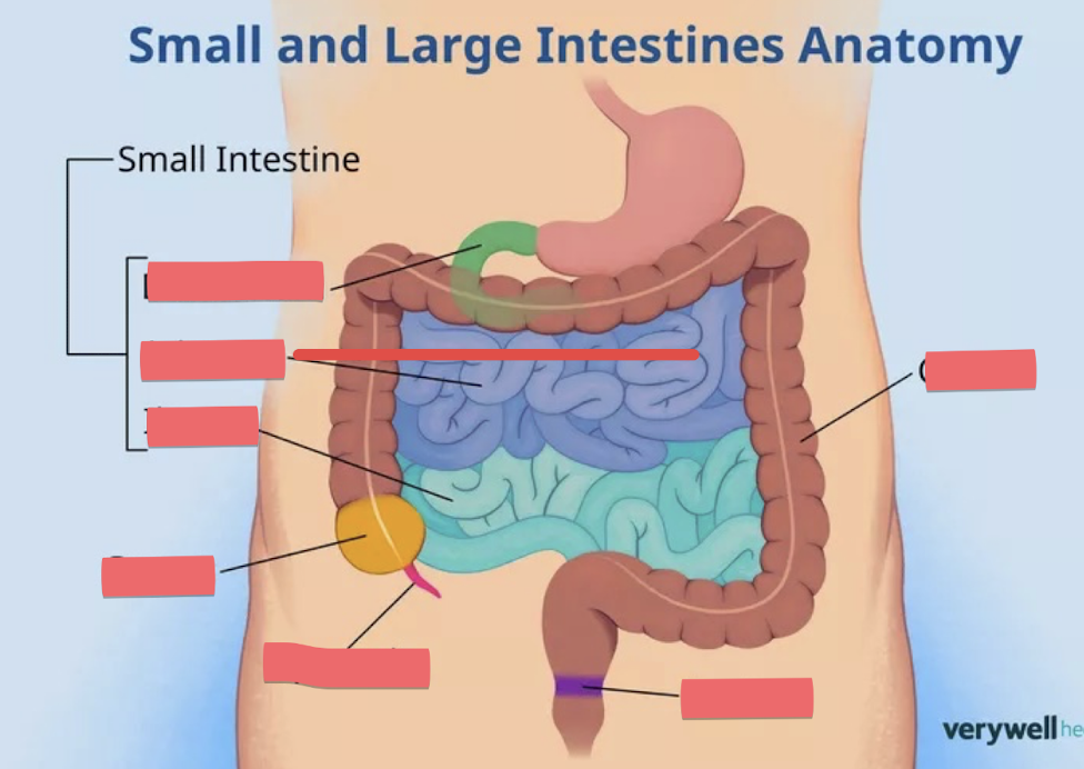
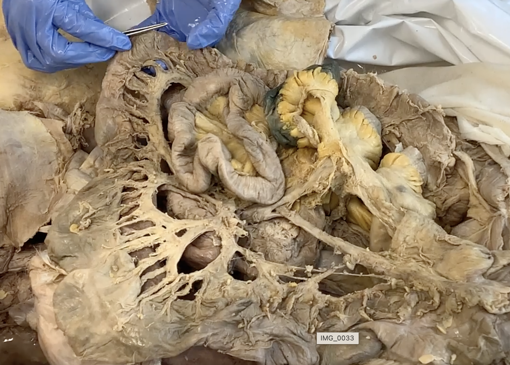
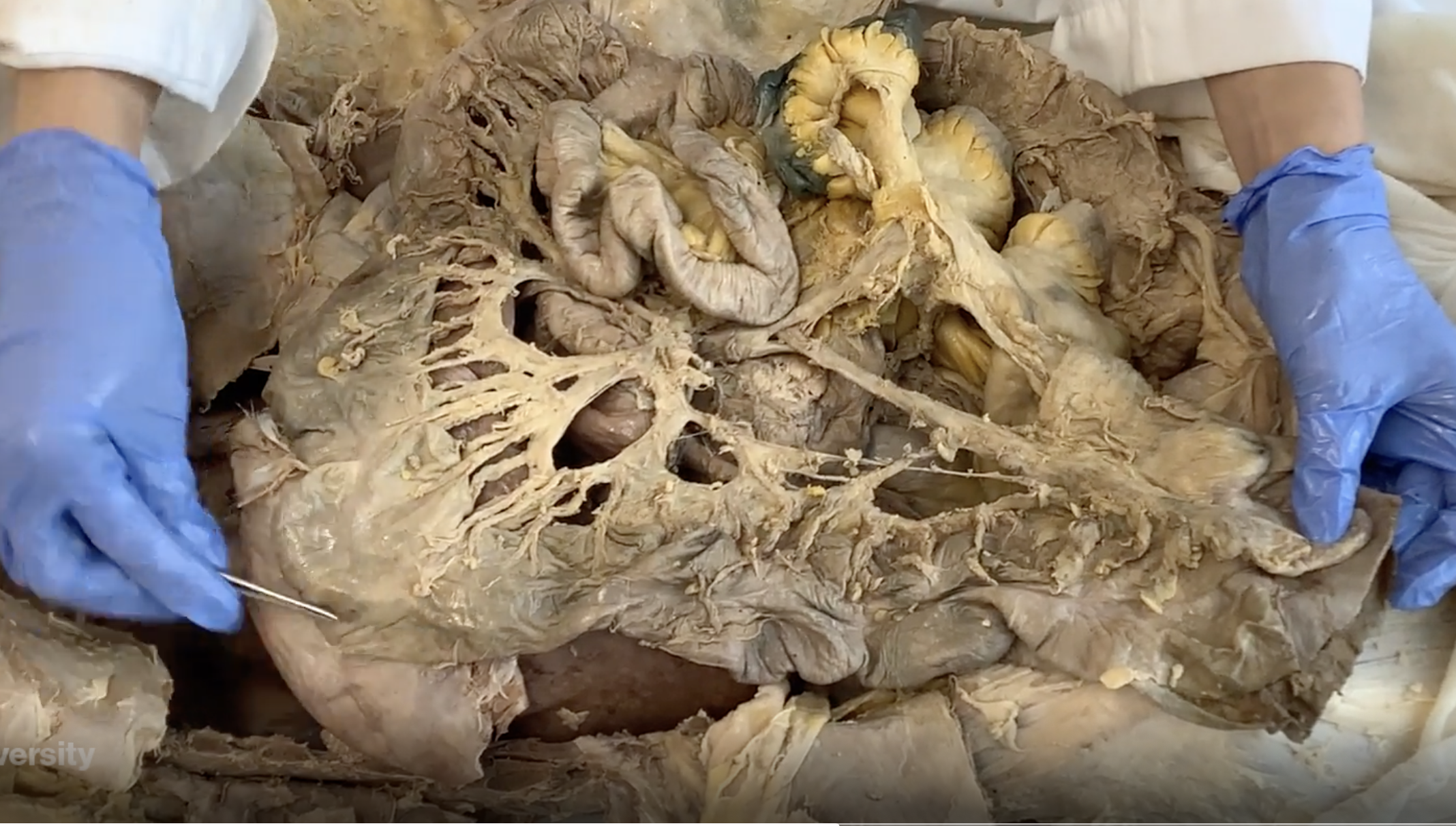
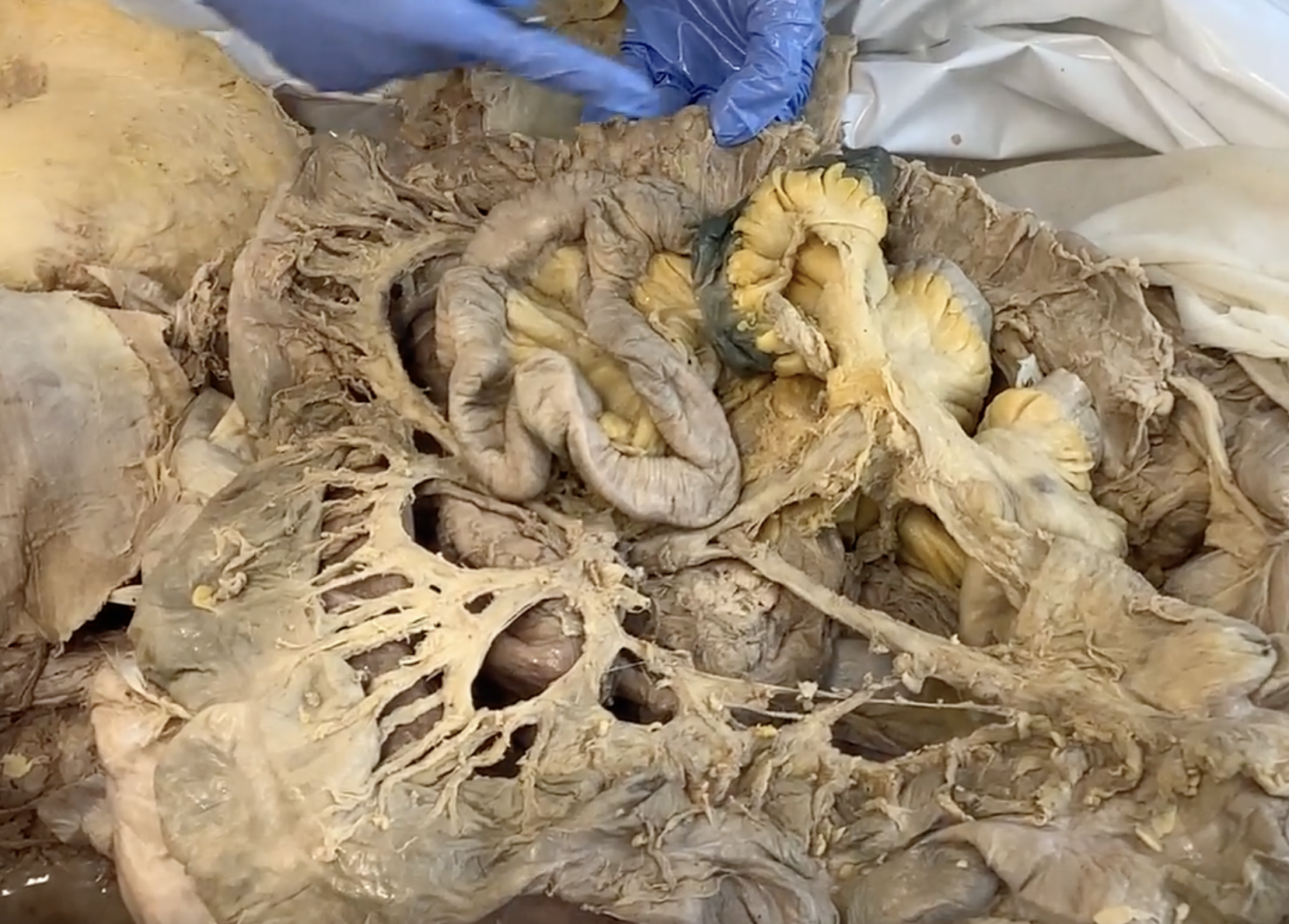
red duodenum
yellow jejunum
purple ascending colon
lt green transverse colon
pink cecum
light blue sigmoid colon
not pictured: ileum, spleen, appendix, descending colon
What structures open into the second part of the duodenum?
Biliary and main pancreatic ducts
jejunum (will be pinned in upper L of abdomen)
ileum (will be pinned in lower R quadrant)
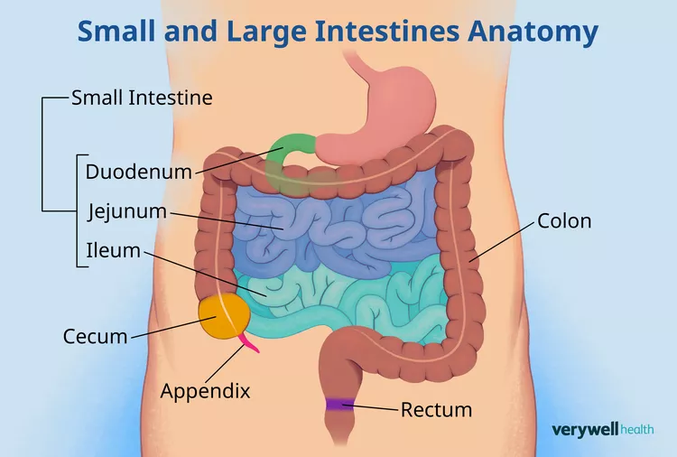
Which vessel courses anterior to the third part of the duodenum?
Superior mesenteric artery (SMA)
From which two "guts" is the large intestine derived?
Midgut and hindgut
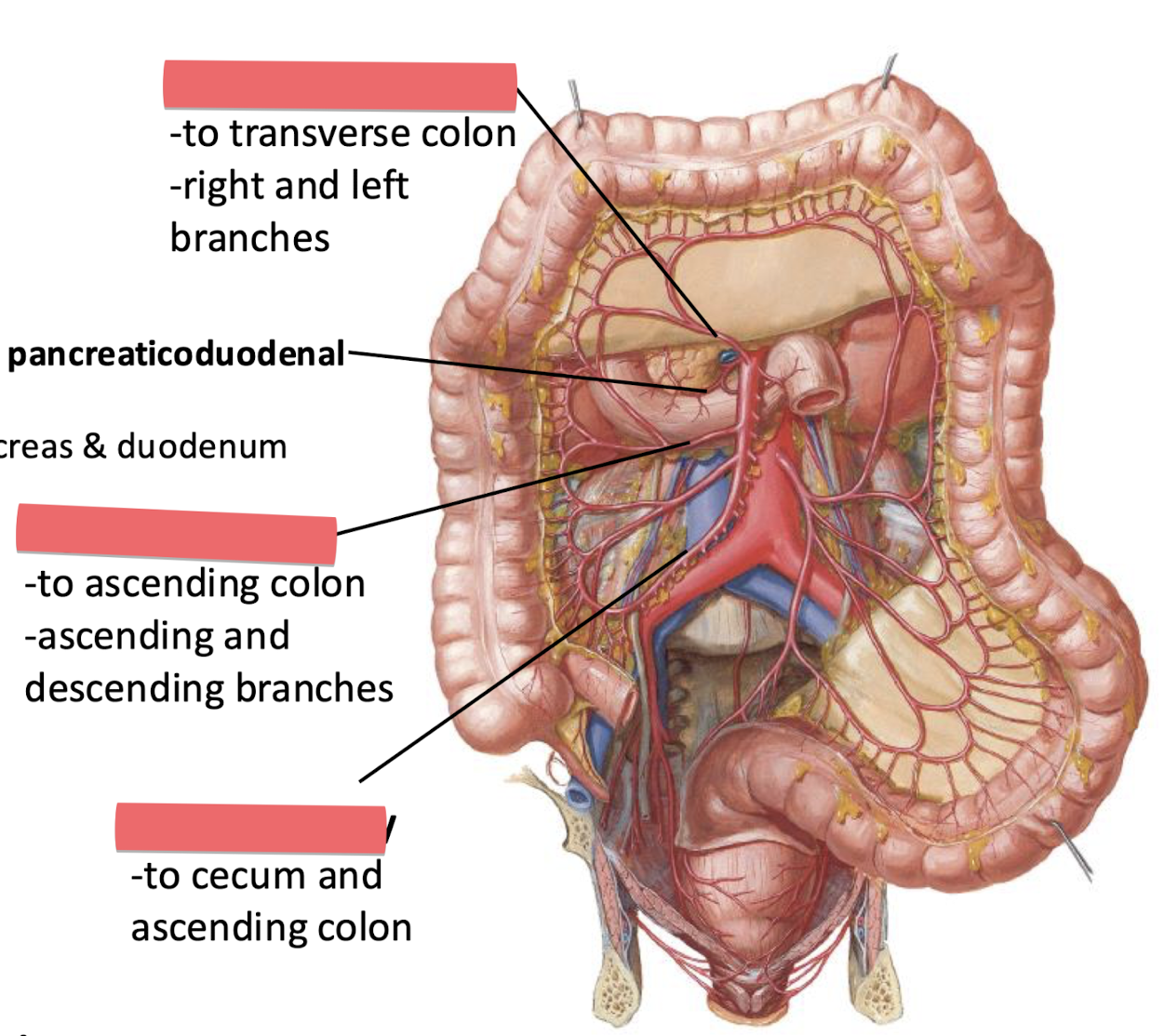
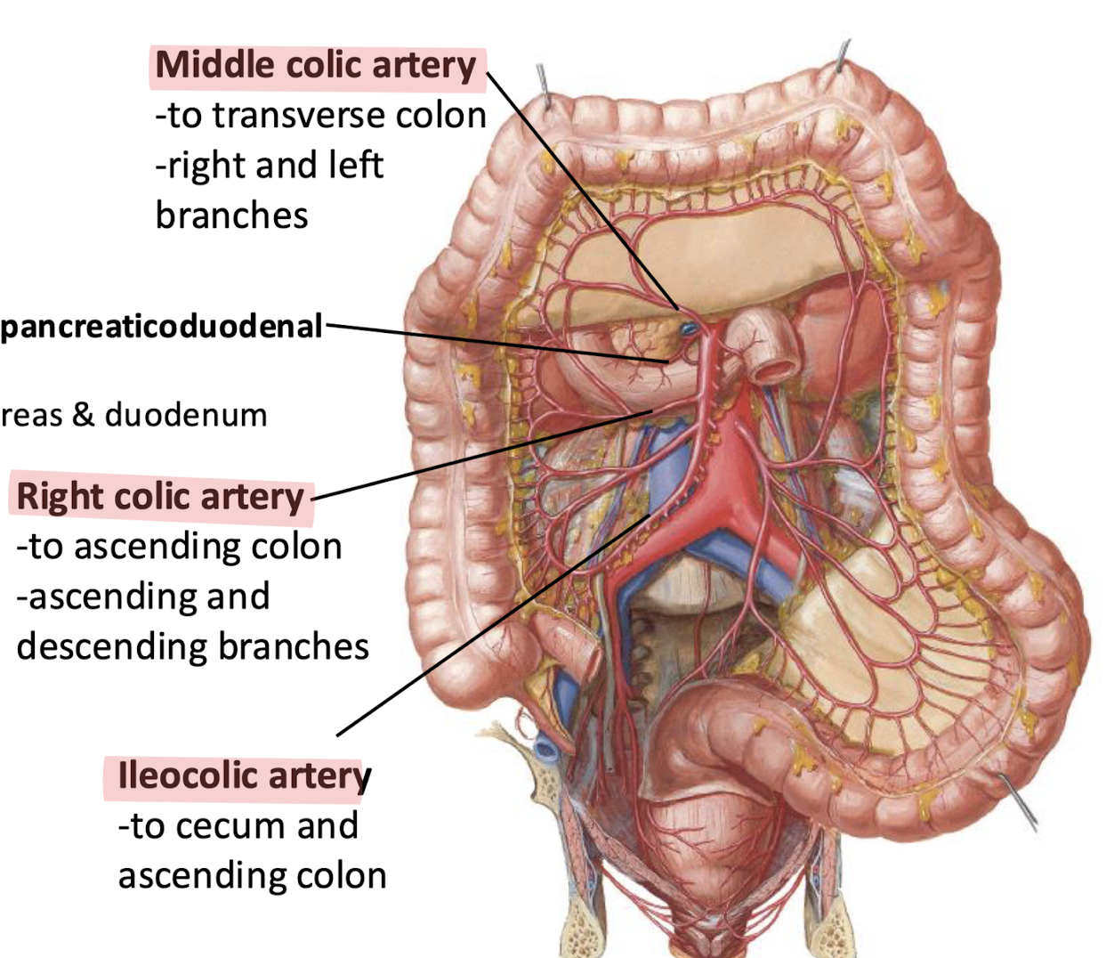
Which artery supplies the ascending colon?
Right colic artery (branch of the SMA)
Which artery supplies the descending colon?
Left colic artery (branch of the IMA)
In which abdominal quadrant does the cecum sit?
Lower right quadrant
Cecum
Appendix
Splenic / Left Colic Flexure
Hepatic / Right Colic Flexure
Descending Colon
Sigmoid Colon
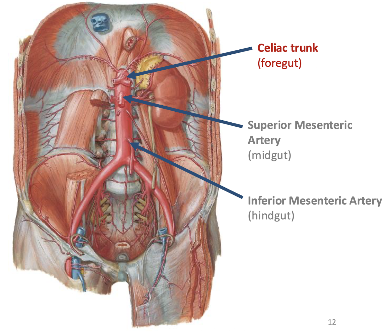
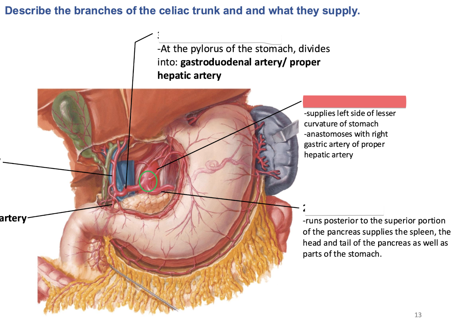
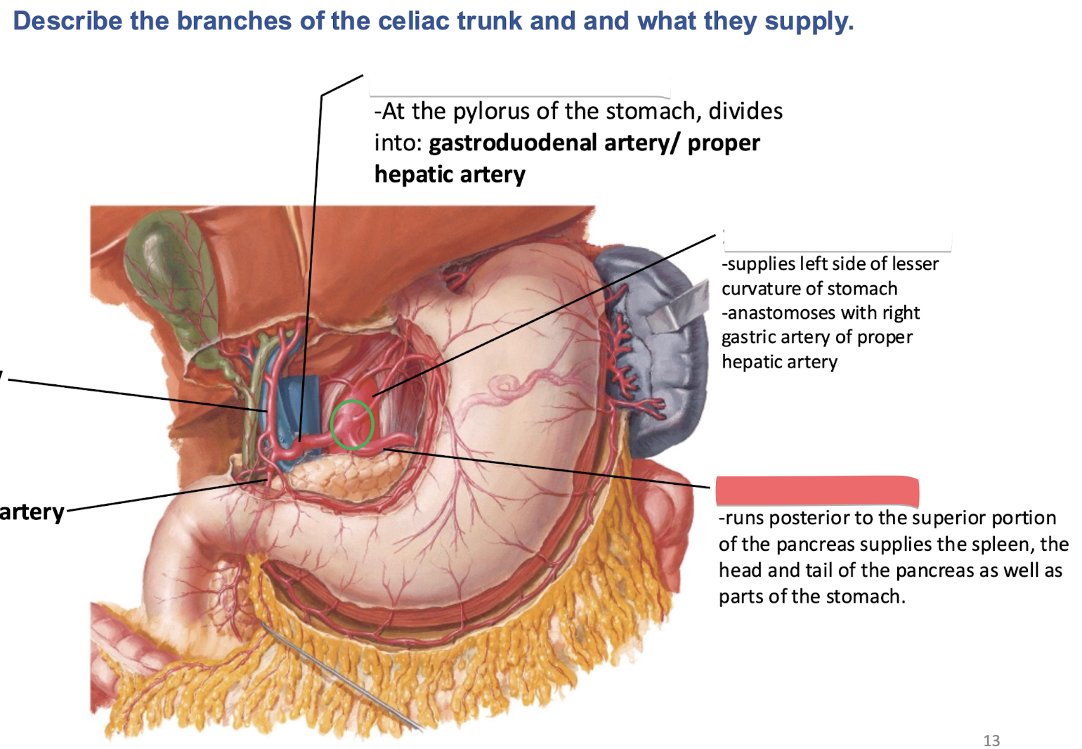
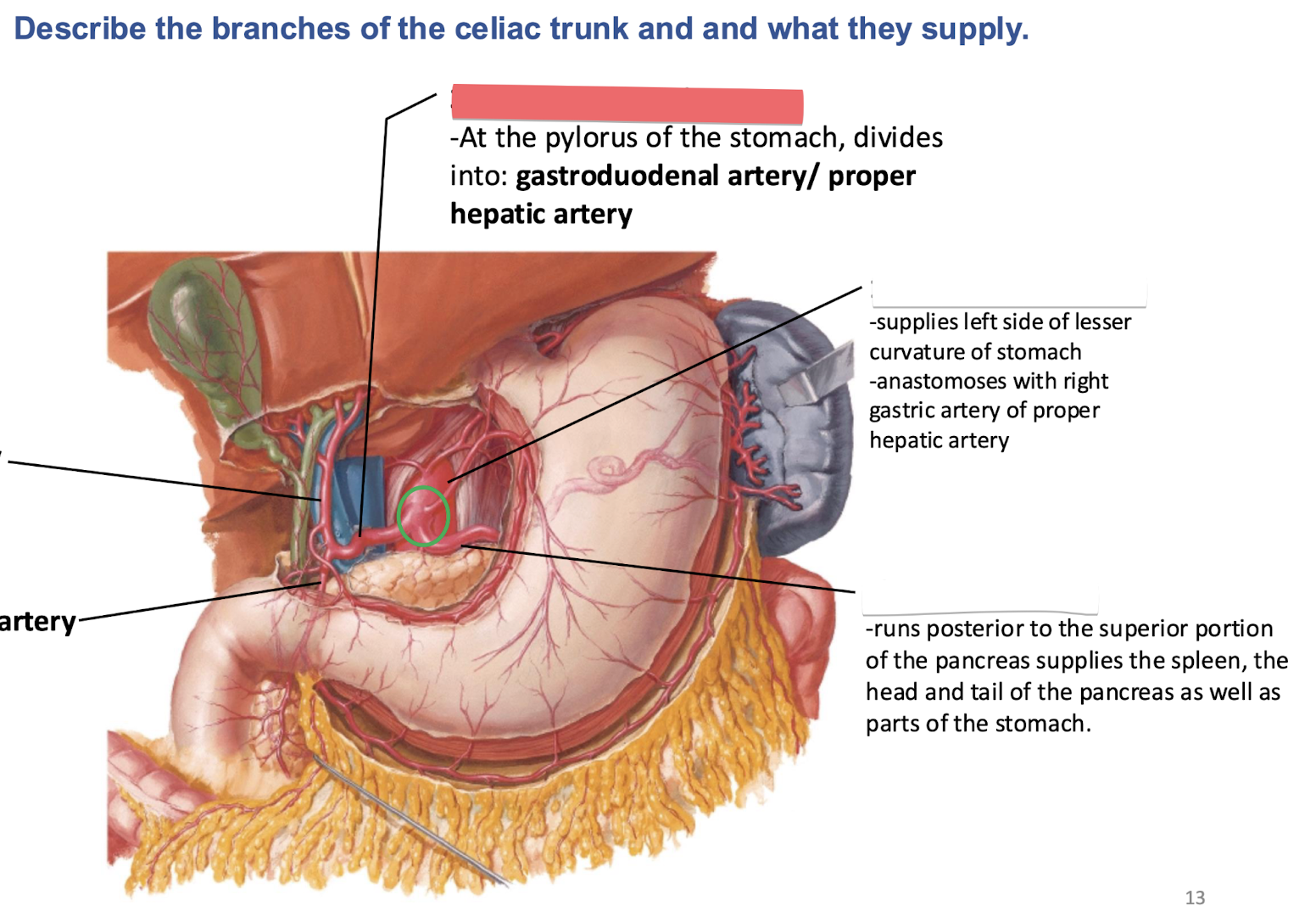
At what vertebral level is the celiac trunk found and which gut does it supply?
T12/L1; it supplies the Foregut
What structures are supplied by the left gastric artery?
Abdominal esophagus and the lesser curvature of the stomach
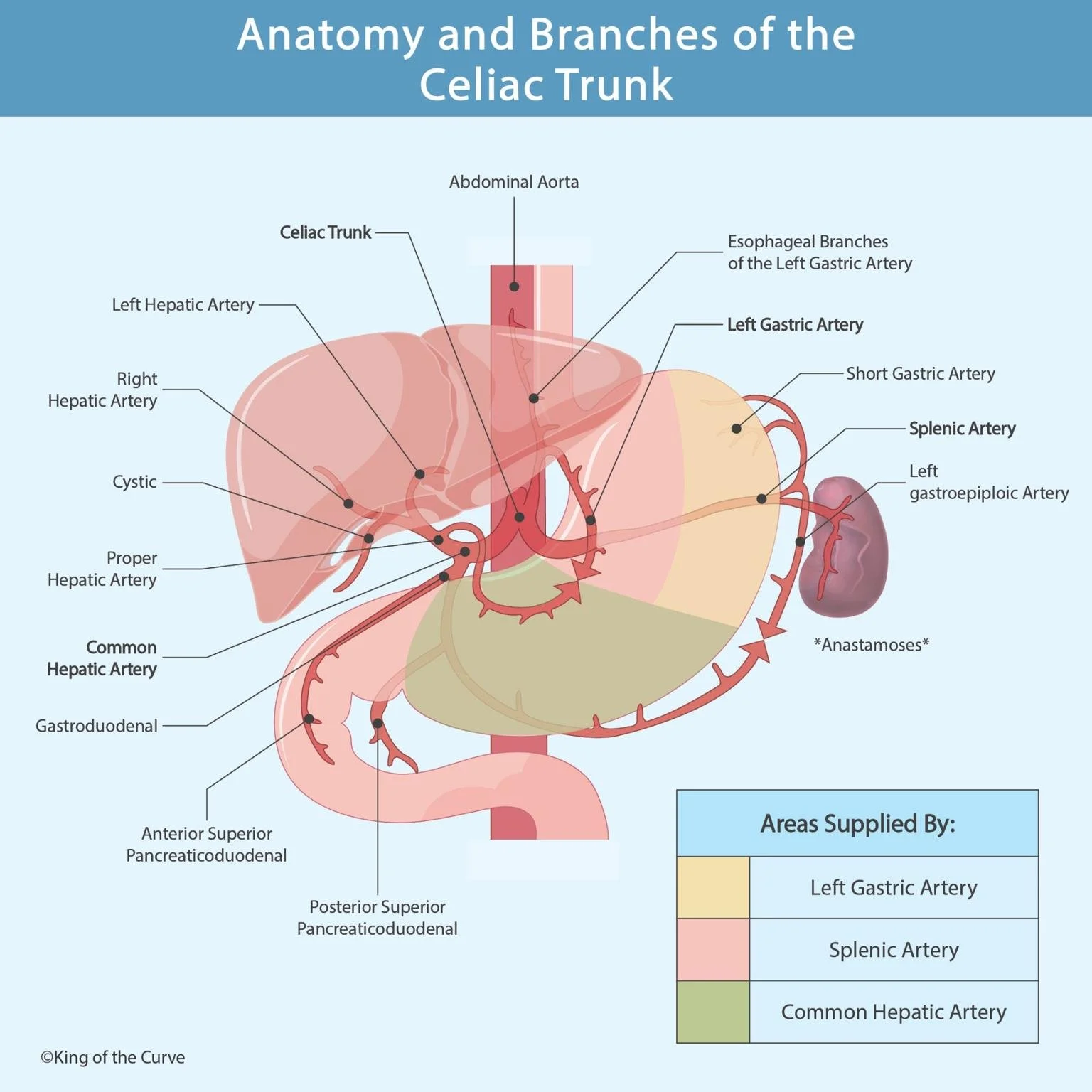
What structures are supplied by the splenic artery?
Pancreas, part of the stomach, and the spleen

What structures are supplied by the common hepatic artery?
Liver, gallbladder, head and uncinate process of pancreas, and the right part of the greater curvature of the stomach

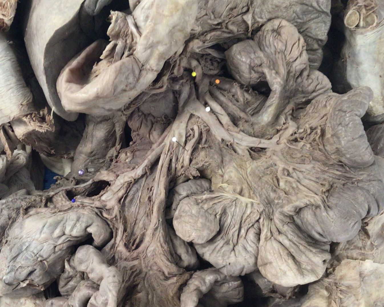
orange - superior mesenteric a.
yellow - middle colic a.
white - intestinal a.
blue - ileocolic a.
purple - right colic a.
SMA and its branches
(not picture: intestinal aa)
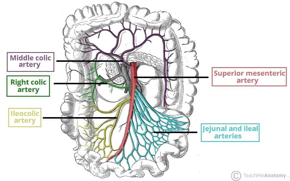
At what vertebral level is the SMA located and which gut does it supply?
L1; it supplies the Midgut
The superior mesenteric artery crosses anterior to which organ?
3rd part of the duodenum
What structures do the intestinal branches of the SMA supply?
Jejunum and ileum
What structures does the ileocolic artery supply?
Terminal ileum, cecum, and appendix
What structure does the right colic artery supply?
Ascending Colon
What part of the transverse colon is supplied by the middle colic artery?
Proximal 2/3 of the transverse colon

white- inferior mesenteric a.
lt blue - left colic a.
yellow - sigmoidal aa.
dk blue - superior rectal a.
At what vertebral level is the IMA located and which gut does it supply?
L3; it supplies the Hindgut
Which parts of the colon are supplied by the left colic artery?
Lateral 1/3 of the transverse colon and the descending colon
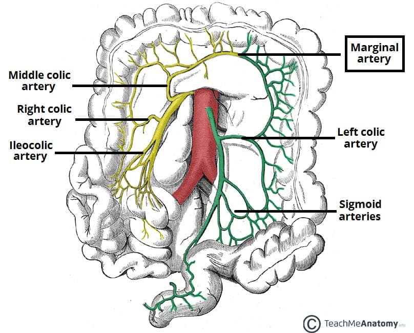
What does the Sigmoidal artery supply?
Sigmoid Colon
What does the superior rectal artery supply?
Rectum and proximal anal canal
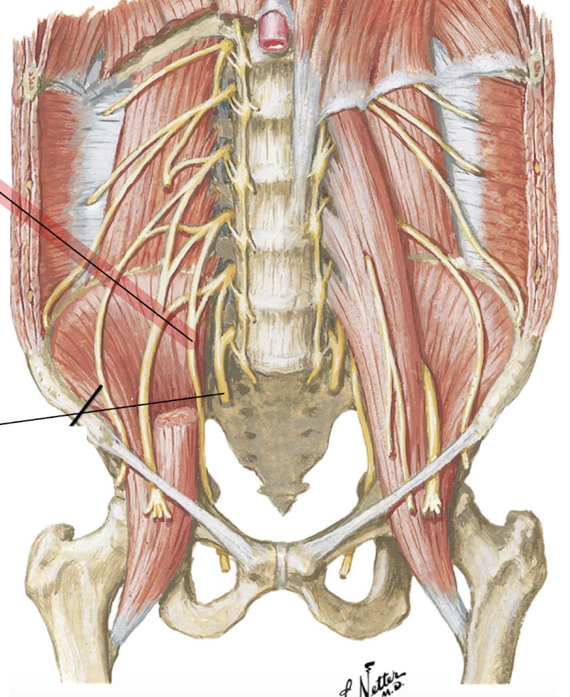
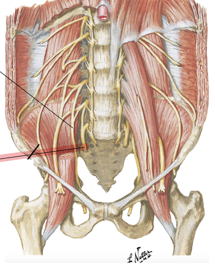
blue - transversus abdominis m.
pink - quadratus lumborum m.
red - psoas major m.
green - iliacus m.
[all paired, all left-sided]
![<p>blue - transversus abdominis m.</p><p>pink - quadratus lumborum m.</p><p>red - psoas major m.</p><p>green - iliacus m.</p><p>[all paired, all left-sided]</p><p></p>](https://assets.knowt.com/user-attachments/382c48ae-9e54-4cee-8319-f16f18126a01.png)
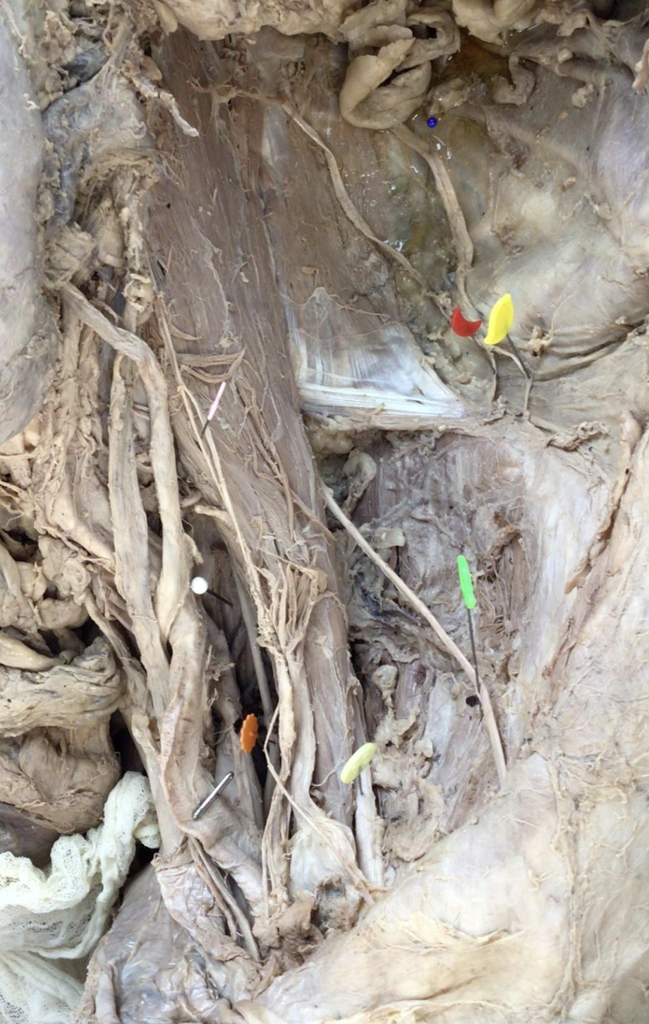
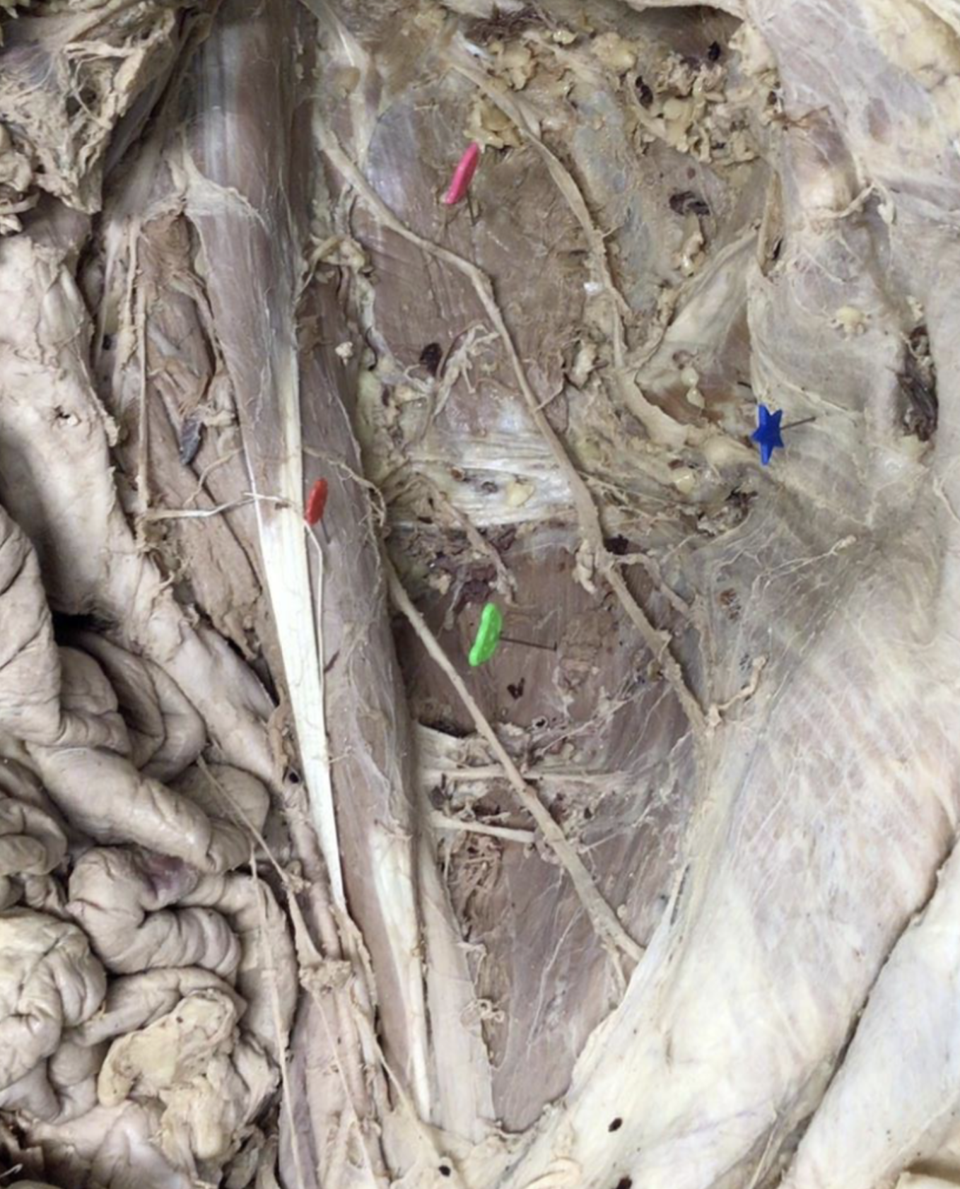
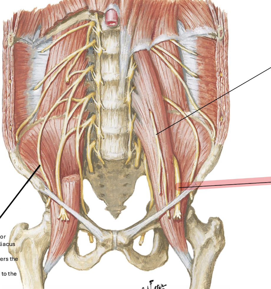
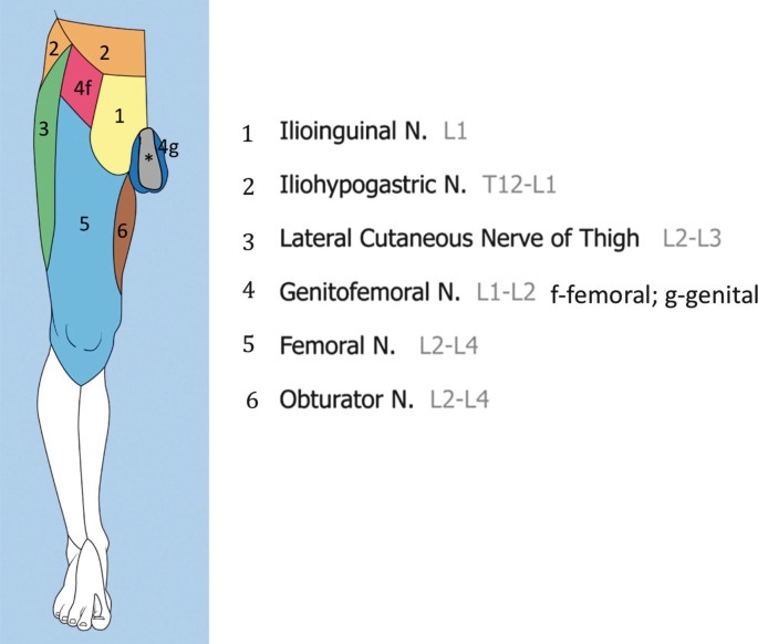
blue - subcostal nerve T12
bright yellow - iliohypogastric nerve L1
red - ilioinguinal nerve L1
pink - genitofemoral nerve L1, L2
green - lateral femoral cutaneous nerve L2, L3
lt yellow - femoral nerve L2, L3, L4
orange - obturator nerve L2, L3, L4
white - lumbosacral trunk L4, L5
[paired nerves, all left-sided]
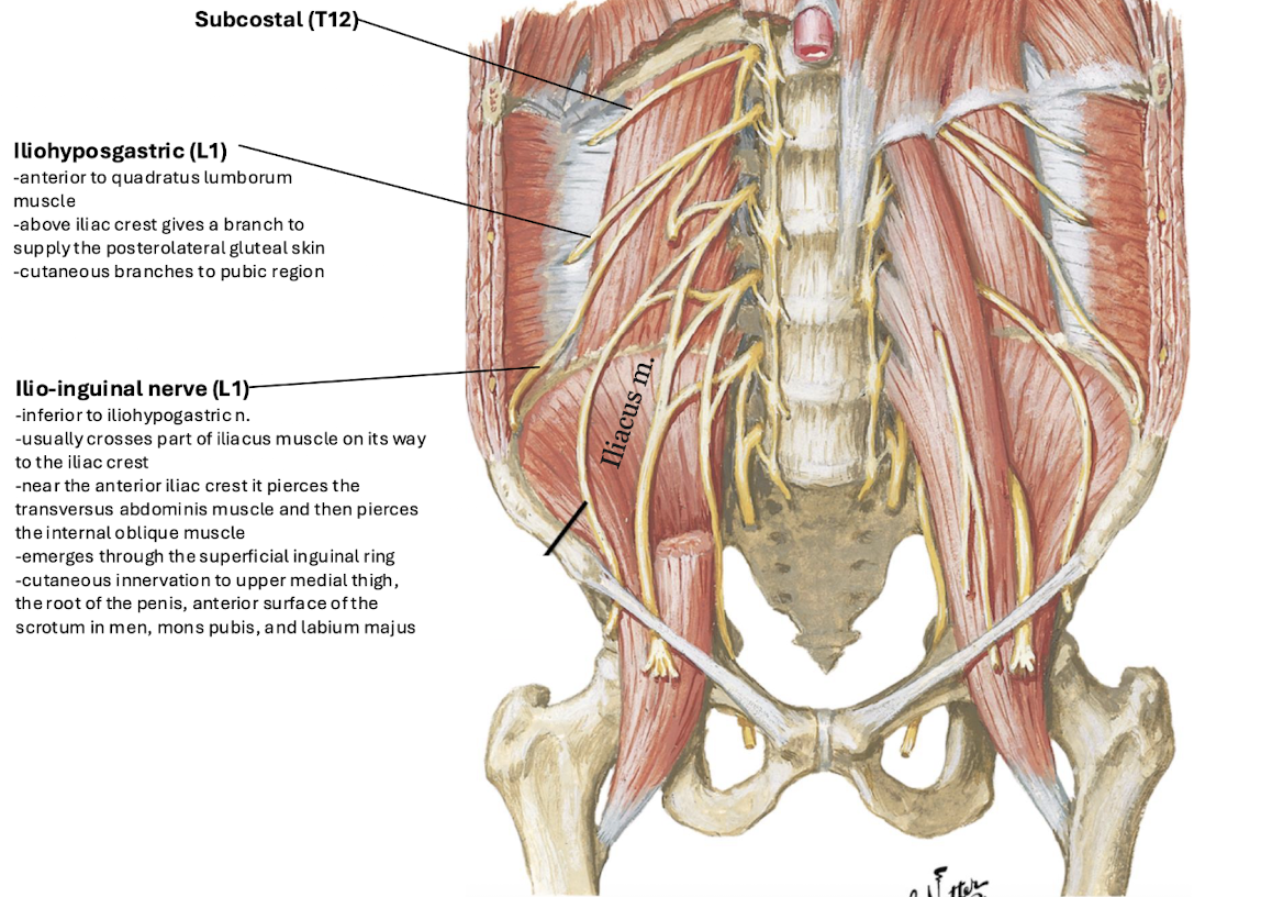
What is the root and location of the subcostal nerve?
T12; located inferior to rib 12.
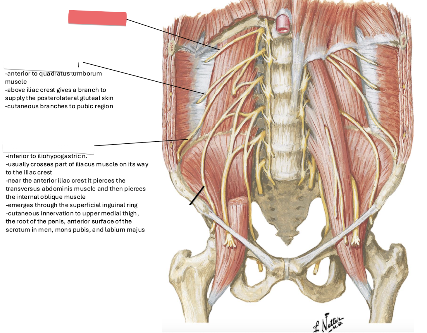
Subcostal Nerve
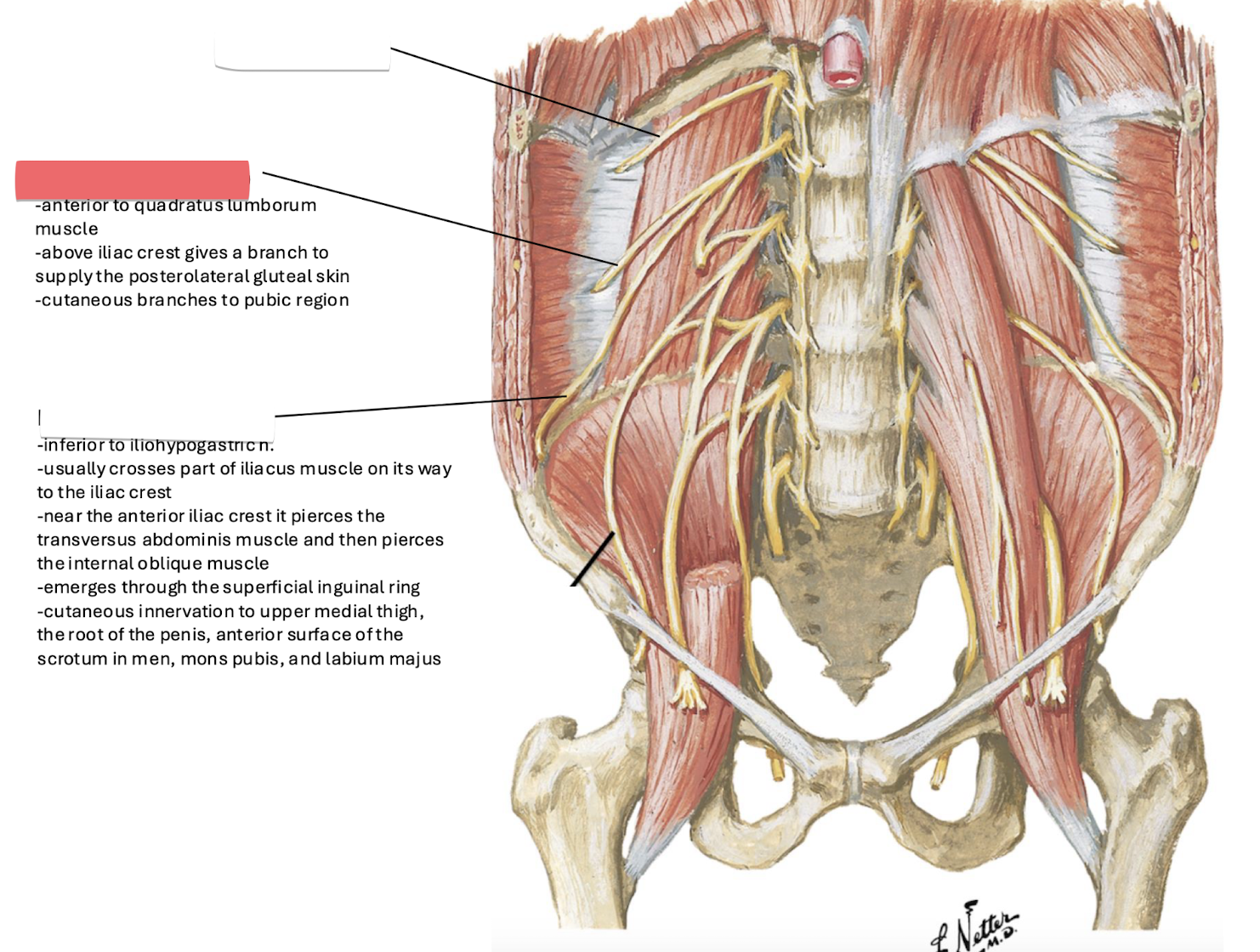
Iliohypogastric
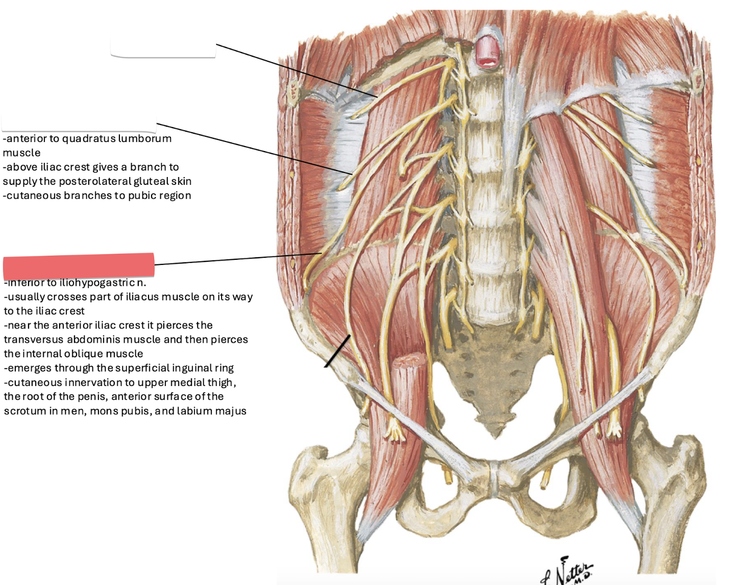
iliolinguinal
What is the root and location of the iliohypogastric nerve?
L1; located anterior to the quadratus lumborum.
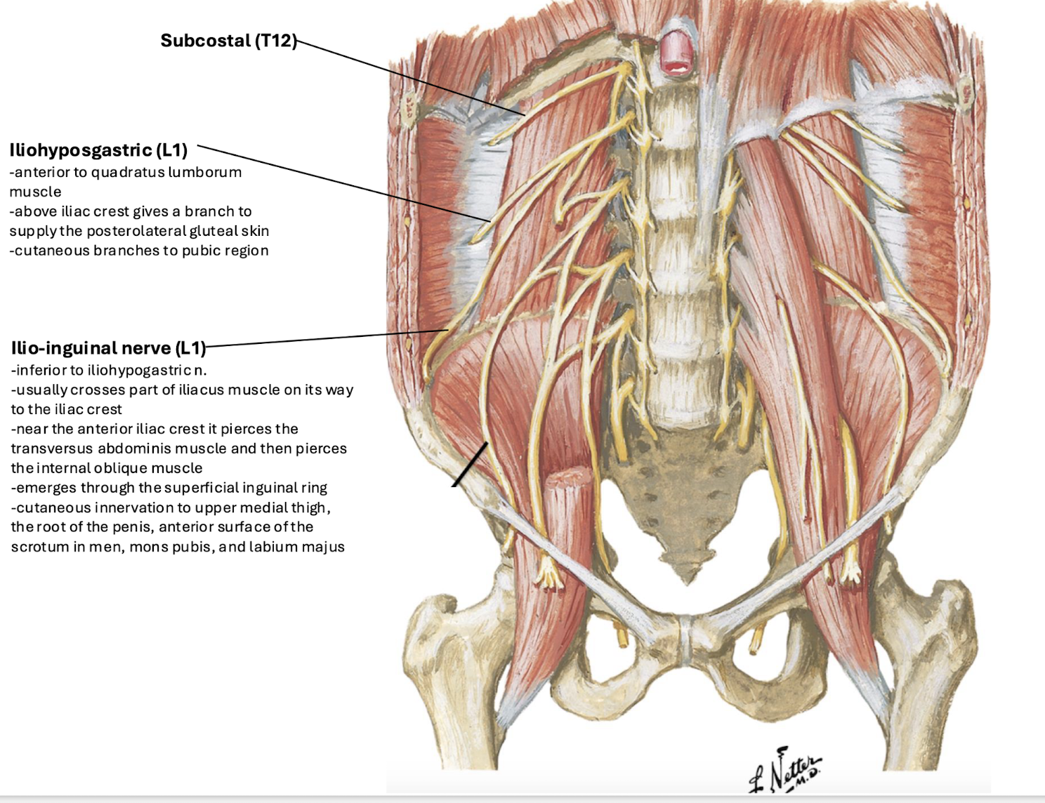
Describe the path of the ilioinguinal nerve (L1).
Usually crosses part of the iliacus muscles toward the iliac crest; near the anterior crest line, it pierces the transversus abdominis then the internal oblique muscle.
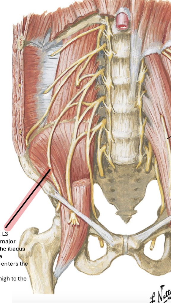
Lateral femoral cutaneous n.
What are the roots and path of the lateral femoral cutaneous nerve?
L2 and L3; emerges from the lateral border of the psoas major, passes obliquely downward across the iliacus toward the ASIS, then passes posterior to the inguinal ligament to enter the thigh.

Femoral Nerve
What are the roots and path of the femoral nerve?
L2 to L4; descends through the psoas major, emerging from its lower lateral border; lies on the lateral border of the psoas major and ASIS; enters the anterior thigh deep to the inguinal ligament.
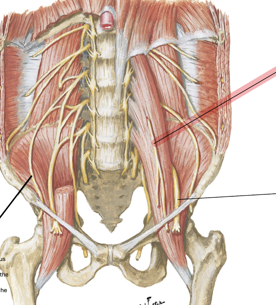
Genitofemoral Nerve
What are the roots and path of the genitofemoral nerve?
L1 and L2; passes downward in and emerges on the anterior surface of the psoas major; passes posterior to the ureter.


Obturator Nerve
What are the roots and path of the obturator nerve?
L2 to L4; emerges from the medial side of the psoas major near the pelvic brim, continues posterior to common iliac vessels, and enters the obturator canal to reach the medial thigh.
Lumbosacral Trunk
What are the roots and destination of the lumbosacral trunk?
L4, L5; passes over the sacrum and descends into the pelvis to participate in the sacral plexus.
What is the motor and sensory function of the subcostal nerve?
Muscles of the anterolateral abdominal wall and overlying skin.
What is the motor and sensory function of the iliohypogastric nerve?
Muscles of the anterolateral abdominal wall; skin of the posterolateral gluteal region and pubic region.
What is the motor and sensory function of the ilioinguinal nerve?
Muscles of the anterolateral abdominal wall; skin of the upper medial thigh, root of the penis and anterior scrotum, or mons pubis and labium majus.
What is the function of the lateral femoral cutaneous nerve?
Sensory only: skin on the anterior and lateral thigh to the knee (no motor function).
What is the motor and sensory function of the femoral nerve?
Motor: Iliacus and anterior compartment of the thigh; Sensory: Anterior thigh and medial surface of the leg.
What is the function of the genitofemoral nerve branches?
Motor: Cremasteric muscle; Sensory (Genital branch): Skin of anterior scrotum or mons pubis and labium majus; Sensory (Femoral branch): Skin of upper anterior thigh.
What is the motor and sensory function of the obturator nerve?
Motor: Medial thigh muscles; Sensory: Medial aspect of the thigh, hip joint, and knee joint.
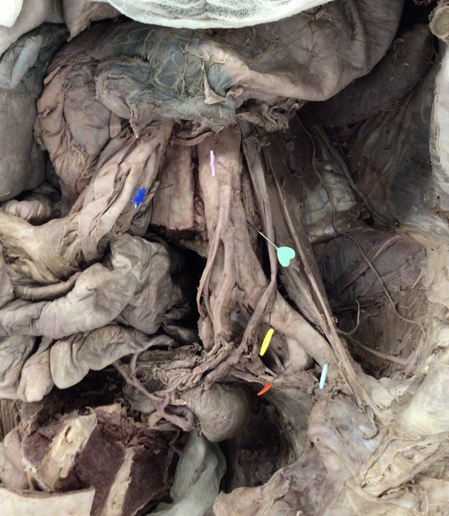
blue - IVC
pink - abdominal aorta
seafoam heart - lumbar arteries (posterior segmental vessels of the abdominal aorta)
yellow - common iliac artery
red - internal iliac artery
baby blue - external iliac arte
Does the inferior vena cava (IVC) sit to the right or left of the abdominal aorta?
Right.
At what vertebral level is the IVC formed and by what vessels?
L5; formed by the common iliac veins.
List the structures that cross anterior to the IVC.
Right common iliac artery, root of mesentery, right testicular/ovarian artery, superior part of duodenum, bile duct, portal vein, and liver.

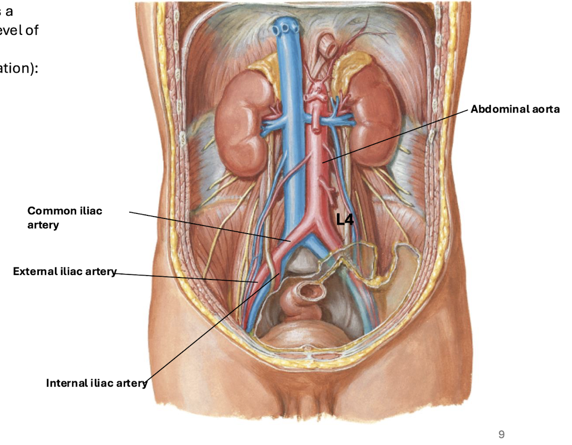
At what vertebral level does the abdominal aorta bifurcate?
L4 (bifurcates into common iliac arteries).
What are the anterior relationships of the abdominal aorta?
Pancreas, splenic vein, left renal vein, and inferior part of the duodenum.
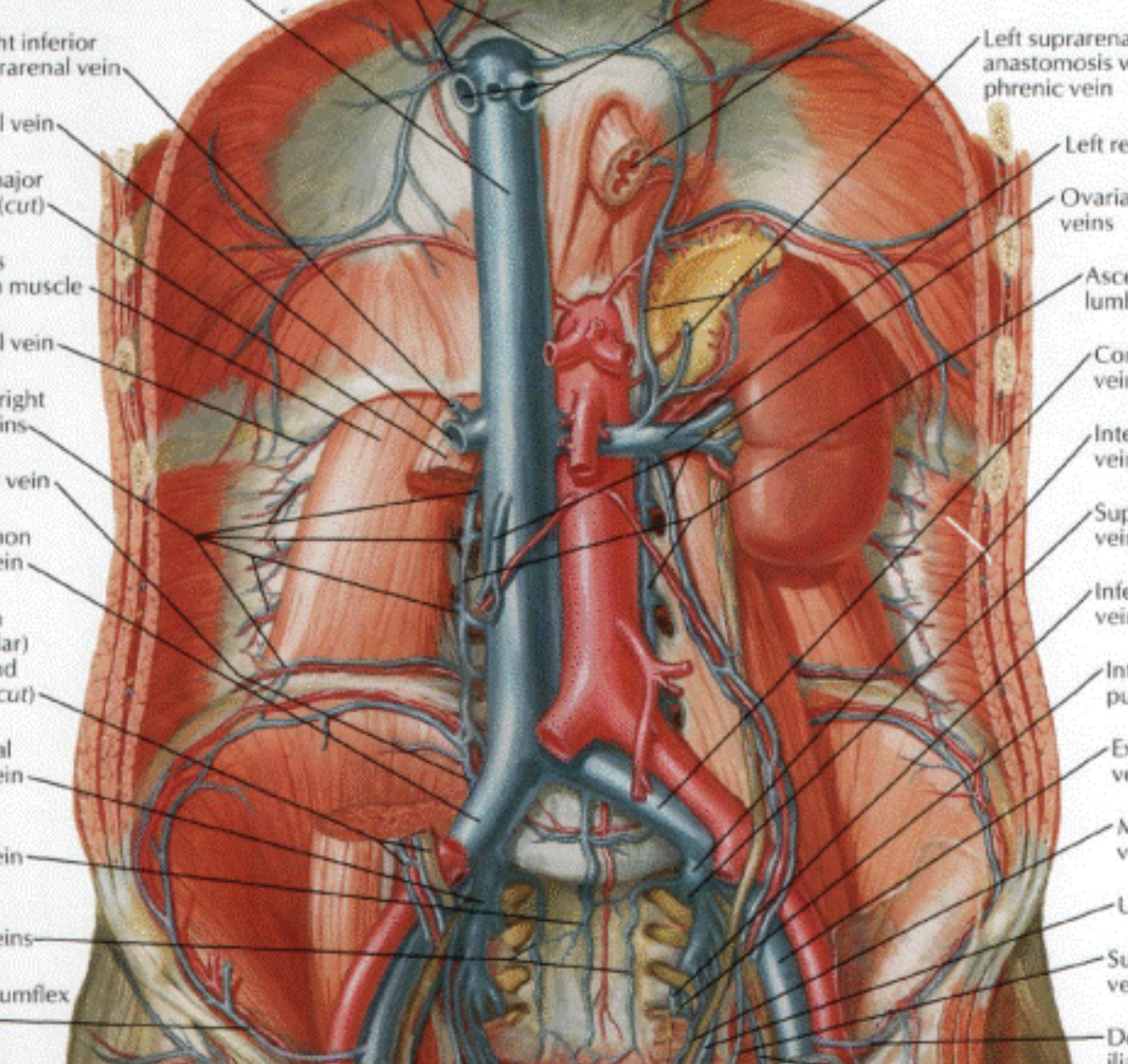
What are the posterior relationships of the abdominal aorta?
Lumbar veins.
What structures are located to the right of the abdominal aorta?
Cisterna chyli, thoracic duct, azygos vein, right crus of the diaphragm, and IVC.
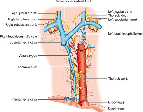
What do the lumbar arteries supply?
Posterior abdominal wall muscles, joints of the lumbar spine, lower 2/3 of the spinal cord, and the lower part of the sympathetic trunk.
What is the origin, insertion, and innervation of the psoas major?
O: Transverse processes of lumbar vertebrae; I: Lesser trochanter of femur; IN: Anterior rami L1–L3.

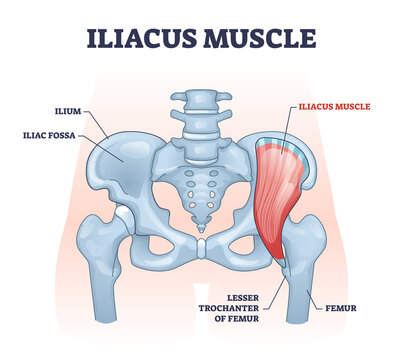
What is the origin, insertion, and innervation of the iliacus?
O: Upper 2/3 of iliac fossa; I: Lesser trochanter of femur; IN: Femoral nerve (L2–L4).
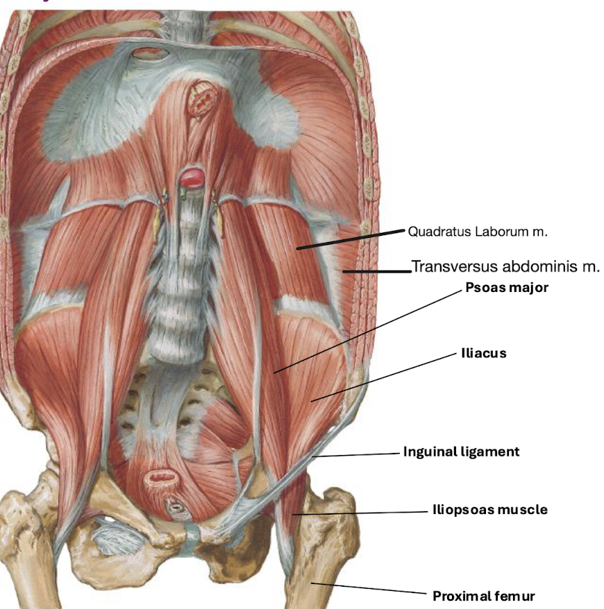
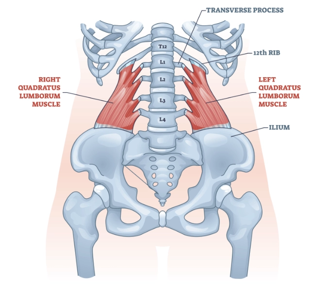
What is the origin, insertion, and innervation of quadratus lumborum?
O: Transverse process of L5, iliac crest; I: Transverse processes of L1–L5 and rib 12; IN: Anterior rami T12 and L1–L4.

What muscle is formed by the union of the psoas major and iliacus?
The iliopsoas muscle (located in the anterior thigh).

What does the anterior thoracolumbar fascia cover?
The anterior aspect of the quadratus lumborum (QL).
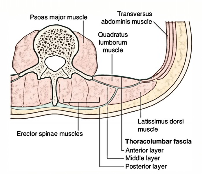
What does the middle thoracolumbar fascia separate?
The quadratus lumborum muscle from the erector spinae muscles.

What does the posterior thoracolumbar fascia surround?
The erector spinae muscles.
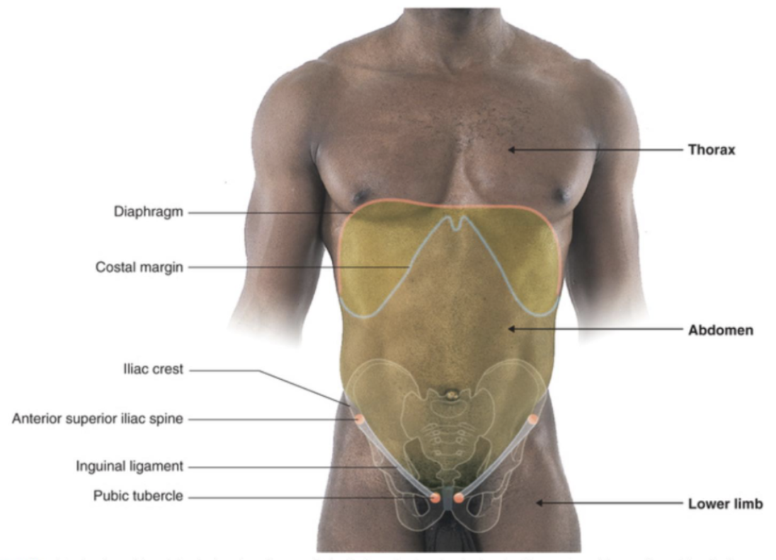
The xiphoid process is the inferior part of which bone?
The sternum
What is the vertebral level of the xiphoid process?
T9 or T10
What is the dermatome level at the xiphoid process?
T5/T6
What regions of the body does the costal margin separate?
The thoracic and abdominal walls
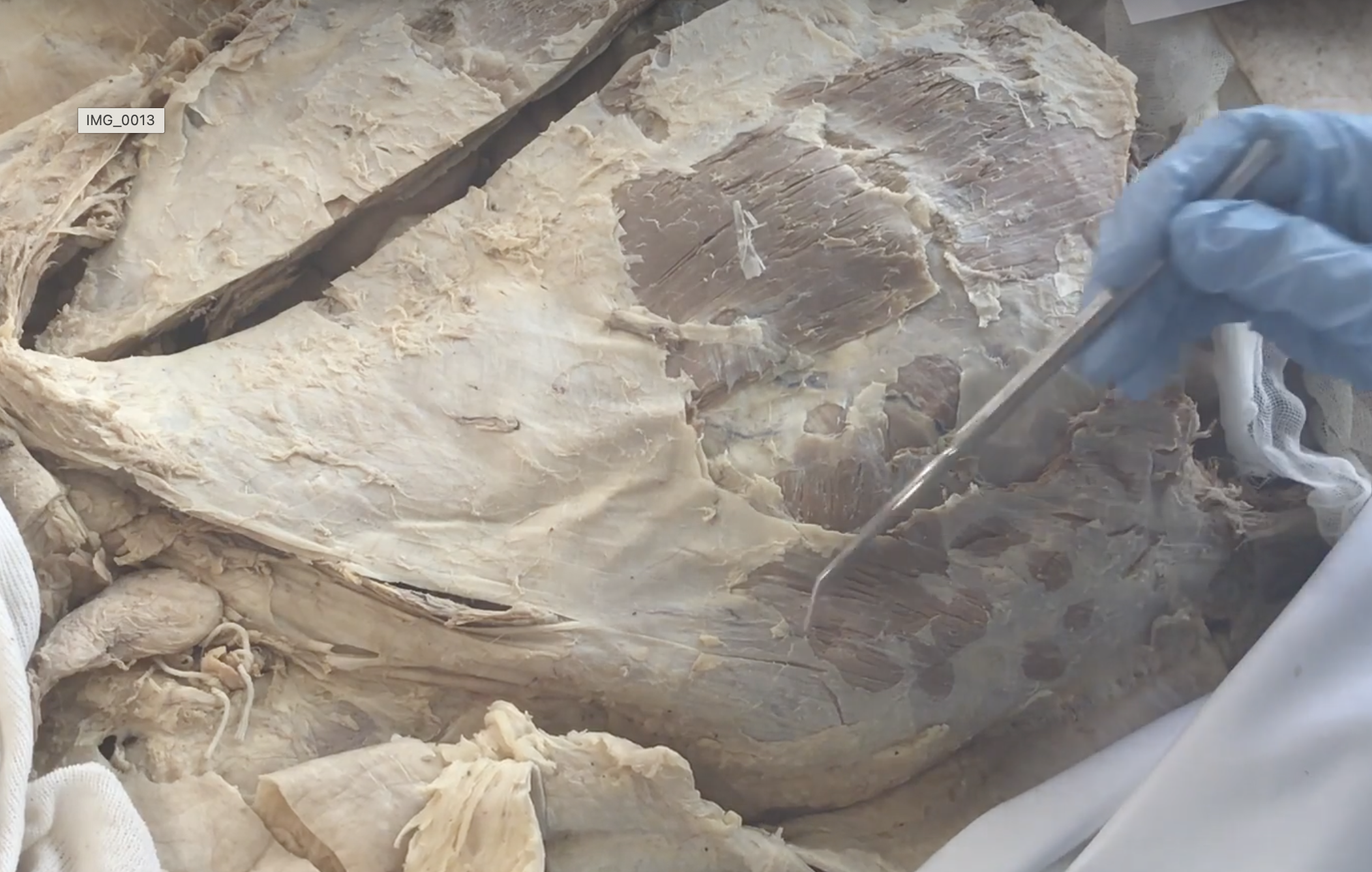
external abdominal oblique m.
"hands in pockets" fiber orientation
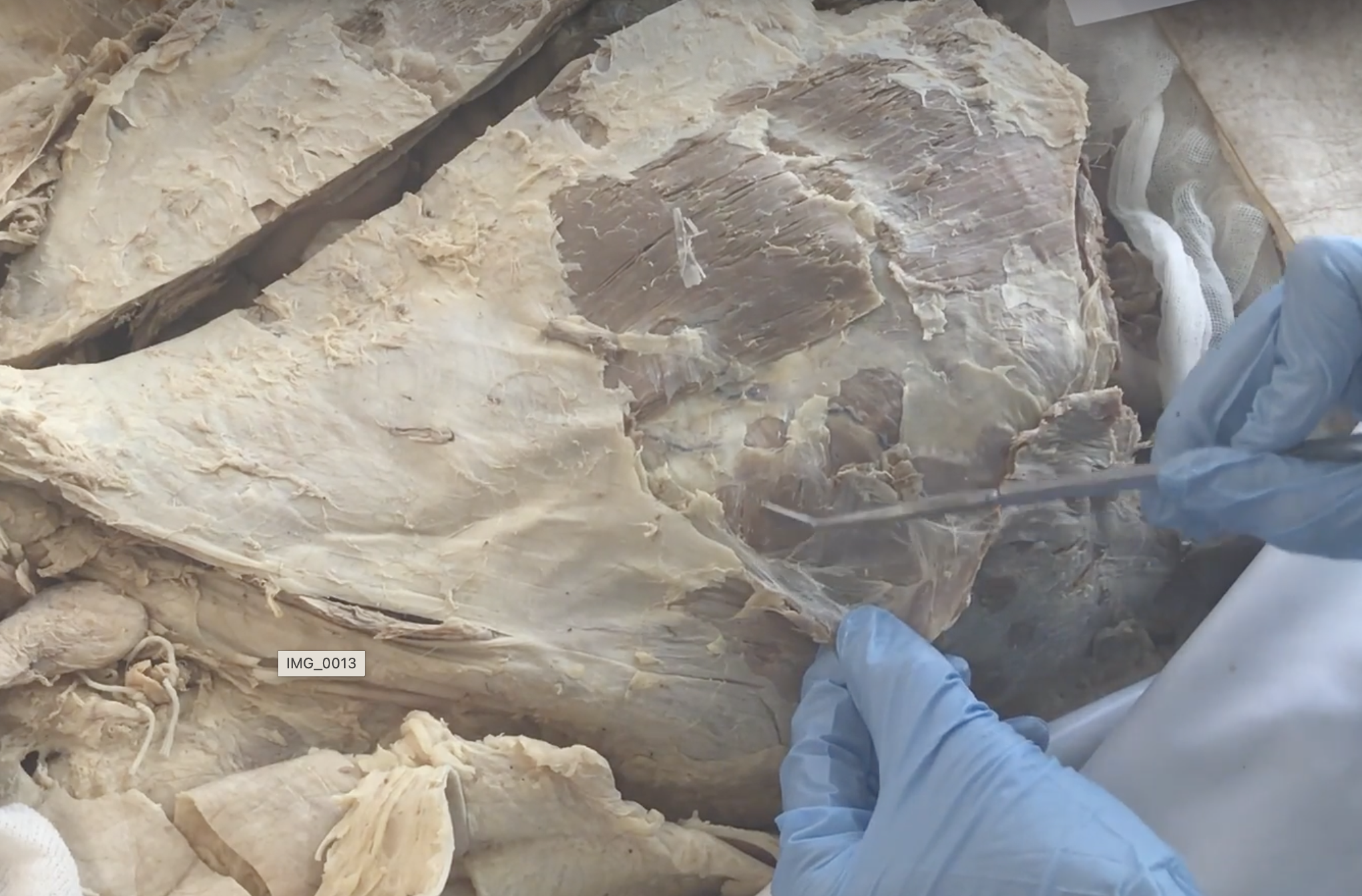
internal abdominal oblique m.
fibers run toward chin: "in toward the chin"
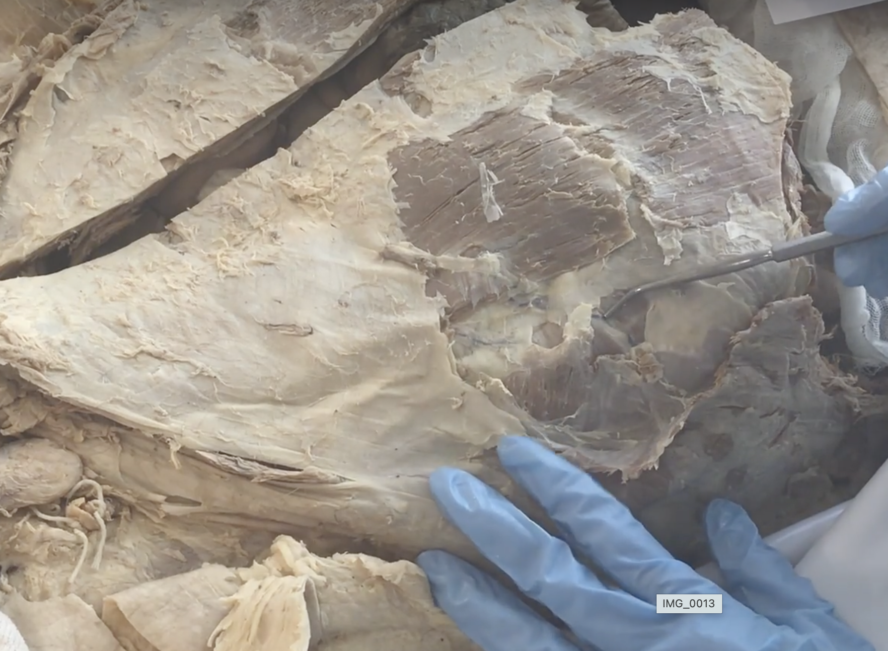
transversus abdominis m.
fibers run horizontally
What innervates the external oblique, internal oblique, and transversus abdominis?
Lower 6 intercostal (T6–T11), subcostal (T12), iliohypogastric (L1), and ilioinguinal (L1) nerves
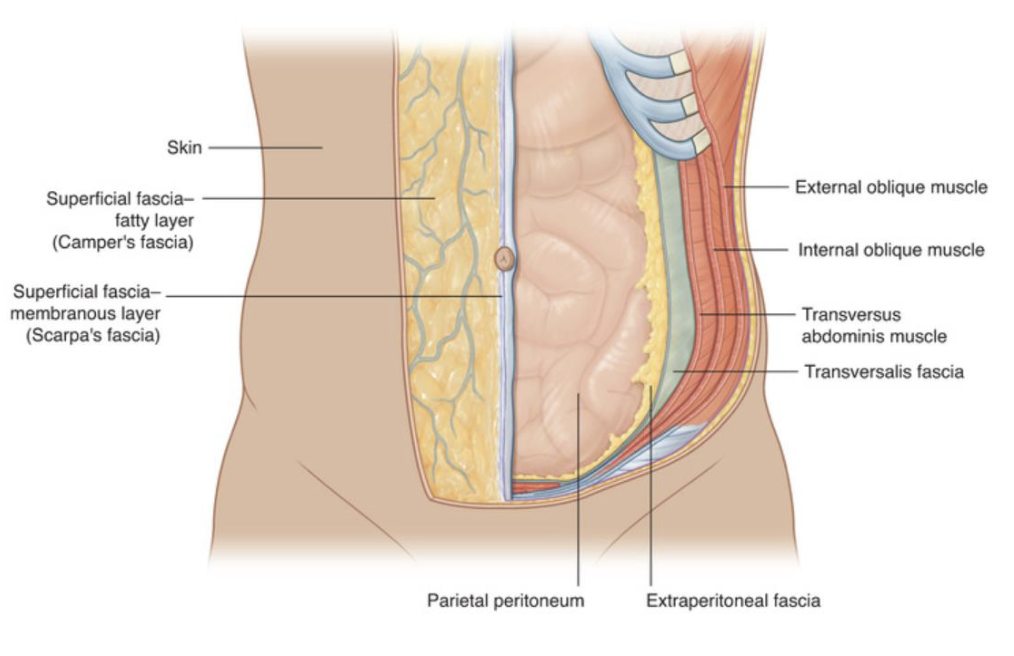
Between which two muscle layers are the abdominal nerves located?
Internal oblique and transversus abdominis

What is the common insertion for all the oblique muscles?
Rectus sheath and Linea alba