Lecture 2: Cytoskeletal Networks
1/31
There's no tags or description
Looks like no tags are added yet.
Name | Mastery | Learn | Test | Matching | Spaced | Call with Kai |
|---|
No analytics yet
Send a link to your students to track their progress
32 Terms
Polarity is caused by cytoskeleton. What do polar microtubules, polarized actin do?
Polar microtubules: transport vesicles/proteins to different ends of the cell.
Polarized actin: define cell shape and behavior
Cytoskeleton has ______ rearragements
Dynamic - always changing!
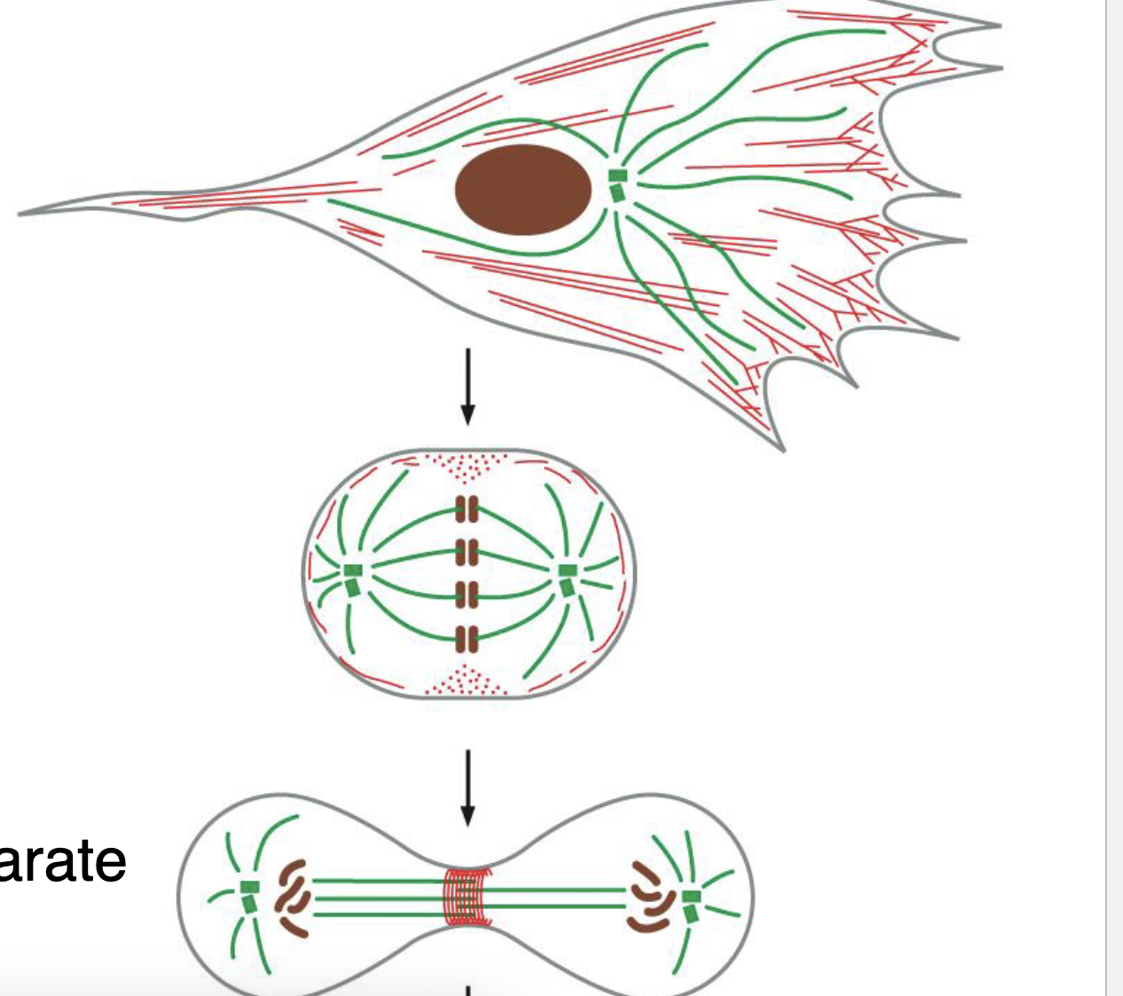
Dynamic rearrangement example in interphase, mitosis, cytokinesis
interphase: microtubules radiate from cell center, a lot of actin at cell cortex
mitosis: microtubule form mitotic spindle
actin at cell cortex disassembles
cytokinesis: microtubule keep cell components separate, actin forms a contractile ring
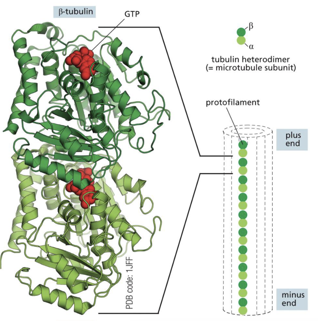
Microtubule is made up of
polar tubulin dimers.
alpha tubulin and beta tubulin forming a dimer
they both have GTP bound to it.
Heterodimers assemble and form ________
make polarized protofilaments
One ends in alpha (minus end), One ends in beta (plus end)
how many protofilaments in one microtubule?
13 parallele protofilaments.
What is a T-form heterodimer?
GTP bound heterodimer. (alpha and beta) → often promotes growht
What is a D-form heterodimer?
GDP-bound heterodimer (only beta) → often promotes shrinking
alpha tubulin is always GTP, never GDP
Depolymerization is much faster at GDP end
Depolymerization process in detail
After some time, GTP will be cut to GDP
Case #1: There is T form heterodimer attached → keeps growing
Case #2: GTP cap is lost, meaning there is only GDP left (beta tubulin left) → depolymerization

What do gamma tubulin do?
binds to the alpha tubulin at the minus end
stabilizes/nucleates the minus end (no more depolymerization)
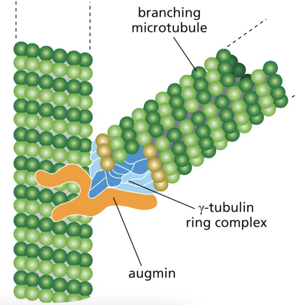
Plant cells have gamma tubulin too! How does that work
Plan cells have branching microtubules. augmin helps the two microtubules to bind, and the gamma tubulin ring complex supports it.
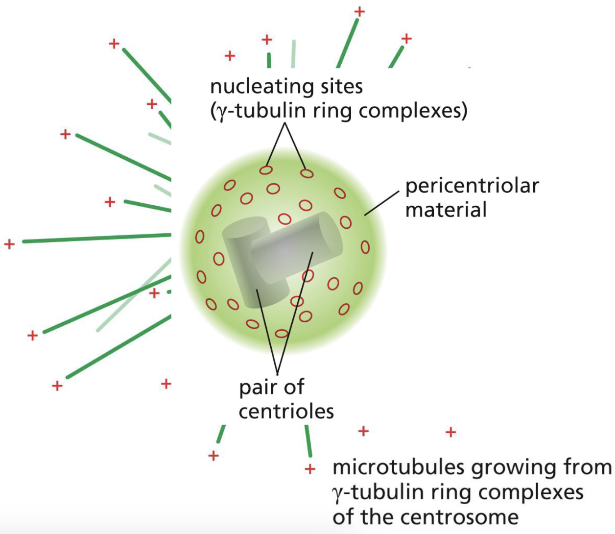
Where is gamma tubulin found in animal cells?
Green sphere: one centrosome
Inside the centrosome → pair of centrioles inside
Green lines (microtubules) sticking out from the green sphere, (centrosome)
Nucleating site (gamma tubulin ring complex): is on the pericentriolar material (green sphere) → NOT ON CENTRIOLE
What are MAPs?
microtubule-associated proteinsW
What are the two category in MAPs?
kinesins: that can walk towards the plus end
dyneins: that can walk towards the minus ends
Similarities of kinesins and dyneins.
Both hold onto vesicles or organelles, and use ATP hydrolysis for energy
What can microtubules aid in?
transporting vesicles and organelles along the microtubules
Example of vesicles/organelles transporting along microtubules: Tilapia fish
Melanin molecules: are dark molecules that make the fish look dark
Dark fish: kinesins and dyneins compete for vesicles → the melanin molecules are dispersed = dark fish
light fish: kinesins inhibited . dyneins walk to (-) ends → to center. Since the dark molecules are not dispersed, it seems lighter.
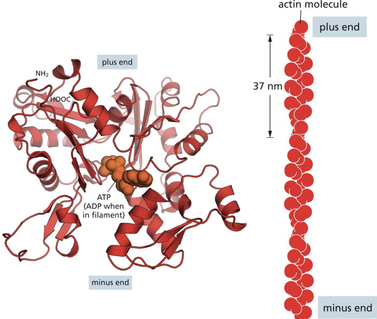
what are the actin monomers
the N and C terminus ends arranges itself. actin monomers are asymmetric and polar
arrange into polarized actin filaments (2 strands twisted together)
Actin monomers have ATP bound to them
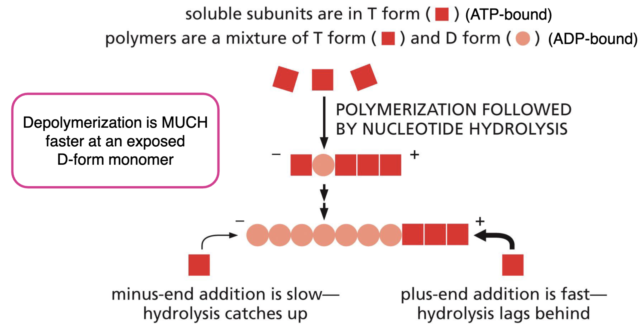
What are T form and D form of actin monomers?
T form: ATP bound, D form: ADP bound
as time passes, ATP is cut to ADP eventually → ADP has much faster depolymerization
Dynamic Instability of Actin Filaments
similar to microtubules
plus end addition of T form monomers is fast.
Addition of T form monomers to (-) side is slow → gets caught up to hydrolysis.. (D forms)
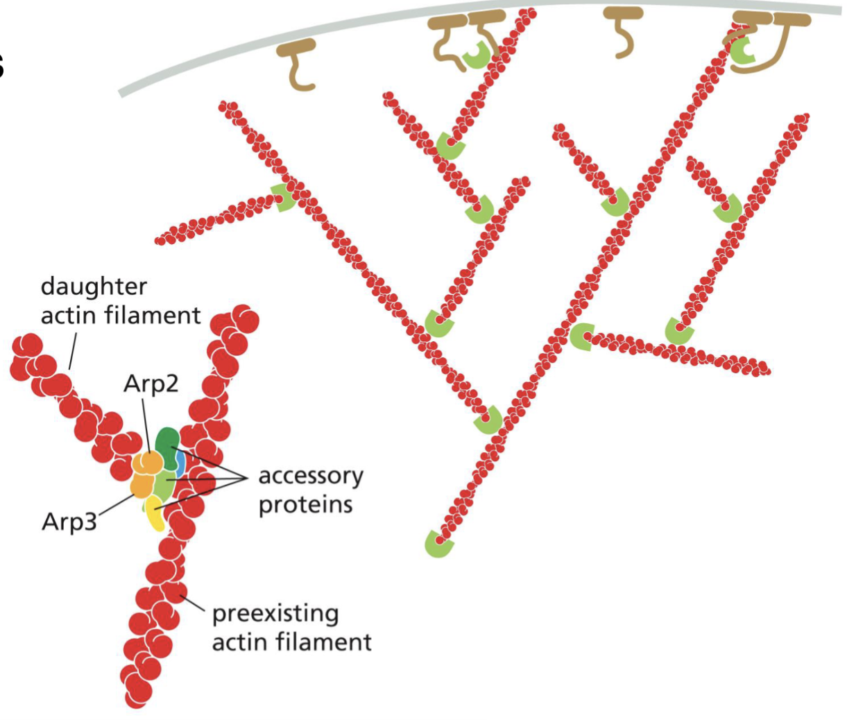
Arp 2/3 and NPF what do they do and what is their mechanism?
inactive Arp 2/3 (Actin related protein) binds with NPF (Nucleation promoting factor) and activates the complex.
These two things bind to the minus end, and stabilize the minus end, and prevent depolymerization

Shaping of the network via treadmilling of actin filaments
generally plus end on edge of cell, minus end deeper in the cell → whole network is polarized
Initially: Arp 2/3 bound to minus end, stable
When protein comes in and cuts the minus end, Arp 2/3 lost, and depolymerization begins
The NPF on the cell membrane binds to actin, and adds more actin monomers.
Treadmilling: one end is continuously cut, and one end is continuously added monomers.
Growth and polymerization on the plus end of the molecule pushes/moves the cell.
If cell is growing to the wrong direction, proteins cap the positive ends.
Leading edge vs Lagging edge
Leading edge: the growing network
Lagging edge: actin myosin contraction, bringing lagging end forward
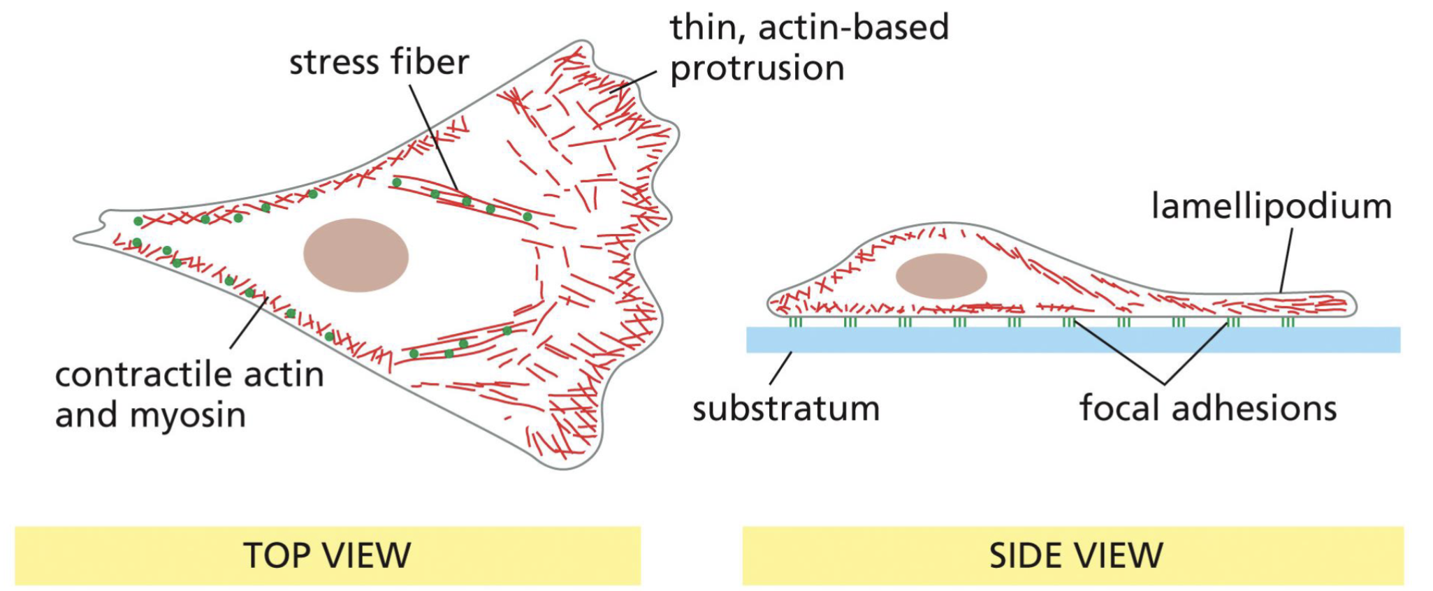
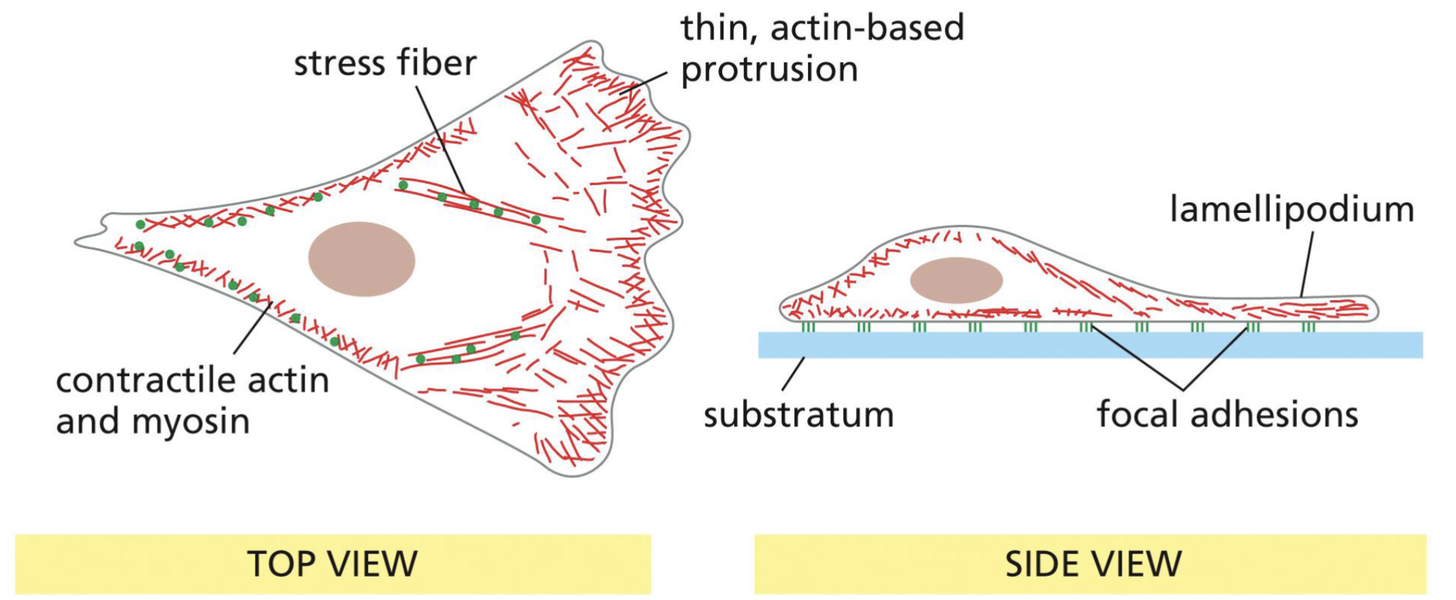
Substratum and focal adhesions?
Substratum = floor
Focal adhesions: Transmembrane proteins that are grabbing onto the floor
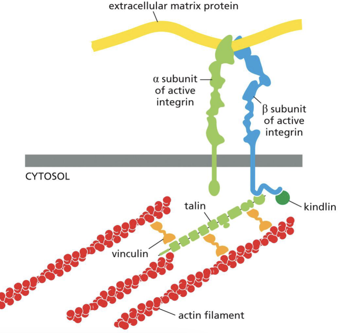
Focal adhesion how it anchors onto the ground
Integrin heterodimers directly bind to extracellular matrix proteins (aka substratum, floor)
indirectly interact with actin filaments via adaptor/anchor proteins
What are the Focal adhesion/integrin heterodimers for?
providing adhesion necessary for migration
How is myosin used for contractile forces?
Myosins motor domain use ATP hydrolysis for energy → holds onto organelles or help cells contract
myosins can walk towards plus end of actin filaments
actin + myosin can generate force → for cell migration
Rho famil small GTPases types
Rho, Rac, CDC42
Over activation of Rac
Leading edge actin network treadmill activation
Overactivation of Rho
Actin myosin contraction of lagging edge
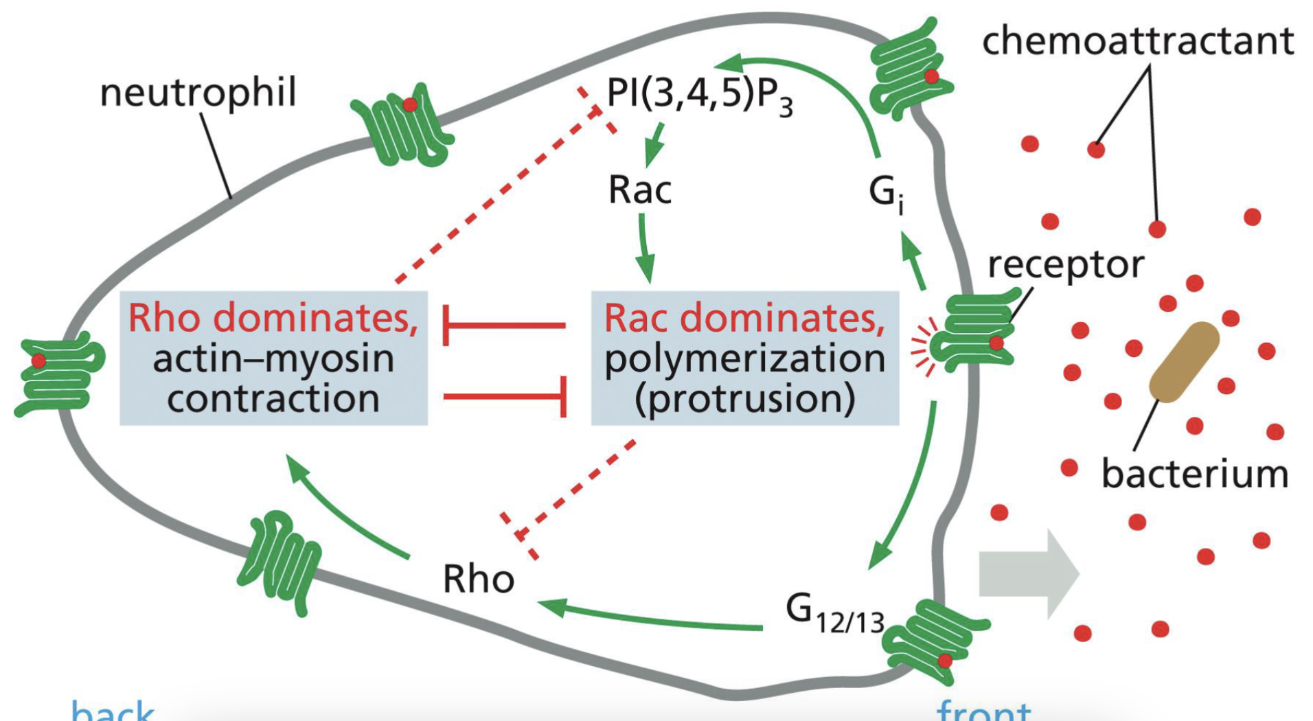
Cell Movement - Example: Neutrophil
Bacteria release chemoattractant
Neutrophil receptors bind to it → activates it.
Area with most activated receptors → most Rac GTP → becomes the leading edge → inhibits Rho GTP on this end
the opposite end gets Rho GTP → inhibits Rac GTP
Neutrophil now has polarity!
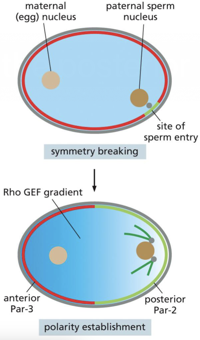
C.elegans defining Cell Polarity
Symmetry breaking → defines posterior and anterior
Sperm entry is the posterior which triggers cytoskeleton polarization
Actin filaments/GEF move away from sperm entry (anterior side)
microtubules are concentrated near centrosome near sperm entry
