Histology of Large Intestine

0.0(0)
studied byStudied by 0 people
learnLearn
examPractice Test
spaced repetitionSpaced Repetition
heart puzzleMatch
flashcardsFlashcards
Card Sorting

1/6

encourage image

There's no tags or description

Looks like no tags are added yet.

Study Analytics
Name
Mastery
Learn
Test
Matching
Spaced

No study sessions yet.

7 Terms

1
New cards

What does the large intestine consist of?

  • Cecum

  • Colon

  • Rectum

  • Anal canal

2
New cards

Characteristics of large intestine

  • Villi?

  • Shape

  • Type of glands

  • Goblet cells?

  • Panneth cells?

  • Lymphatic nodules

  • Compare surface of tunica mucosa to TM in small intestine

  • Why is mucosa layer is thicker?
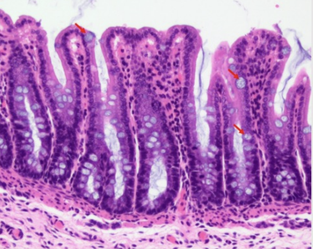
Villi: Absent

Shape: Less coiled

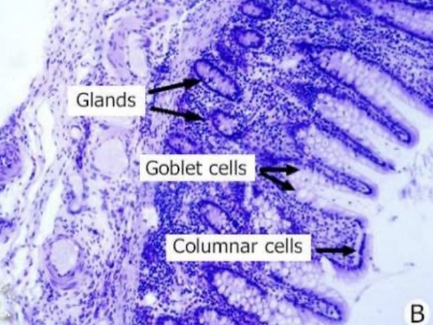
Type of glands: Simple tubular intestinal glands

Goblet cells: Lots

Panneth cells: Absent

Lymphatic nodules: Increased number

Surface of tunica mucosa compared to it in SI: More uniform and flatter

Mucosa layer is thicker because: Increase length of intestinal glands

<p><strong><u>Villi:</u> </strong><span style="color: green"><strong>Absent</strong></span></p><p><strong><u>Shape:</u> </strong><span style="color: green"><strong>Less coiled</strong></span></p><p><strong><u>Type of glands:</u></strong><span style="color: green"><strong> Simple tubular intestinal</strong></span><strong> </strong>glands</p><p><strong><u>Goblet cells:</u> </strong><span style="color: green"><strong>Lots</strong></span><strong> </strong></p><p><strong><u>Panneth cells:</u> </strong><span style="color: green"><strong>Absent</strong></span></p><p><strong><u>Lymphatic nodules:</u></strong><span style="color: green"><strong> Increased number </strong></span></p><p><strong><u>Surface of tunica mucosa compared to it in SI:</u> </strong><span style="color: green"><strong>More uniform</strong></span><strong> </strong>and <span style="color: green"><strong>flatter</strong></span><strong> </strong></p><p><strong><u>Mucosa layer is thicker because:</u></strong><span style="color: green"><strong> Increase length</strong></span><strong> </strong>of<span style="color: green"> <strong>intestinal glands</strong></span></p>
3
New cards
<p><span style="color: green"><strong><mark data-color="green" style="background-color: green; color: inherit">Histology of Large Intestine:</mark></strong></span><strong> </strong><span style="color: red"><strong>Tunica Mucosa </strong></span></p><ul><li><p>Epithelium </p><ul><li><p>Type of glands</p></li><li><p>Type of lining </p></li><li><p>Type of cells </p></li></ul></li><li><p>Lamina propria </p><ul><li><p>Consist of </p></li></ul></li><li><p>Muscularis mucosae </p><ul><li><p>Location </p></li><li><p>Function</p></li></ul></li></ul><p></p>

Histology of Large Intestine: Tunica Mucosa

  • Epithelium

    • Type of glands

    • Type of lining

    • Type of cells

  • Lamina propria

    • Consist of

  • Muscularis mucosae

    • Location

    • Function

Epithelium:

  • Type of glands: Straight tubular glands

  • Type of lining: Absorptive columnar cells

  • Type of cells:

    • Goblet cells

    • Stem cells

    • Lymphocytes

Lamina propria:

  • Consist of:

    • Loose CT

    • Blood and lymphatic vessels

    • Collagen

    • Lymphocytes

    • Plasma cells

Muscularis mucosae:

  • Location: Beneath base of the crypts

  • Function: Undergoes rhythmic contractions

<p><span style="color: red"><strong><mark data-color="red" style="background-color: red; color: inherit"><u>Epithelium:</u></mark></strong></span></p><ul><li><p><strong><u>Type of glands:</u> </strong><span style="color: red"><strong>Straight tubular glands</strong></span></p></li><li><p><strong><u>Type of lining:</u> </strong><span style="color: red"><strong>Absorptive columnar cells</strong></span></p></li><li><p><strong><u>Type of cells:</u> </strong></p><ul><li><p><span style="color: red"><strong>Goblet cells </strong></span></p></li><li><p><span style="color: red"><strong>Stem cells </strong></span></p></li><li><p><span style="color: red"><strong>Lymphocytes</strong></span></p></li></ul></li></ul><p><span style="color: red"><strong><mark data-color="red" style="background-color: red; color: inherit"><u>Lamina propria:</u></mark></strong></span></p><ul><li><p><strong><u>Consist of:</u> </strong></p><ul><li><p><span style="color: red"><strong>Loose CT </strong></span></p></li><li><p><span style="color: red"><strong>Blood </strong></span>and <span style="color: red"><strong>lymphatic vessels </strong></span></p></li><li><p><span style="color: red"><strong>Collagen </strong></span></p></li><li><p><span style="color: red"><strong>Lymphocytes </strong></span></p></li><li><p><span style="color: red"><strong>Plasma cells</strong></span></p></li></ul></li></ul><p><span style="color: red"><strong><mark data-color="red" style="background-color: red; color: inherit"><u>Muscularis mucosae:</u></mark></strong></span></p><ul><li><p><strong><u>Location:</u> </strong><span style="color: red"><strong>Beneath base</strong></span><strong> </strong>of the <span style="color: red"><strong>crypts</strong></span><strong> </strong></p></li><li><p><strong><u>Function:</u> </strong><span style="color: red"><strong>Undergoes rhythmic contractions</strong></span></p></li></ul><p></p>
4
New cards

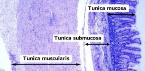
Histology of Large Intestine: Submucosa

  • Describe

  • Typical

  • Glands extend to submocosa

5
New cards

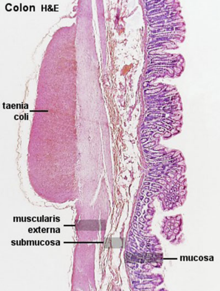
Histology of Large Intestine: Tunica Muscularis #ffb300

  • 2 layers

  • What separates the second layer

    • Function

2 layers:

  • Inner circular

  • Outer longitudinal

Separates second layer: 3 longitudinal bands of taenia coli (smooth muscle)

  • Function: Movement of more solid waste to rectum (aid in contraction)

<p><strong><u>2 layers:</u></strong></p><ul><li><p><span style="color: #ffb300"><strong>Inner circular </strong></span></p></li><li><p><span style="color: #ffb300"><strong>Outer longitudinal </strong></span></p></li></ul><p><strong><u>Separates second layer:</u> </strong><span style="color: #ffb300"><strong>3 longitudinal bands </strong></span>of<span style="color: #ffb300"> <strong>taenia coli </strong></span><span style="color: #000000">(<em>smooth muscle</em>)</span></p><ul><li><p><strong><u>Function:</u> </strong><span style="color: #ffb300"><strong>Movement</strong></span><strong> </strong>of <span style="color: #ffb300"><strong>more solid waste</strong></span><strong> </strong>to <span style="color: #ffb300"><strong>rectum</strong></span><strong> </strong>(<em>aid in contraction</em>) </p></li></ul><p></p>
6
New cards

Histology of Large Intestine: Tunica Serosa

  • Describe

Typical

7
New cards
<p>Between A and B which <span style="color: blue"><strong>small intestine</strong></span><strong> </strong>and<span style="color: green"> <strong>large intestine</strong></span>?</p>

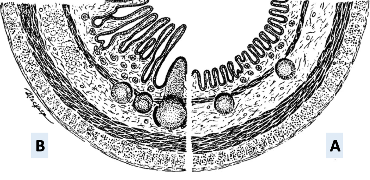
Between A and B which small intestine and large intestine?

Small intestine: B

Large intestine: A

Because: Tunica mucosa is more uniform and flat

<p><strong><u>Small intestine:</u> </strong><span style="color: blue"><strong>B</strong></span></p><p><strong><u>Large intestine:</u> </strong><span style="color: green"><strong>A</strong></span></p><p><strong><u>Because:</u> </strong><span style="color: purple"><strong>Tunica mucosa </strong></span>is more <span style="color: purple"><strong>uniform</strong></span><strong> </strong>and <span style="color: purple"><strong>flat</strong></span><strong> </strong></p><p></p>