Lec 2: Cell structure and components
1/78
There's no tags or description
Looks like no tags are added yet.
Name | Mastery | Learn | Test | Matching | Spaced | Call with Kai |
|---|
No analytics yet
Send a link to your students to track their progress
79 Terms
What is Cell Theory?
All organisms are made up of cells.
Cells are like building blocks to make more complicated structures
cells are specialized to carry out different functions
The cell is the fundamental unit of life.
cells are important unit of life
cell is the simplest entity that we can define as living.
Cells come from preexisting cells
arise from preexisting cells through the process of cell division.
When a single parent cell divides, it produces daughter cells. Of course
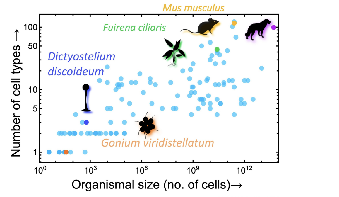
What is the evolution of cellular differentiation theory?
Theory: Size of organism scales with it's complexity
Left corner: simple form of life --> gradually become more complicated
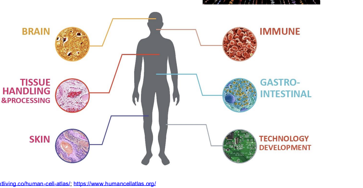
What is the human cell atlas (HCA)?
Trying to build comprehensive map of human cells
structure and function are closely related
red blood cell looks and functions very differently from the long, slender muscle cells shown in that contract to exert force
Serve as basis for understand human biological process --> used to diagnose, monitor, and treat diseases
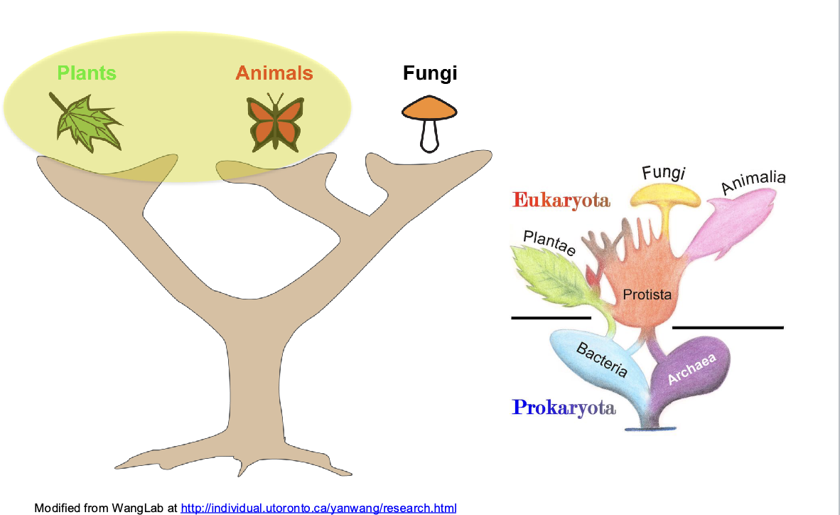
Prokaryotes vs Eukaryotes
Prokaryotes
bacteria, archaea
lack a nucleus
First forms of life
genetic material is organized in one circular chromosome with many loops.
Instead of a nucleus, this genetic material is concentrated in a discrete region of the cell known as the nucleoid
cells smaller than eukaryotes
Eukaryotes
animals, plants, fungi, protists
have a nucleus: houses the vast majority of the cell’s DNA
transcription takes place in the nucleus first, and translation takes place later in the cytoplasm
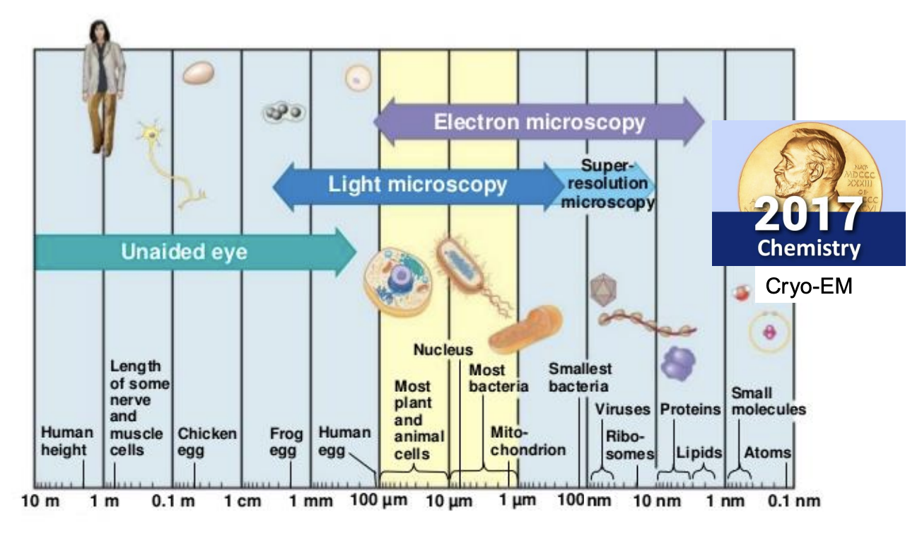
What is microscopy?
method of studying cells that can’t be seen by the human eye
below 100 fancy m meters
What are the types of micrscopy?
unaided eye: anything above 100 fancy ums
light microscopy: 100 - 1
most plant and land animals
nucleus
most bacteria
mitochondria
Electron microscopy: 100 -1 nm
light microscopy +
smallest bacteria
viruses
ribsoomes
proteins
lipids
super resolution microscopy: 1 fancy u - 10 nm
Cryo-EM: 1 - 0.1 nm
small molecules
atoms
What is needed to view microscopic things?
different kinds of microscopes
How can a fungal culture be seen in a microscope?
Left: fungal culture
Can see with naked eye
Microbial fungi --> colony formed
Middle: microscope
Colony on slide --> higher structure of fungal spores and fungal hierarchy
630: what spores look like (aquatic microbes that infect hosts, has felangees)
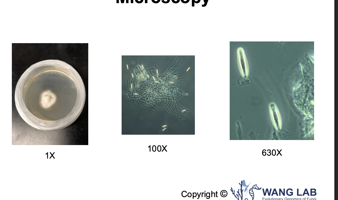
How can a mushroom be seen in a microscope?
See fine details on surface and inside the cell
Right: study content inside the cell
How nutrients transported
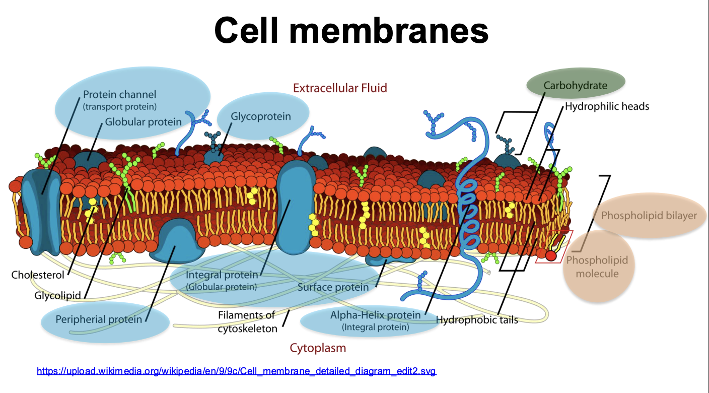
What is the cell membrane?
The membrane that surrounds the cytoplasm of the cell, separating the inside of the cell from the outside of the cell; also called the plasma membrane. (book)
composed of lipids, proteins, and carbohydrates.
What are the parts of the cell membrane?
Phospholipid bilayer
phospholipid molecule
protein chanel (transport protein)
Globular protein (integral protein)
peripheral protein
glycoprotein
Alpha-Helix protein (Integral protein)
filaments of cytoskeleton
cholesterol
carbohydrate
hydrophilic tails
hydrophobic tails
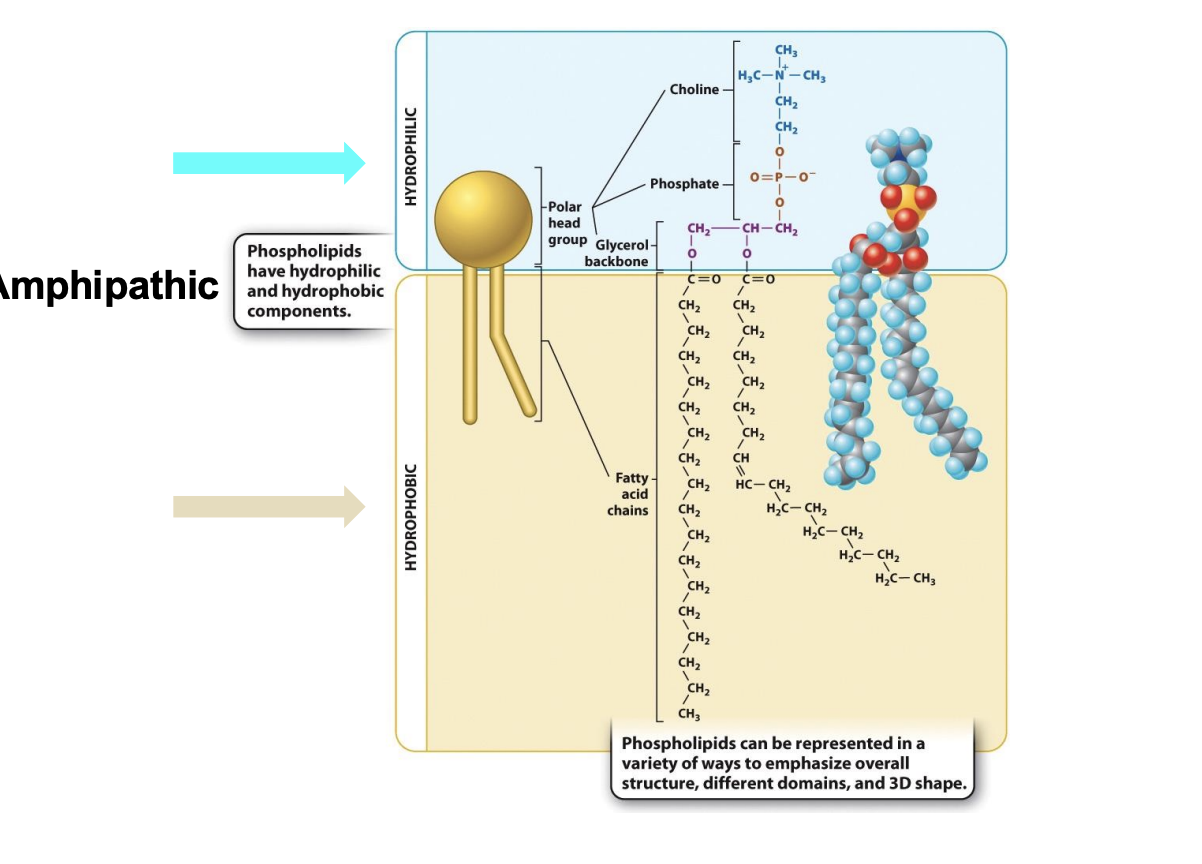
What is the structure of phospholipids?
Amphipathic: both hydrophilic and hydrophobic
Hydrophilic head (polar):
Choline + phosphate + glycerol backbone
Hydrophobic tail
Fatty acid chain
nonpolar → do not form hydrogen bonds with water
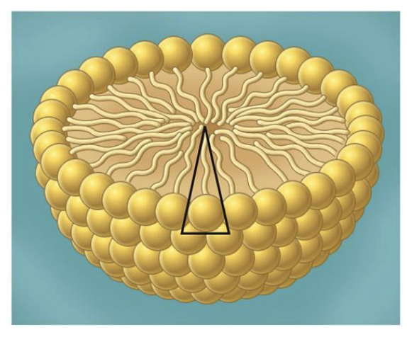
Way to build stable larger structure --> assemble with peers
spontaneously arrange themselves into various structures in which the polar head groups on the outside interact with water and the nonpolar tails come together on the inside away from water.
results from the tendency of polar molecules like water to exclude nonpolar molecules or nonpolar groups of molecules.
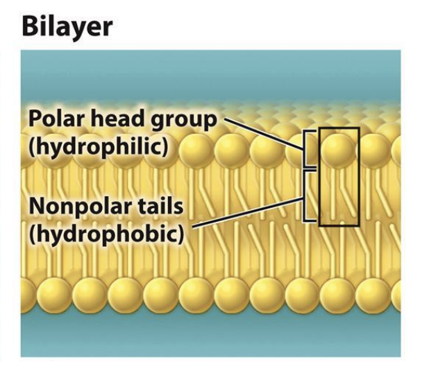
What are the lipid structures?
Micelle
bilayer
liposome
What is a micelle?
Head large
phospholipids can form circular structure (tails inside)
Head is large and bulky with one hydrophobic tail buried.
What is a bilayer lipid?
Heads small: form bilayer structure
Typical cell membrane structure
two hydrophobic tails stuck inside layers
What is a liposome?
Bilayers can further form inclosed structure Liposome
surround central space
Liposome have two layers --> formed by small head phospholipids
Phospholipids spontaneously form enclosed bilayers
hydrophilic core and hydrophobic bilaye
Can place medicine in liposome and it will deliver to body and release into cell
Book: Forms spontaneously (when pH is 7 and concentration of high free phospholipids are high)
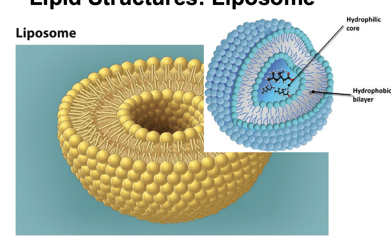
Liposome vs micelle?
Liposome have two layers --> formed by small head phospholipids
Micelle: just single layer and large head structure
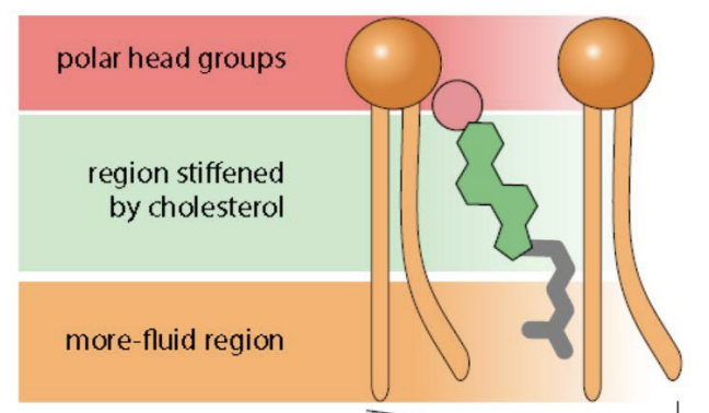
What is cholesterol?
a component of animal cell membranes.
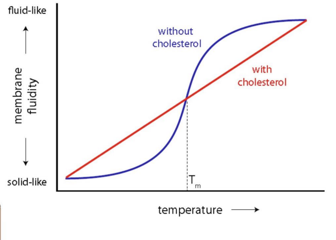
Cholesterol acts as a buffer to lessen the impact of temperature on membrane fluidity.
What important role does cholesterol play?
Influence membrane fluidity
Forms strong interactions with phospholipids
Between the phospholipids
cholestoral acts as a buffer to lessen the impact of temp of membrane fluidity
What is cell membrane fluidity?
membrane lipids are able to move in the plane of the membrane, the membrane
Lipids freely associate with one another because of the extensive van der Waals forces between their fatty acid tails
These weak interactions are easily broken and re-formed, so lipid molecules are able to move within the plane of the membrane
book: The longer the fatty acid tails, the less fluid the membrane
book: fewer the number of carbon–carbon double bonds, the less fluid the membrane
Saturated fatty acid tails, which have no carbon–carbon double bonds, are straight and tightly packed, reducing mobility
Carbon–carbon double bonds in unsaturated fatty acids introduce kinks in the fatty acid tails, reducing the tightness of packing and enhancing lipid mobility in the membrane
How does cholesterol work in high temps?
High temp, rigid cholesterol interacts with phosolipid tail and reduce fluidity of membrane (lower tail) → more stable
High temp phosphos separate from each other --> cholesterol and drags them together --> stability

How does cholesterol work in low temps?
Cholesterol prevent phospholipids from packing tighlty--> inc. Fluidity
Is a rigid structure --> packs or separates them
Creates stable line (red line)
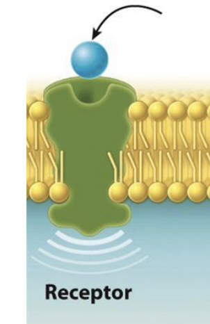
What are the proteins in the membrane?
Receptors, Enzymes, and Anchors
Protein is catagorized based on function
What are receptors?
allow the cell to receive signals from the environment.
responds in certain ways --> receives signal and decides how to response
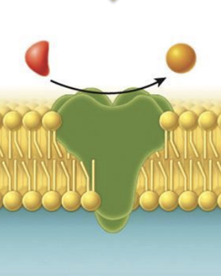
What are enzymes?
catalyze chemical reactions
change one thing to another to serve biological functions
A protein that functions as a catalyst to accelerate the rate of a chemical reaction; enzymes are critical in determining which chemical reactions take place in a cell (book)
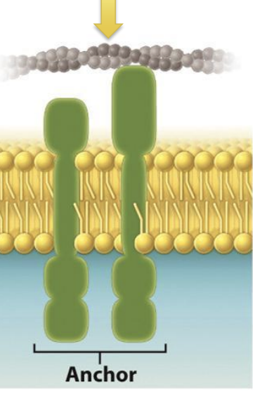
What are anchors?
attach to other proteins that help maintain cell structure and shape
A membrane protein that attaches to other proteins and helps to maintain cell structure and shape.
act as backbone
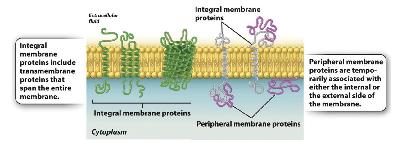
What are integral membrane proteins?
green color
permanently associated with cell membranes
cannot separate them without destroying cell membrane structure

What do transmembrane proteins include?
most integral membrane proteins are transmembrane proteins
span entire lipid bilayer
There are two hydrophilic regions, one protruding from each face of the membrane in contact with the aqueous environment inside and outside of the cell.
one hydrophobic region that spans the hydrophobic interior of the membrane
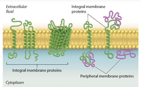
What are peripheral membrane proteins?
temporarily associated with the lipid bilayer or with integral membrane proteins through weak noncovalent interactions
Remove them or isolate them and the membrane can still be in tact
temporarily associated with either the internal or external side of the membrane
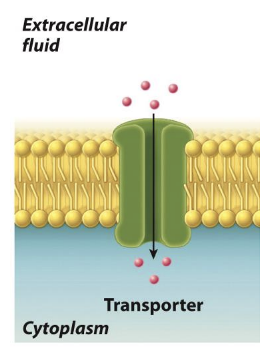
What are transporters?
Integral membrane protein
move ions or molecules across the membrane.
Show Whole lipid bilayer --> how they can transport molecules inside and outside the cell
What is the plasma membrane?
a selective barrier that controls the movement of molecules between the inside and the outside of the cell (partially impermeable - semipermeable)
hydrophobic interior of the lipid bilayer prevents ions and charged polar molecules from moving across it (B)
boundaries that define space of cell
Water can move in and out freely
Others under control through propane channels (ex: ions, water, nutrients)
Some items not allowed to move into cel b/c of dmg
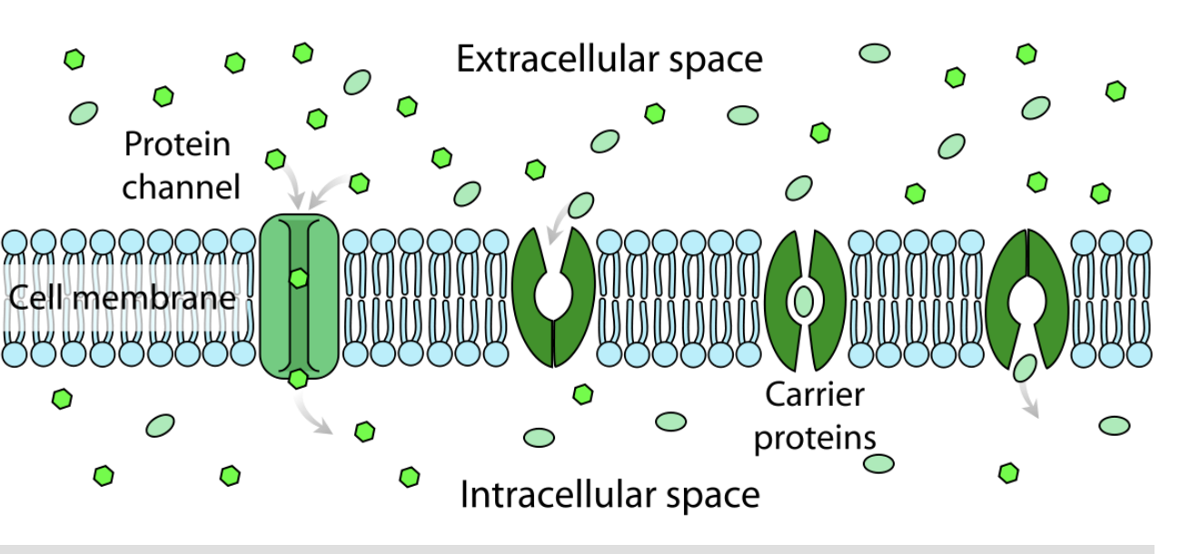
What is the protein channel?
molecules move directly through the lipid bilayer, while in facilitated diffusion, molecules move through a membrane transporter.
provides an opening between the inside and outside of the cell through which certain molecules can pass, depending on their shape and charge.
Some membrane channels are gated, which means that they open in response to some sort of signal, which may be chemical or electrical
What are carrier proteins?
A transporter that facilitates movement of molecules across a cell membrane.
binds to and then transports specific molecules.
Membrane carriers exist in two conformations:
open to one side of the cell,
open to the other side of the cell.
Binding of the transported molecule induces a conformational change in the membrane protein, allowing the molecule to be transported across the lipid bilayer,

What is the fluid mosiac model (book)?
A model that proposes that the lipid bilayer is a fluid structure that allows molecules to move laterally within the membrane and is a mosaic of different types of molecules, including lipids, proteins, and carbohydrates.
ex: protiens move across the bilaer
(Book) What is homeostasis?
A model that proposes that the lipid bilayer is a fluid structure that allows molecules to move laterally within the membrane and is a mosaic of different types of molecules, including lipids, proteins, and carbohydrates.
What is diffusion?
Molecules are in constant, random motion in most environments.
The net movement of molecules from areas of higher to lower concentration as a result of their random thermal motion.
no cost of energy (passive transport)
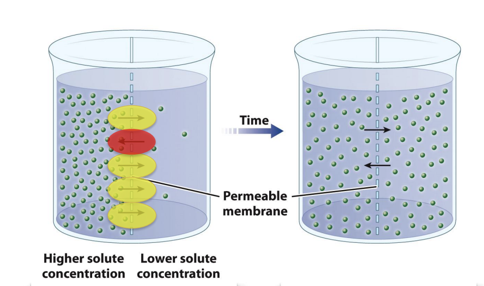
What does this image show?
left: net movement of solute from area of higher solute concen to area of lower solute concen
Right: no net movement of solute but diffusion continues
Molecules are always moving backwards as well but not comparable (total volume from high to low)
What is passive transport?
Movement of substances across a cell membrane by diffusion
The simplest movement into and out of cells is passive transport, which works by diffusion
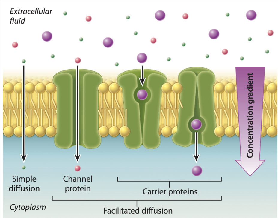
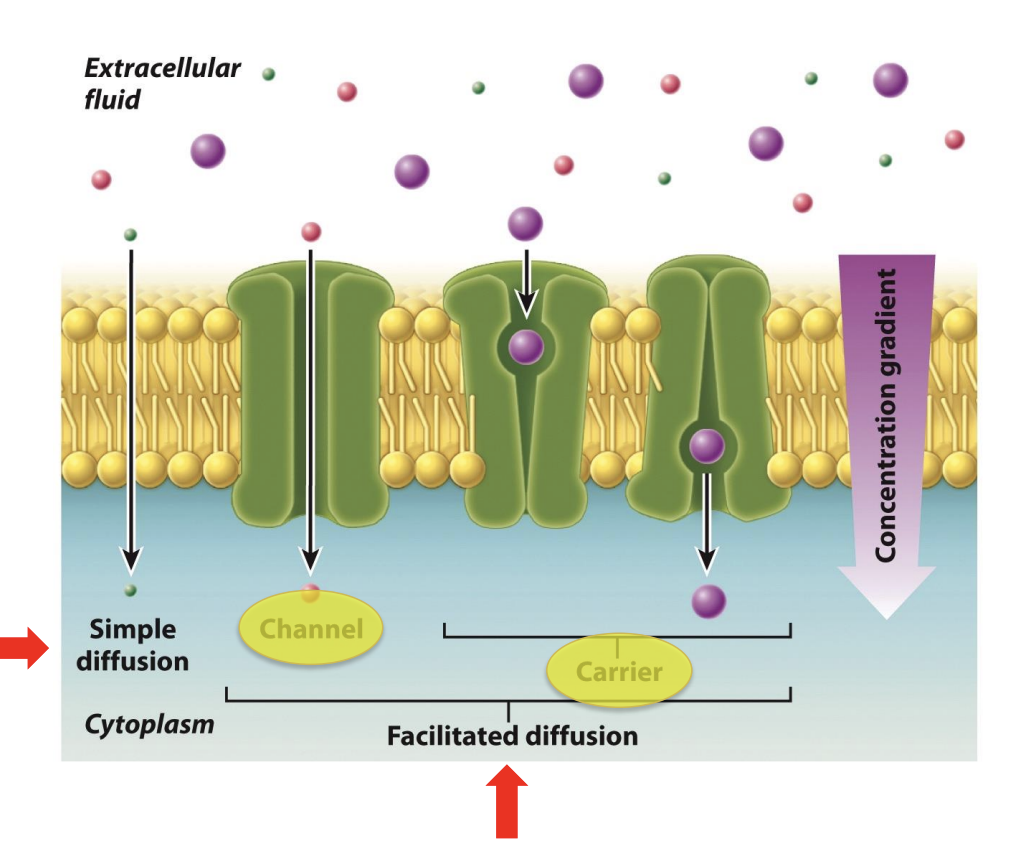
What is facilitated diffusion?
Diffusion across a cell membrane through a transmembrane protein, such as a channel or carrier.
Large: cannot pass through it --> too large
Need door to open channel or have a carrier
What uses simple and facilitated diffusion?
both passive diffusion
Simple: Small molecules can move across lipid bilayers through simple diffusion
diffuse directly through the cell membrane
usually hydrophobic molecules (lipid bilayer also hydrophobic)
Larger: need help of facilitated diffusion through membrane protein (channel or carrier)
Depends on how they are moved
molecules move directly through the lipid bilayer, while in facilitated diffusion, molecules move through a membrane transporter.
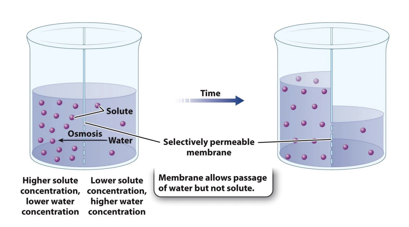
What is osmosis?
The net movement of a solvent, such as water, across a selectively permeable membrane toward the side of higher solute concentration.
diffusion with water
Higher solute concentration = lower water concentration
lower solute concentration = higher water concentration
Selectively permeable membrane allows passage of water but not solute
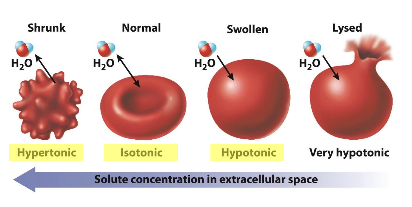
What is hypertonic?
A solution will be hypertonic to a cell if its solute concentration is higher than that inside the cell
What is isotonic?
a solution that contains the same concentration of water and solutes —> no net water movement
What is hypotonic?
If the solute concentration outside the cell is lower than inside the cell
How does osmosis impact blood cells?
Animals cells do not have cell wall--> cells are fragile and only have control of osmosis
Some cells burst or become compressed
Human blood cells
Red blood cell placed in hypertonic solution
Higher concentration outside than inside of cell
Water leave cell with osmosis --> cell shrinks in extreme env
Maintain same inside and outside concentration --> isotonic
Ideal for cels
Typical red blood cell
Placed in hypotonic sol: higher solute concentration inside than outside
Water moves into cell --> cell have no cell wall --> keeps adding water --> swells --> bursts in extreme situations
What are contractile vacuoles?
organelles that take up excess water from inside the cell and then, by contraction, expel it into the external environment
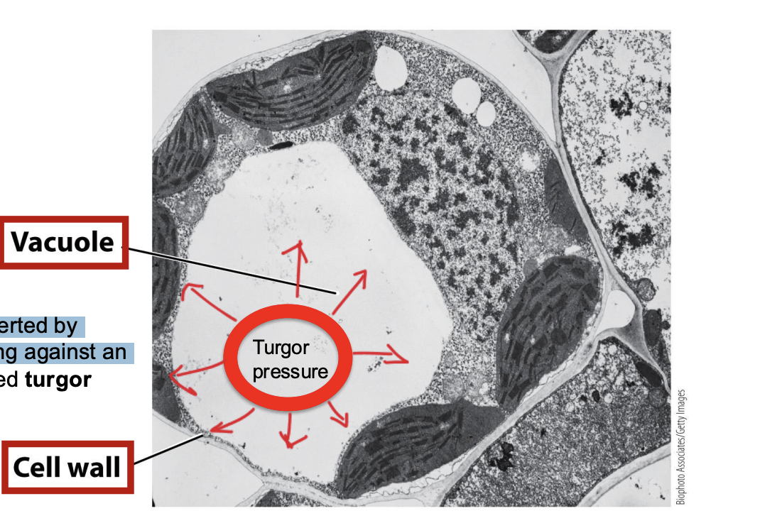
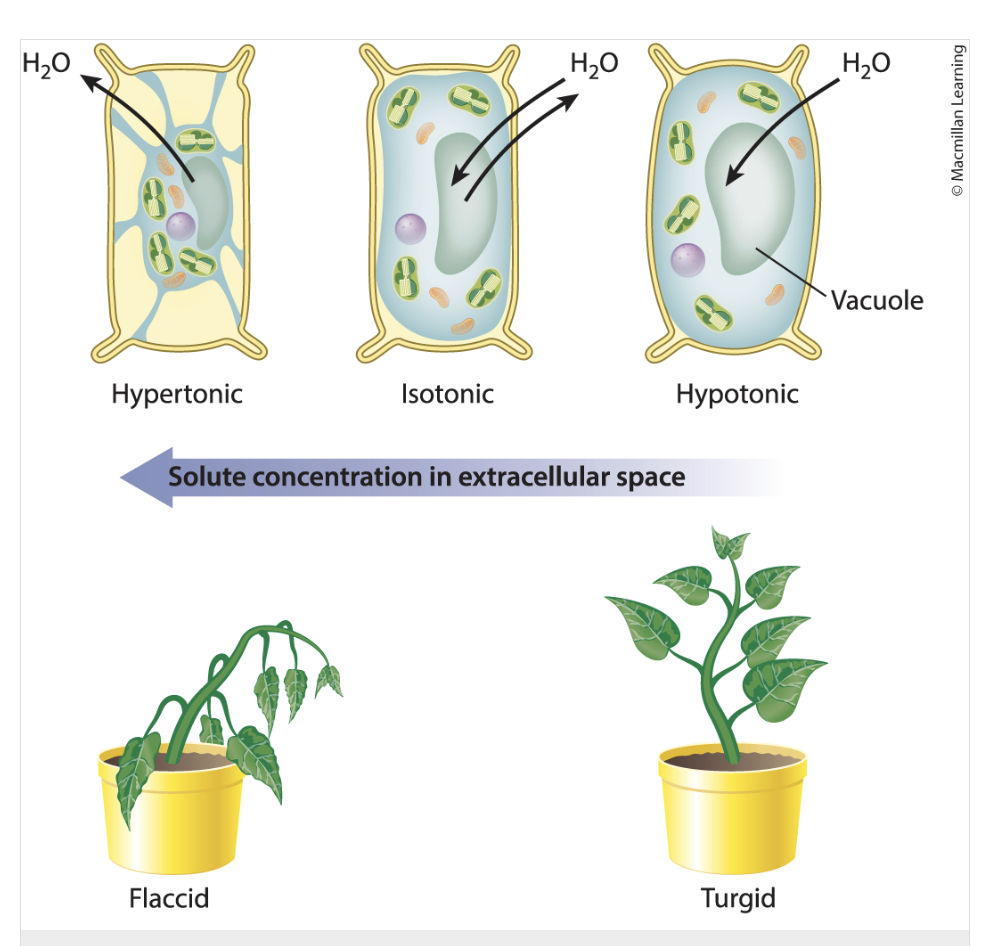
What is a vacuole?
absorbs water and contributes to turgor pressure.
plant and fungal cells
What is the cell wall?
organisms include bacteria, fungi, many protists, most algae, and all plants
provides structural support and protection for the cell.
Because the cell wall is rigid and resists expansion, it allows pressure to build up when water enters a cell.
What is turgor pressure?
The force exerted by water pressing against an object
Pressure within a cell resulting from the movement of water into the cell by osmosis and the tendency of the cell wall to resist deformation.
Inside plant cell have vacuole
Turgor pressure builds as a result of water moving by osmosis into cells surrounded by a cell wall.
Vacuole absorbs water from env and form turgor pressure
Turgor pressure: press against the cell wall --> vacuole will absorb water from env and contribute to turger pressure
promote cell volume expansion
the higher the turgor pressure is, the larger the leaf expansion will be
Why it turgor pressure important?
Cells contain high concentrations of solutes
when a plant cell is placed in a hypotonic solution, water enters the cell by osmosis until the turgor pressure created by the cell wall increases to a level to stop osmosis
Turgor pressure develops because the cell wall resists being stretched and pushes back on the interior of the cell → provides structural support
High turgor pressure means that a plant cell is full of water and is firm, rigid, and turgid
loss of water from vacuoles → triggers turgor pressure → cells no longer maintain shape within cell wall
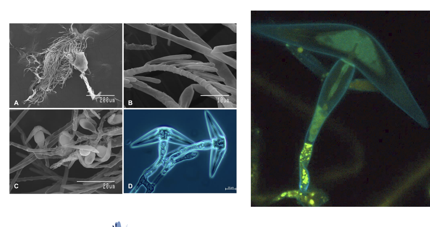
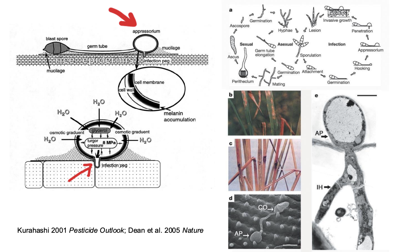
How do pathogens use turgor pressure?
Magnaporthe grisea: pathogens use turgor pressure to complete life cycle
Use turgor pressure to invade/infect host tissue (plants)
Fungal spore attach to host surface --> release enzyme from body -->increase turgor pressure in plant cell with specialized compounds (melanin) --> increase concentration in cell --> start absorb water and increase turgor pressure
Cell uses turgor pressure to penetrate plants surface (pressure used to break cell epidermis) --> creates infection path --> gets into cell and finish cycle in cell
Absorb and use hots nutrients
What is passive transport?
Movement of substances across a cell membrane by diffusion
only works when the move is consistent with concentration gradient from high to low
What is active transport?
The movement of substances across a cell membrane against an electrochemical gradient, requiring an input of energy (ATP)
Cell moves substance against constant gradients (low concentration -->high): use active transport
cells move substances through transport proteins embedded in the cell membrane
Ex:
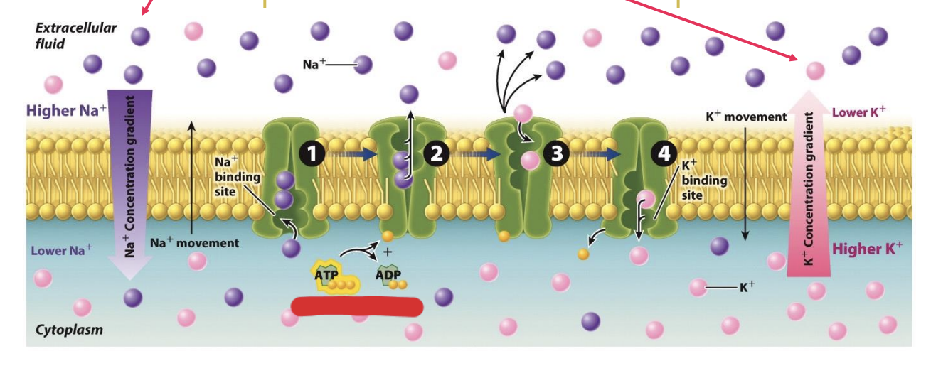
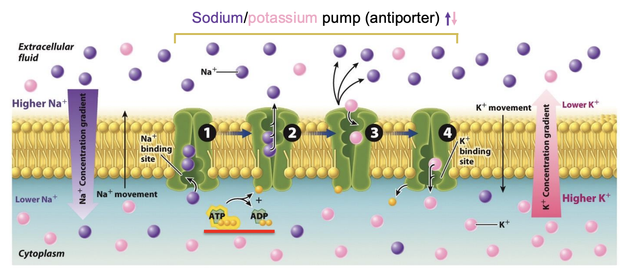
sodium–potassium pump
Within cells, sodium is kept at concentrations much lower than in the external environment; the opposite is true of potassium.
both sodium and potassium have to be moved against a concentration gradient.
The sodium–potassium pump actively moves sodium out of the cell and potassium into the cell

How does primary active transport work?
uses energy stored in ATP to move sodium and K+ against concentration gradient
Energy is directly used to support transportation
The sodium and potassium ions move in opposite directions, so the pump is an antiporter. .
3 Na+ out of, 2 K+ in
What symporotrs (co-transporters)?
The transporters that move two molecules in the same direction are called symporters or co-transporters
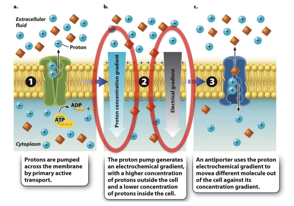
What is secondary active transport?
Indirect (does not use energy direclty)
Because the movement of the coupled molecule is driven by the movement of protons and not by ATP directly,
Rely on consequence of primary active transportation and created concentration gradient
small ions cannot cross the lipid bilayer → many cells have transport proteins that build up the concentration of a small ion on one side of the membrane.
Result: concentration gradient stores potential energy that can be harnessed to drive the movement of other substances across the membrane against their concentration gradient.
How does secondary active transport work?
There are more protons (H⁺ ions) outside the cell than inside → Primary pumps protons across cell membrane using ATP --> transport protected(?) creates proton concentration gradient (protons charged) --> proton concentration gradient is also electron gradient --> movement of proton move concentration gradient to drive the movement of other molecules (ex: square molecules against their won gradient)
Some cells actively pump protons ( H + ) across a membrane using ATP
protons pumped across membrane by primary active transport
proton pump generates electrochemical gradient, with higher concentration of protons outside the cell and lower concen of protons inside the cell
concentration differences favor the movement of protons back to the other side of the membrane.
blocking the movement of protons to the other side → the lipid bilayer creates a store of potential energy
Transporter: move one square molecules requires two protons to move from outside to inside
can use the movement of protons to drive the movement of other molecules against their concentration gradient
The movement of protons is always from regions of higher to lower concentration,
movement of the coupled molecule is from regions of lower to higher concentration.
anitporter uses the proton electrochemical gradient to move different molecules of of the cell against the concentration gradient
Video in Bio book 3.3
What is the endomembrane system?
is an interconnected system of membranes that includes the nuclear envelope, endoplasmic reticulum, Golgi apparatus, lysosomes, vesicles, and plasma membrane.
isolated entities and they communicate with each other
Transporter substance: connects between organelles --> creates endomembrane system
Vesicles buds off from enodomembrane system -→ fuse with plasma membrane and deliver process into intercellular space (exocytosis)
How do things move throughout the ER (book)?
endomembrane system divides the interior of a cell into two distinct spaces, one inside the compartments defined by these membranes and one outside these compartments.
A molecule within the interior of the endoplasmic reticulum (ER) can stay in the ER, in the interior of the Golgi apparatus, or even outside the cell by the budding off and fusing of a vesicle between these organelles.
a molecule associated with the ER membrane can move to the Golgi membrane or the cell membrane by vesicle transport. Molecules in the cytosol are in a different physical space, separated by membranes of the endomembrane system.
physical separation allows specific functions to take place within the spaces defined by the membranes and within the membrane itself.
What are vesicles?
small membrane-enclosed sacs that transport substances within a cell or from the interior to the exterior of the cell.
form by budding off an organelle, taking with them a piece of the membrane and internal contents of the organelle from which they derive.
They then fuse with another organelle or the cell membrane, re-forming a continuous membrane and unloading their contents.
What is exocytosis?
vesicle fuses with the cell membrane
provides a way for a vesicle to empty its contents to the extracellular space or to deliver proteins embedded in the vesicle membrane to the cell membrane
a vesicle that has budded off from the endomembrane system can fuse with the plasma membrane and deliver its contents into the extracellular space.
transport stuff from inside to outside?
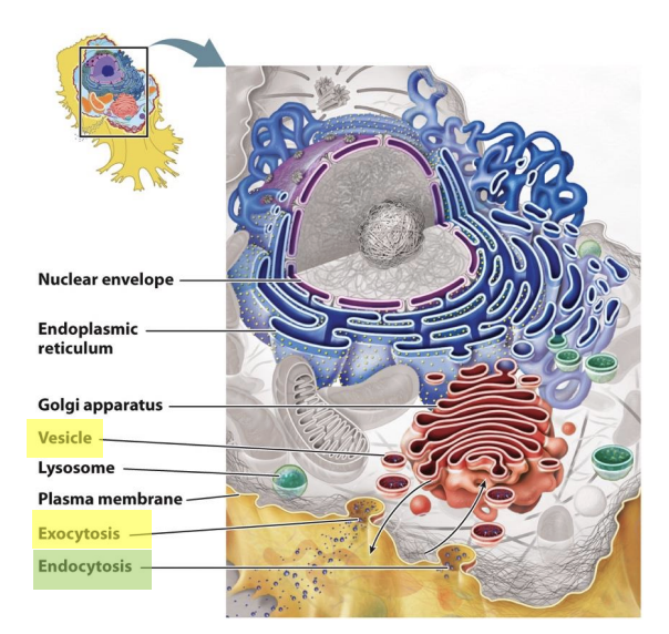
What is endocytosis?
material from outside the cell is brought into a vesicle that can then fuse with other organelles
a vesicle can bud off from the cell membrane, enclosing material from outside the cell and bringing it into the cell interior
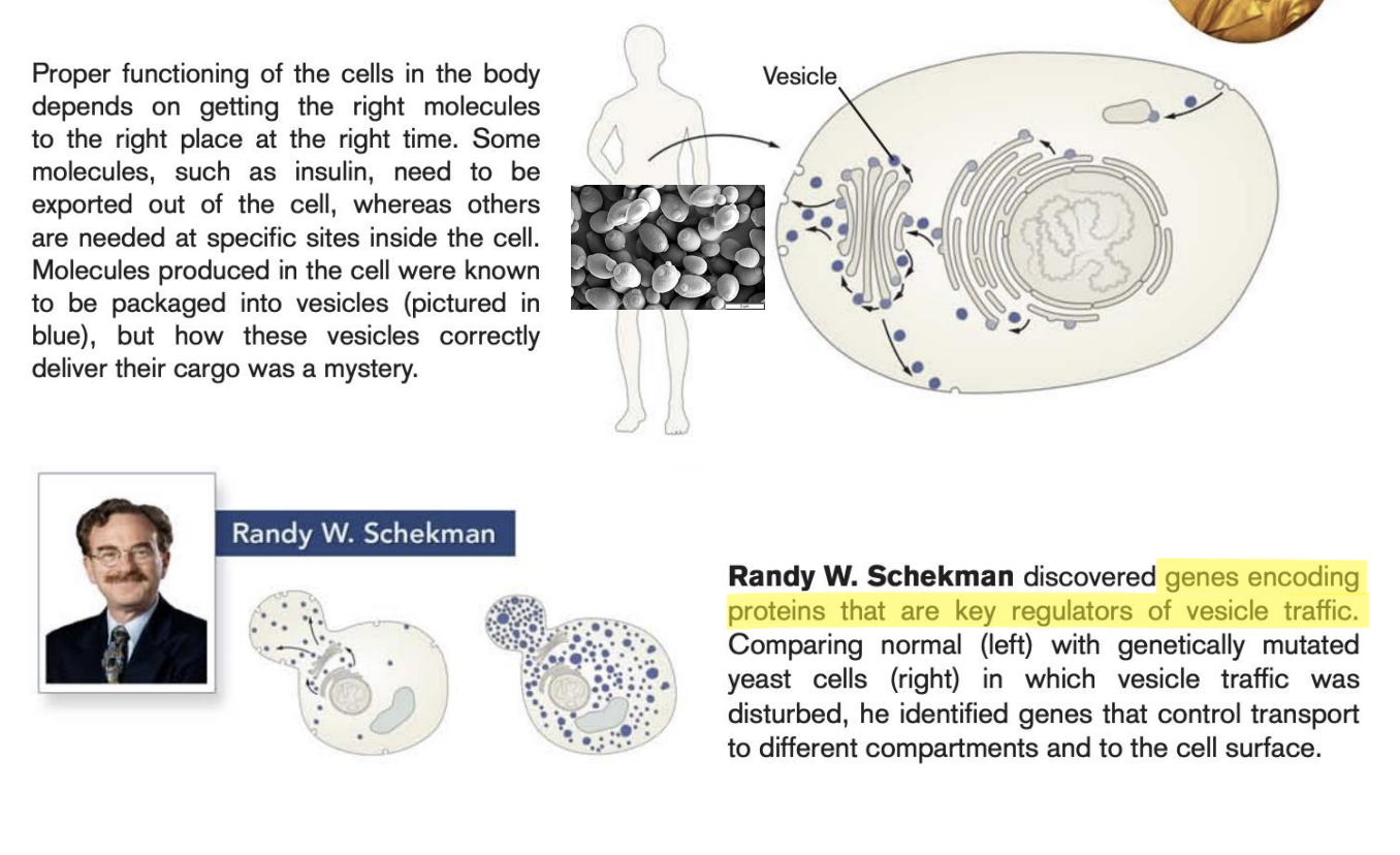
What did Randy W. Schekman discover?
genes encoding proteins are key regulators of vesicle traffic
Dude discovered the vesicles
Uses yeast (fungal spores)
Discovered genes that encode protein
If you destory the gene
Organized delivery of produce in cell come to a mass and is not working?
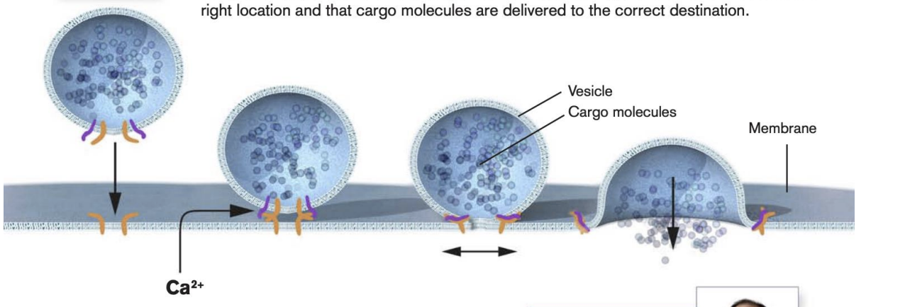
What did James E. Rothman and Thomas Sudhof discover?
protein complex (orange) enables vesicles to fuse with target membranes
proteins on vesicles bind to specific complementary proteins on the target membrane, ensuring that vesicle fuses at the right location and that cargo molecules are delivered to the correct destination
Molecular machinery (purple) senses calcium ions (Ca2+) and triggers vesicle fusion
how temporal percision is achieved and how signaling substances can be released from the vesicles on command
Membrane and surface of vesicle have protienes that recongize eachtoher
Find spot where they land --> fuse with bilipid layer and release content within the cell
Liposome: put medicine in liposome and release the medicine into the human cell
efficient
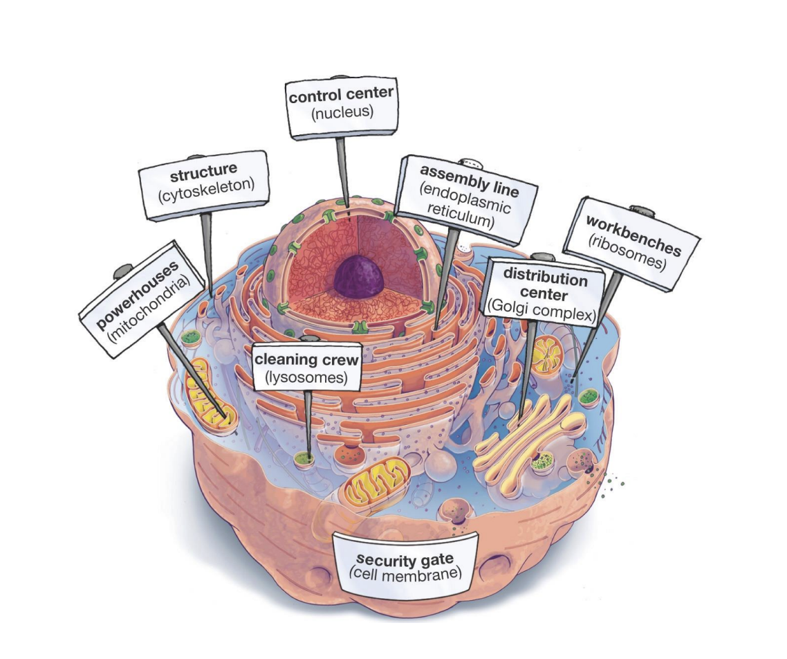
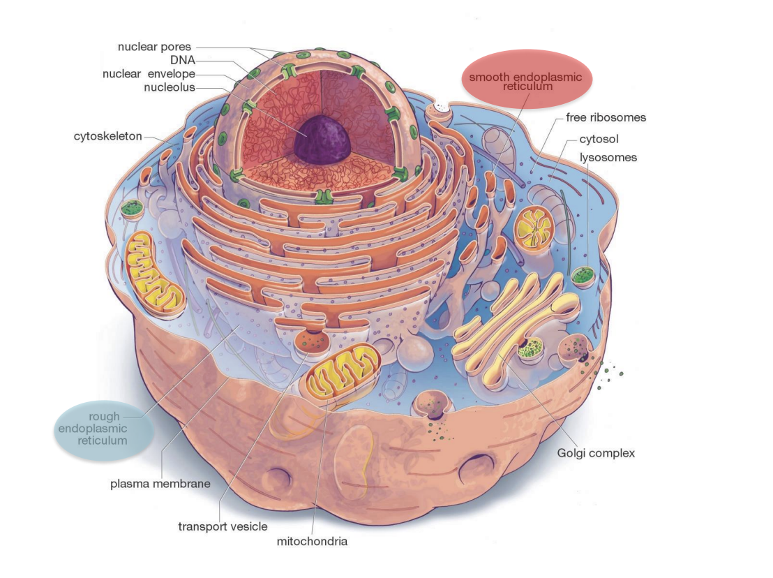
What are the jobs of the cell?
cytoskeleton: structure
Mitochondria: powerhouse
Lysosomes: breaking down and recycling waste materials and old cell parts
Golgi complex: modifies, sorts, and packages proteins and lipids for transport to different destinations in and out of the cell
ribosomes: workbenches
Endoplasmic reticulum: assembly line
nucleus: control center
Cell membrane: security gate
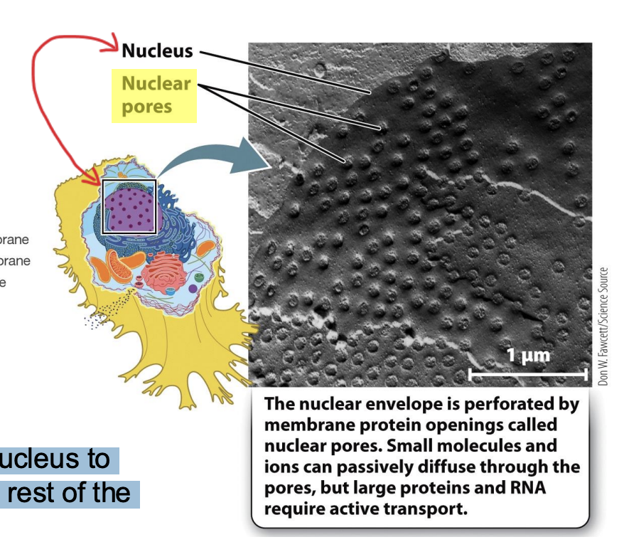
What is the nuclear envelope?
Nucleus: stores cell DNA
Nuclear envelope: defines boundary of nucleus
Have protein opening (nuclear pores) --> how small molecules can travel outside of the nucleus envelope
Needed to communicate with the rest of the cell (especially mRNA)
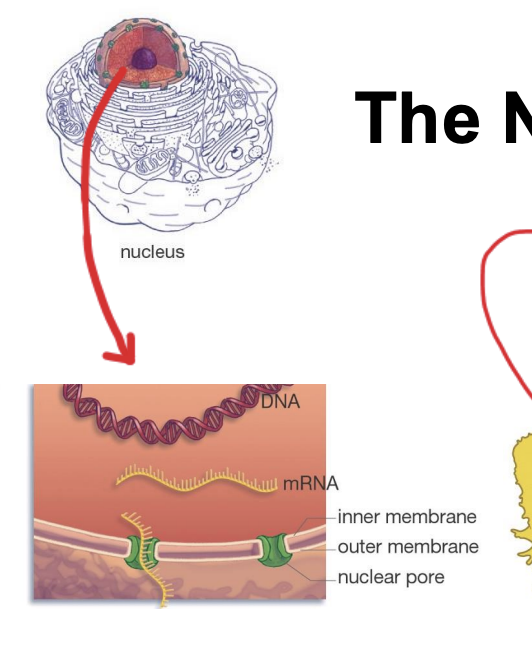
What are nuclear pores?
are essential for the nucleus to communicate with the rest of the cell.
envelope perforated by membrane protein openings (pores)
small molecules and ions can passively diffuse through the pores, but large proteins and RNA require active transport
What is the endoplasmic reticulum (ER)?
a large organelle in most eukaryotic cells, and it produces many of the lipids and proteins used inside and outside the cell.
outer membrane of the nuclear envelope is physically continuous with the ER
Its interior is continuous throughout and is called the lumen
involved in protein and lipid synthesis and site of production of most of the lipids that make up the various cell membranes
two catogries
Rough ER (RER)
Smooth ER
What are ribosomes?
the sites of protein synthesis, where amino acids are assembled into polypeptides.
round particles and exposed to cytosol --> where proteins are synthesized
Amino acids assembled into peptides that are folded later to become proteins
What is the RER?
associated with ribosomes (on surface)
many proteins, including those destined for secretion, are synthesized by ribosomes associated with RER
synthesizes transmembrane proteins, proteins that end up in the interior of organelles, and proteins destined for secretion

What is smooth ER?
lacks ribosomes
site of fatty acid and phospholipid biosynthesis. → primary site of lipid synthesis
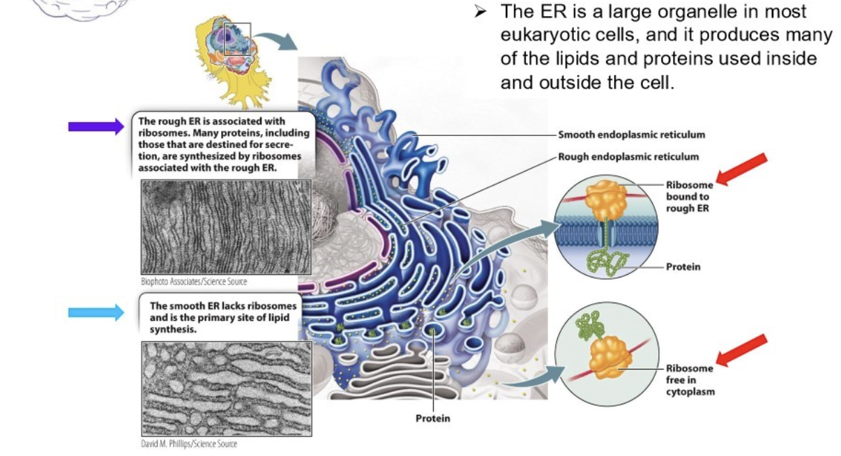
What is predominantly synthesized at the ERs?
Light blue: cytosol
RER ribosomes are exposed to cytosol --> main sides of synthesizes of proteins dominant
Smooth ER:
Predominate that synthesize lipids
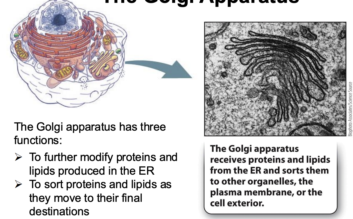
What is the Golgi apparatus?
receives proteins and lipids from the ER and sorts them to other organelles, the plasma membrane, or the cell exterior
Not physically attached to ER
Next stop for vesicle that bud from ER
Functions:
To further modify proteins and lipids produced in the ER
To sort proteins and lipids as they move to their final destinations
To synthesize the cell’s carbohydrates
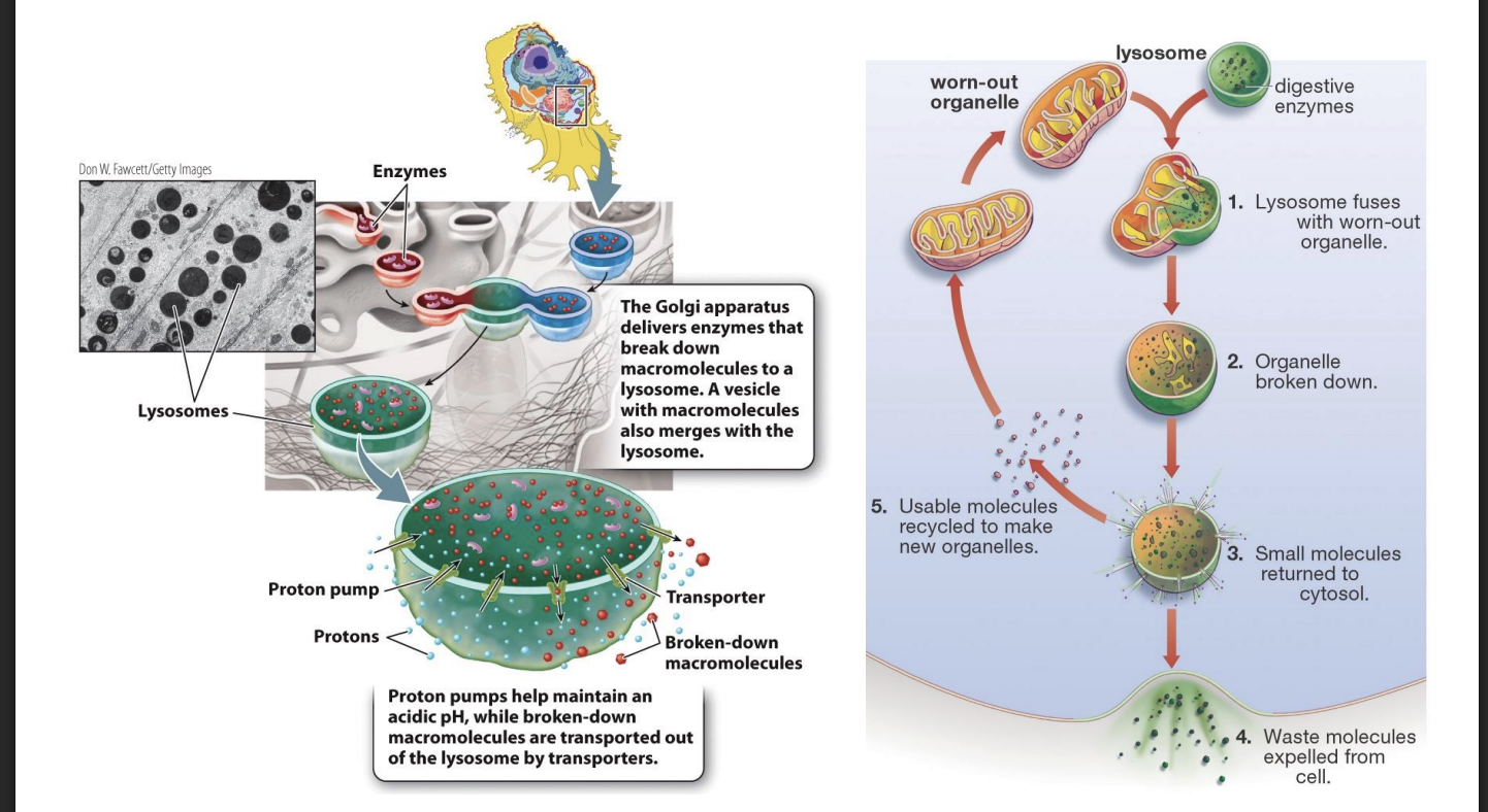
What are lysosomes?
can degrade proteins, nucleic acids, lipids, and complex carbohydrates
Proteins can be synthesized or degraded
Damaged or malfunctions --> cells need a way to recycle them
Lysosomes: specialized vesicle from the Golgi apparatus that degrade damaged or uneeded macromolecules
Can degrade proteins, nucleic acids, lipiss, and complex carbohydrates
Lysosome Fuses with vesicle that contain micromolcules --> degraded and recycles
Needed for healthy cells to remain functional
How do lysosomes do their job?
Golgi apparatus delivers enzymes that break down macromolecues to lysosomes: enzymes inside the lysosomes are synthesized in the rough ER, sorted in the Golgi apparatus, and then packaged into lysosomes.
vesicles with macromolecules also merges with the lysosome
proton pumps help maintain acidic pH
while broken down macromolecules are transported out of the lysosome by transporters
lysosomes fuses with worn out organelle
organelle is broken down
small molecules returned to cytosol
waste molecules expelled from cell
usable molecules recycled to make new organelles
Lysosomal enzymes cannot function in the normal cellular environment, which has a pH of about 7
many of a cell’s enzymes and proteins would unfold and degrade if the entire cell were at the pH of the inside of a lysosome.
By restricting the activity of these enzymes to the lysosome, the cell protects proteins and organelles in the cytosol from degradation.
What are Mitochondria and chloroplasts?
organelles involved in harnessing energy and likely evolved from free-living prokaryotes.
Not part of Endomembrane system
Mitochondria and chloroplast: harvest energy for the cell and hold genomes
Can grow and multiply independently --> once bacteria that were captures by ancestor or eukaryotic cell
Over time evolved current function in eukaryotic cells
Genes sometimes change: endosymbiosis theory
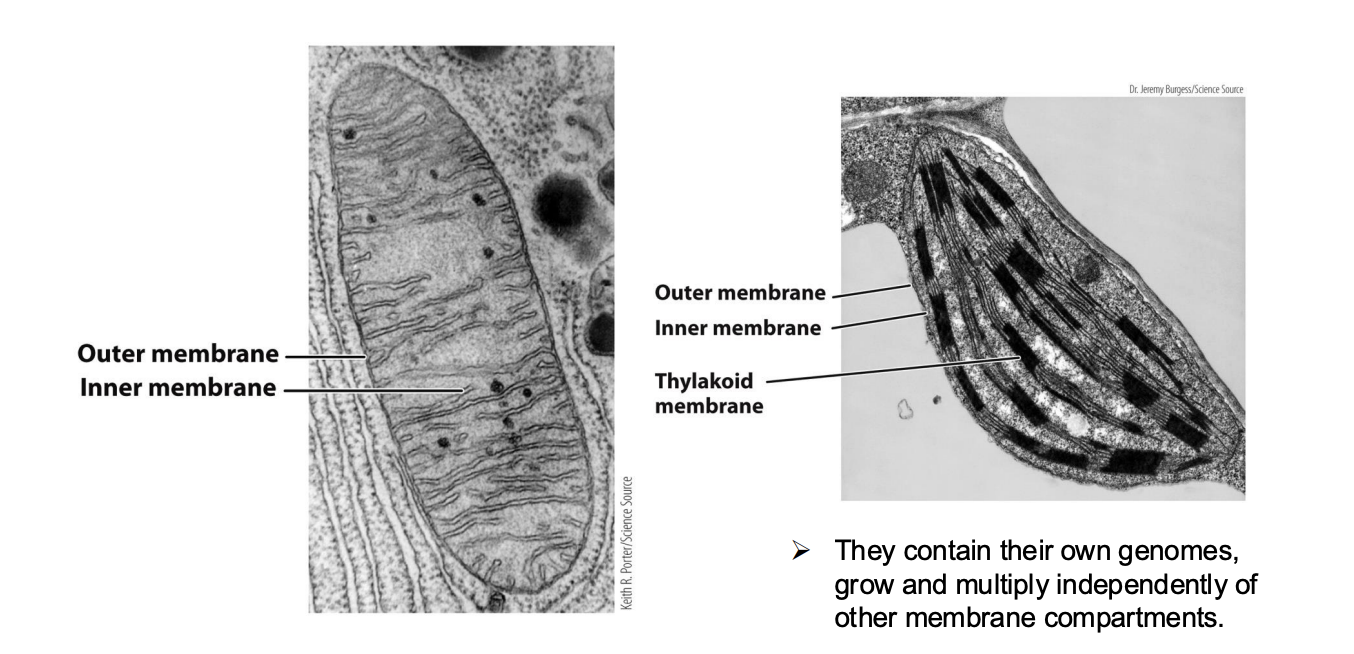
How does the mitochondria function (book) ?
an outer membrane and a highly convoluted inner membrane whose folds project into the interior A proton electrochemical gradient is generated across the inner mitochondrial membrane, and the energy stored in the gradient is used to synthesize ATP for use by the cell. The presence of folds of the inner mitochondrial membrane increases the surface area available for the biochemical machinery that pumps protons and then synthesizes ATP. The more folds there are, the more surface is available and the more ATP is synthesized. This is another example of structure and function coming together.
In the process of breaking down sugar and synthesizing ATP, oxygen is consumed and carbon dioxide is released.
How does the chloroplast function (book)?
plant cells and green algae have organelles called chloroplasts that capture the energy of sunlight to synthesize simple sugars
This process, called photosynthesis, results in the release of oxygen as a waste product. Like the nucleus and mitochondria, chloroplasts are surrounded by a double membrane.
They also have a third, internal membrane, called the thylakoid membrane.
membrane defines a separate internal compartment called the thylakoid. The thylakoid membrane contains specialized light-collecting molecules called pigments, of which chlorophyll is the most important.
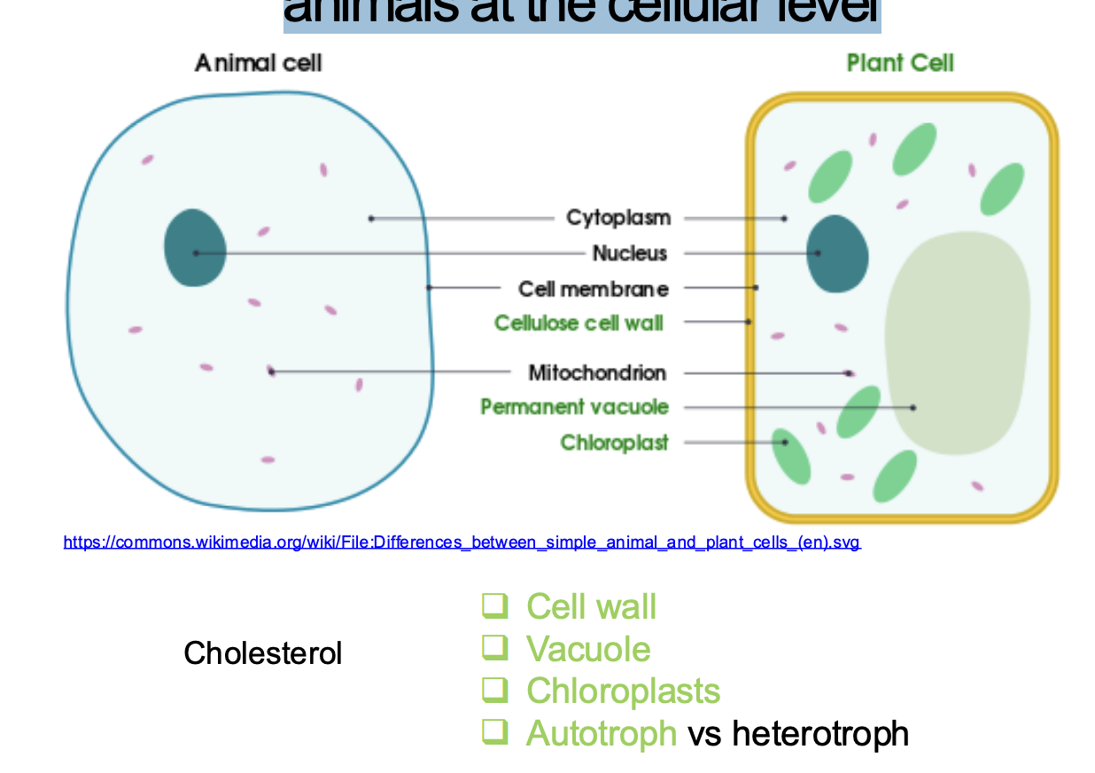
What are the major differences between plants and
animals at the cellular level?
Share common structures, but some unique
Plant cell
Chloroplast
Vacuole
Cell wall
Animal
Cholesterol only in animal cells
Diff leads to diff ecological roles in nature
Plant cell uses energy to produce food in chloroplast (autotroph)
Animal cells cannot produce food themselves (prey on other --> heterotroph)