bio 15 Control and coordination
5.0(1)
5.0(1)
Card Sorting
1/35
Earn XP
Study Analytics
Name | Mastery | Learn | Test | Matching | Spaced |
|---|
No study sessions yet.
36 Terms
1
New cards
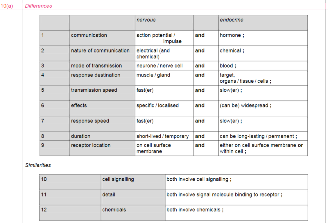
compare the features of the nervous system and the endocrine system
2
New cards
describe the features of the endocrine system
-a system of endocrine glands, which produce hormones that affect target organs
-a hormone is a chemical messenger that travels in the blood so Endocrine glands have a good blood supply
- hormones are either polypeptide chains (ADH, glucagon and insulin) or steroids (estrogen and progesterone)
-a hormone is a chemical messenger that travels in the blood so Endocrine glands have a good blood supply
- hormones are either polypeptide chains (ADH, glucagon and insulin) or steroids (estrogen and progesterone)
3
New cards
The mammalian nervous system is made of
the brain and spinal cord, which form the central nervous system, and the cranial and spinal nerves, which form the peripheral nervous system
4
New cards
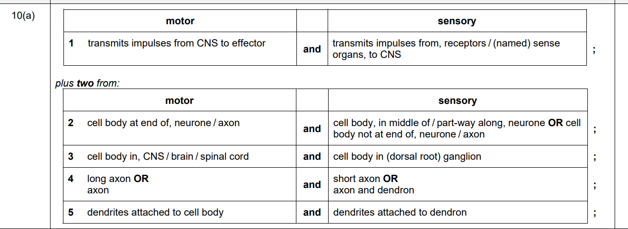
What are the three main types of neurone and where do they transmit impulses?
Sensory: carry impulses from receptors to the CNS (brain or spinal cord)
Relay (intermediate): connect sensory and motor neurones
Motor: carry impulses from the CNS to effectors (muscles or glands)
Relay (intermediate): connect sensory and motor neurones
Motor: carry impulses from the CNS to effectors (muscles or glands)
5
New cards
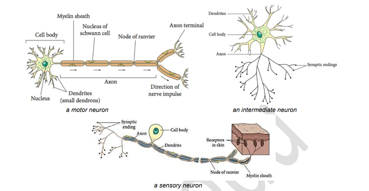
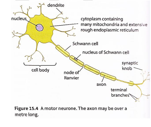
describe the structure of a motor neurone
6
New cards
what is myelin sheath made up of
Schwann cells which wrap themselves around the axon all along its length.
7
New cards
what are the gaps between schwann cells called
- nodes of ranvier
- which occur every 1-3 mm in human neurons
- The nodes are very small, around 2-3 µm long.
- which occur every 1-3 mm in human neurons
- The nodes are very small, around 2-3 µm long.
8
New cards
function of myelin sheath
protect and provide insulation to the axon, prevent leakage of impulse from the axon, increase the speed of transmission of nerve impulse
9
New cards
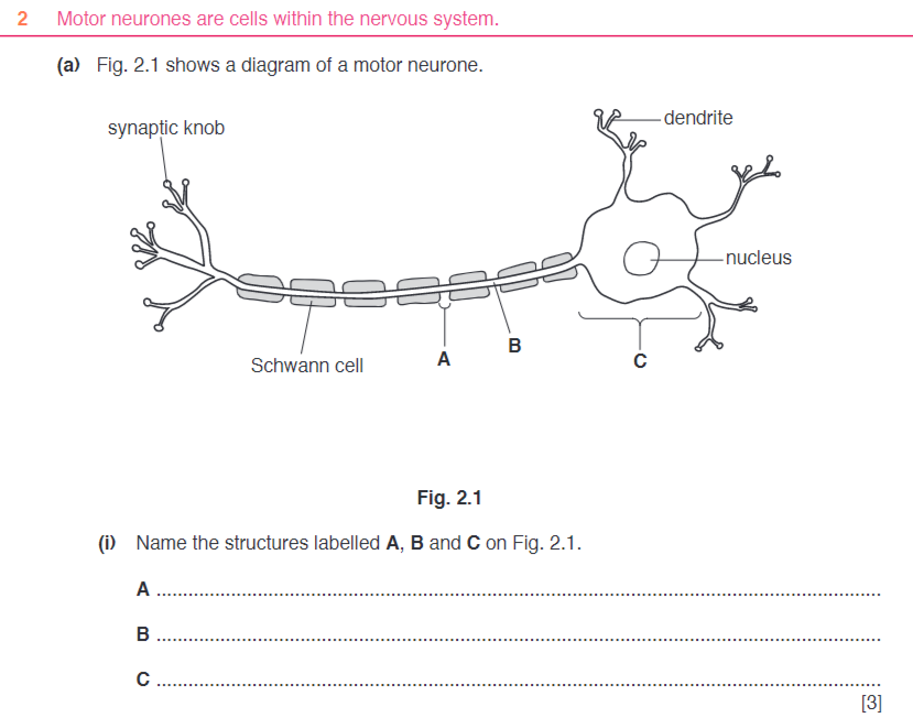
A node of Ranvier ;
B axon ;
C cell body
B axon ;
C cell body
10
New cards
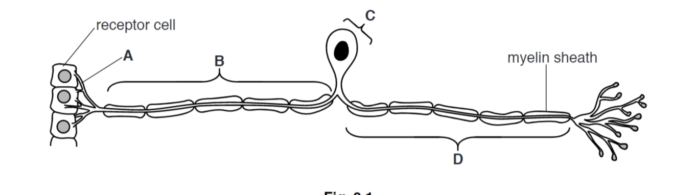
structure of a sensory nuerone
A – dendrite(s) ;
B – dendron / (sensory) axon ;
C – cell body (of neurone) / soma / centron ;
D – axon (membrane) ; A terminal axon
B – dendron / (sensory) axon ;
C – cell body (of neurone) / soma / centron ;
D – axon (membrane) ; A terminal axon
11
New cards
role of sensory receptor cells
- converting stimulus into an electrical impulse
- detecting stimuli and stimulating the transmission of impulses in sensory neurones
- detecting stimuli and stimulating the transmission of impulses in sensory neurones
12
New cards
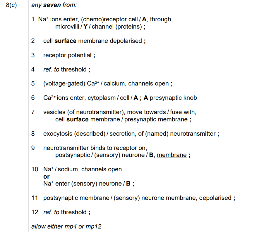
Sequence of events that results in an action potential in a sensory neurone, using a chemoreceptor cell in a human taste bud as an example
- Na+ diffuse through highly selective channel proteins in the CSM of the microvilli which leads to depolarization of the membrane
- The increase in positive charge inside the cell is the receptor potential.
- The receptor potential/depolarization stimulates the opening of voltage-gated calcium ion channel proteins.
- increased conc of Ca2+ stimulates movement of vesicles containing neurotransmitter to release the neurotransmitter by exocytosis.
- The neurotransmitters stimulate an action potential in the sensory neurone
13
New cards
Describe how a resting potential is maintained in an axon
1. axon phospholipid bilayer impermeable to K+ / Na+ ;
2. sodium – potassium pump ;
3. detail of sodium-potassium pump ; e.g. transmembrane / globular / ATP binding site
4. active process / ATP used / energy needed ;
5. 3 Na+ (pumped) out / 2 K+ (pumped) in ;
6. K+ diffuse out / Na+ diffuse in ;
7. through, protein channels transport proteins ;
8. more K+ channels open than Na+ channels ;
9. therefore, membrane more permeable to K+ or more K+ leave than Na+ enter (axon) ;
10. inside relatively more negative than outside ;
11. –65mV ; A –70mV
14
New cards
define resting potential
The pd that is maintained across a neuron when it is not transmitting an action potential
15
New cards
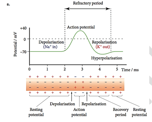
describe the events that occur during an action potential
\-sodium channels in the axon membrane open fully. But the potassium channels are closed
* influx of Na+ in to the axon
* which removes the charge difference across the membrane and the membrane is depolarized.
\-the charge on the inside of the axon membrane goes from -70 to +40mV
* influx of Na+ in to the axon
* which removes the charge difference across the membrane and the membrane is depolarized.
\-the charge on the inside of the axon membrane goes from -70 to +40mV
16
New cards
outline how the resting potential is restored during the refractory period
1 reference to closing Na+ channels;
2 opening of K+ channels;
3 K+ diffuses out (across axon membrane);
4 (charge on the K+) restores the membrane/resting potential;
5 reference to slight overshoot/hyperpolarisation;
6 reference K+ channels close
-the time during which hyperpolarization occurs is called recovery period
2 opening of K+ channels;
3 K+ diffuses out (across axon membrane);
4 (charge on the K+) restores the membrane/resting potential;
5 reference to slight overshoot/hyperpolarisation;
6 reference K+ channels close
-the time during which hyperpolarization occurs is called recovery period
17
New cards
label a full potential against time diagram during an action potential
18
New cards
Describe the importance of the refractory period in the transmission of action potentials.
1 limits / controls, (maximum) frequency of action potentials ;
2 (action potentials / impulses) travel in one direction
- every action potential is separated and action
potentials cannot overlap
-this separation of action potentials allow them to be distinguished from one another
2 (action potentials / impulses) travel in one direction
- every action potential is separated and action
potentials cannot overlap
-this separation of action potentials allow them to be distinguished from one another
19
New cards
Describe how action potentials are transmitted along a myelinated axon
1 local circuit / movement of ions from positive to negative region ;
2 (causes) opening of Na+ channels ;
3 at next, node (of Ranvier) / gap in myelin sheath ;
4 causing next / new, action potential / depolarisation ;
5 saltatory conduction ;
6 one-way transmission ;
7 AVP ; e.g. myelin (sheath) / Schwann cells, insulate axon / prevent (named) ion movement / impermeable to (named) ions / speed up transmission / lengthens local circuits
20
New cards
Explain what is meant by saltatory conduction and describe its effect on the transmission of a
nerve impulse.
nerve impulse.
1 action potential / impulse, ‘jumps’ from node to node ;
2 local circuits, set up between nodes / longer ; A local currents
3 fast / increases speed (of transmission of impulse) ;
2 local circuits, set up between nodes / longer ; A local currents
3 fast / increases speed (of transmission of impulse) ;
21
New cards
what is the refractory period
period of time following the start of an action potential when another stimulus will not result in a second action potential
22
New cards
Factors affecting the speed of transmission of action potentials:
diameter of the axon:
\-thick axons transmit impulses faster than thin ones.
myelin sheath:
\-speeds up transmission
Temperature:
\- the higher the temp the faster the nerve impulse (uptill the optimum temp)
\-thick axons transmit impulses faster than thin ones.
myelin sheath:
\-speeds up transmission
Temperature:
\- the higher the temp the faster the nerve impulse (uptill the optimum temp)
23
New cards
describe the relationship between the strength of the
stimulus and the resulting action potential.
stimulus and the resulting action potential.
1 only depolarisation / receptor potential / potential difference, that reaches threshold produces an action potential
2 the action potential is the same size no matter how strong the stimulus
3 the size of action potentials resulting from a strong and a weak stimulus remains the same but only its frequency changes
2 the action potential is the same size no matter how strong the stimulus
3 the size of action potentials resulting from a strong and a weak stimulus remains the same but only its frequency changes
24
New cards
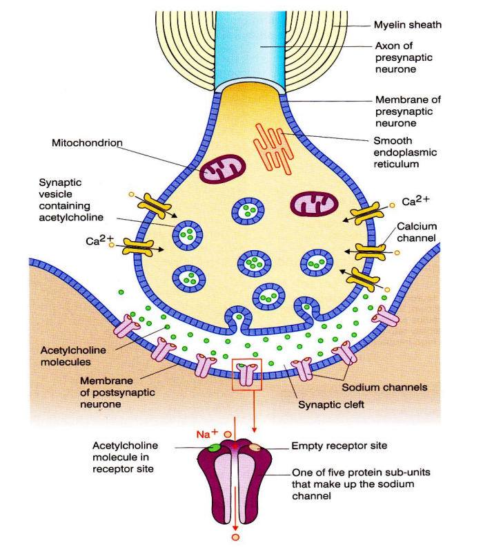
structure of a cholinergic synapse
25
New cards
role of calcium ions in cholinergic synapse
The arrival of an action potential at the presynaptic membrane stimulates voltage-gated calcium ion channel proteins to open
Calcium ions diffuse into the cytoplasm of the presynaptic neurone
This stimulates ACh-containing vesicles to fuse with the presynaptic membrane, releasing ACh molecules into the synaptic cleft
Calcium ions diffuse into the cytoplasm of the presynaptic neurone
This stimulates ACh-containing vesicles to fuse with the presynaptic membrane, releasing ACh molecules into the synaptic cleft
26
New cards
function of acetylcholinesterase
- To prevent the sodium ion channels staying permanently open
- to stop continuous depolarisation/action potentials at the postsynaptic membrane, the ACh molecules are broken down/hydrolysed into acetate and choline and recycled
- to stop continuous depolarisation/action potentials at the postsynaptic membrane, the ACh molecules are broken down/hydrolysed into acetate and choline and recycled
27
New cards
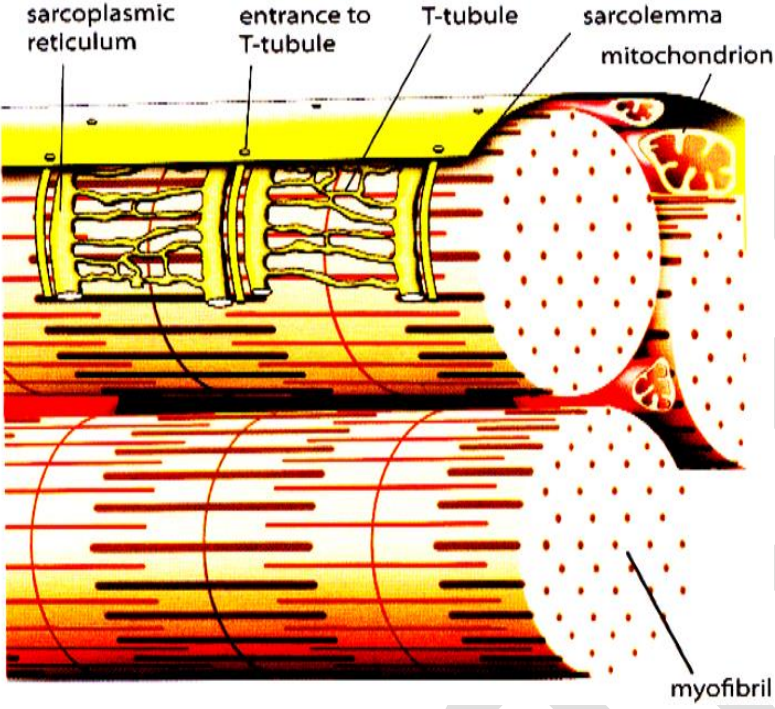
Structure of a muscle fiber:
cell surface membrane called sarcolemma
cytoplasm called sarcoplasm
large concentration of mitochondria and ER called sarcoplasmic reticulum (SR)
sarcolemma has many deep infoldings into the interior of the muscle fiber, called T-tubules
Each muscle fiber is made up of many parallel myofibrils
cytoplasm called sarcoplasm
large concentration of mitochondria and ER called sarcoplasmic reticulum (SR)
sarcolemma has many deep infoldings into the interior of the muscle fiber, called T-tubules
Each muscle fiber is made up of many parallel myofibrils
28
New cards
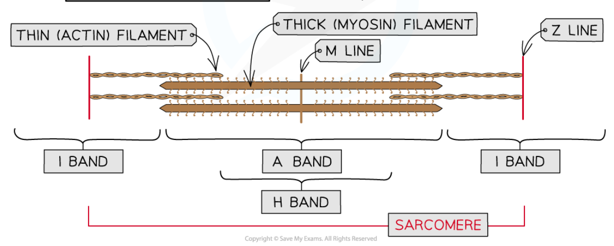
Detailed structure of a myofibril:
- made up of components called protein filaments
- thick filaments made of myosin protein and thin ones made of actin protein
H band: only thick myosin
I band: only thin actin
A band: only thick myosin + parts where actin and myosin overlap
M line: attachment for myosin filaments
Z line: attachment for actin filaments
Sarcomere: between two Z lines
29
New cards
what two (other) proteins are found in a muscle and what do they do
Tropomyosin, which forms a fibrous strand twist around the actin filament
Troponin, a globular protein attached to the actin chain at regular intervals
Troponin, a globular protein attached to the actin chain at regular intervals
30
New cards
changes that occur to a sarcomere when a muscle contracts
-the I-band becomes narrower
-the Z-lines more closer together (sarcomere shortens)
-the H-zone becomes narrower
-the A-band remains the same width.
-the Z-lines more closer together (sarcomere shortens)
-the H-zone becomes narrower
-the A-band remains the same width.
31
New cards
Sliding filament model of muscular contraction:
1 calcium ions released from sarcoplasmic reticulum ;
2 calcium ions bind to troponin ;
3 troponin changes shape and moves tropomyosin ;
4 exposes binding site on actin ;
5 myosin head, binds to site / forms cross bridge ;
6 myosin head tilts ;
7 pulls actin / power stroke ;
8 myosin head, has ATPase / hydrolyses ATP ;
9 myosin head lets go of actin ;
10 myosin head goes back to previous orientation / myosin head re-cocks ;
11 process repeated ;
12 sarcomere shortens ;
32
New cards

33
New cards

34
New cards

35
New cards
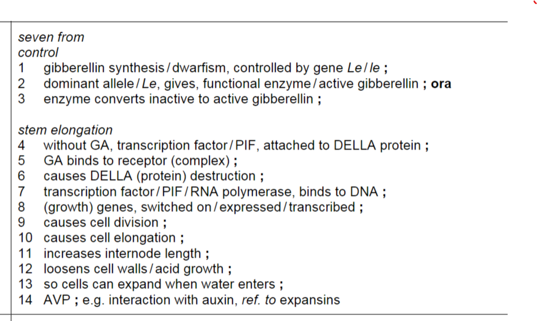
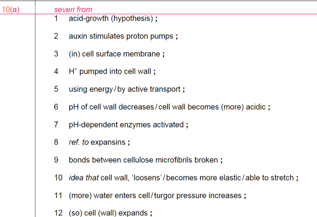
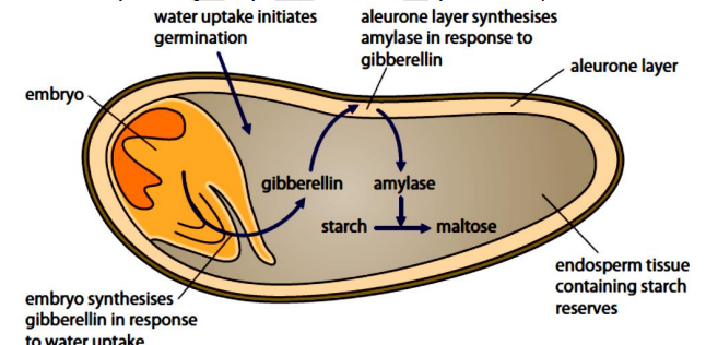
\n Describe the role of gibberellin in the germination of barley.
1. DELLA proteins inhibit germination ;
2. seed absorbs water ;
3. stimulates production of gibberellin ;
4. by embryo ;
5. gibberellin causes breakdown of DELLA proteins ;
6. leads to transcription of mRNA coding for amylase ;
7. in aleurone layer ;
8. (amylase) hydrolyses starch to maltose ;
9. ref. maltose converted to glucose ;
10. glucose respired by embryo during germination ;
36
New cards
Contrast the structure and function of sensory neurones and motor neurones.