Unit 3: Ankle/Foot Region
1/108
There's no tags or description
Looks like no tags are added yet.
Name | Mastery | Learn | Test | Matching | Spaced | Call with Kai |
|---|
No analytics yet
Send a link to your students to track their progress
109 Terms
How does the sufficiently pliable structure of the ankle and foot affects its function?
allows for absorbance of impact, adaptation to uneven surfaces, and allows motion in multiple directions around the planted surface, however still has adequate rigidity to transfer high forces
why are there additionally applied movement definitions in the ankle and foot?
movements purely based on the plane perpendicular to the three standard axes of rotation are inadequate to describe all the motions possible
which applied movement terminology refers to the movement that occurs about an oblique axis of rotation?
pronation (consisting of abduction, dorsiflexion, and eversion: ADE is PRO), and supination (adduction, and plantarflexion, and inversion)
which bone is common to both the ankle and the rearfoot?
the talus
which bones and joints make up the rearfoot?
calcaneous and talus, and the subtalar joint
which bones and joints make up the ankle?
the tibia, fibula, and talus, and the proximal tibiofibular, distal tibiofibular, and the talocrural
which bones and joint make up the midfoot?
navicular, cuboid, and cuneiforms (medial, intermedial, and lateral), and the transverse tarsal joints, the distal intertarsal
which bones and joints make up the forefoot?
metatarsals, phalanges, and the tarsometatarsals, the intermetatarsals, the metatarsophalangeal, and the interphalangeal joints
which structures bind the fibula to the tibia?
all of the above
how much motion of anterior and posterior translation is available at the proximal tibiofibular joint?
1-3 mm
what does the stability of the proximal tibiofibular joint offer the ankle?
ensures force transfer from the fibula to the tibia
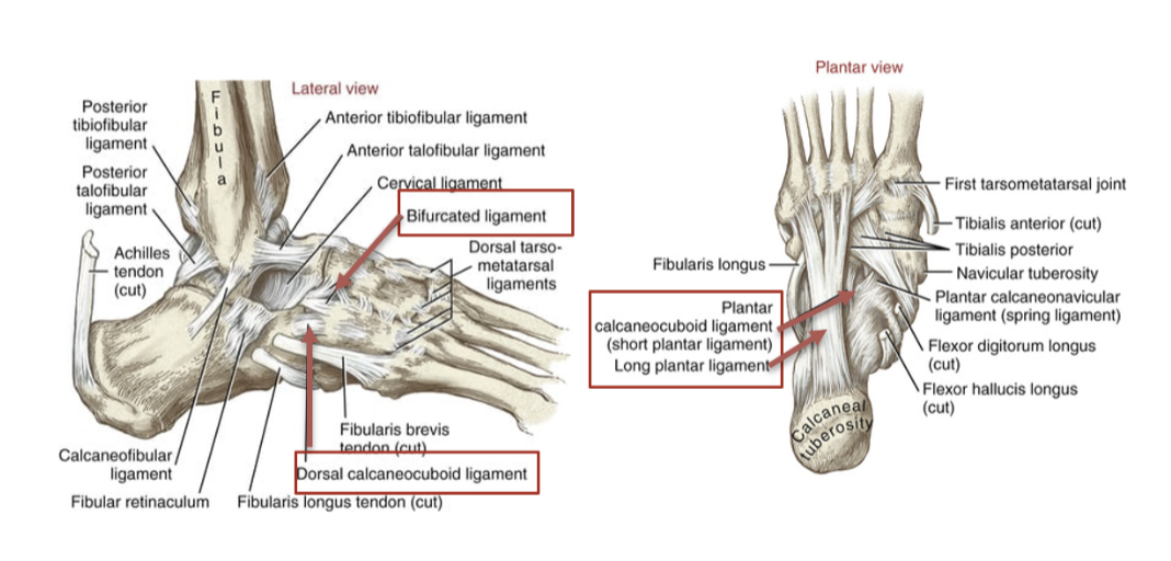
which ligaments attach at the distal tibiofibular joint?
the interosseus, anterior tibiofibular, and posterior tibiofibular
which joint, the distal or proximal tibiofibular joint, allows for slight movement?
the distal tibiofibular joint, but the two are associated to reinforce the talocrural joint (both joints are considered ankle joints)
what is the one functional articulation of the ankle?
the talocrural joint
describe the shape and function of the talocrural joint
is shaped like a carpenter mortise joint, accepts the forces that pass between leg and foot
how much cartilage is at the talocrural joint, allowing for load absorption?
3 mm of articular cartilage
which ligaments provide stability and propioceptive inputs at the talocrural joint?
medially: the medial collateral (deltoid ligament), and laterally: the lateral collateral consisting of the anterior talofibular, the posterior talofibular, and the calcaneofibular, as well as the inferior transverse ligament
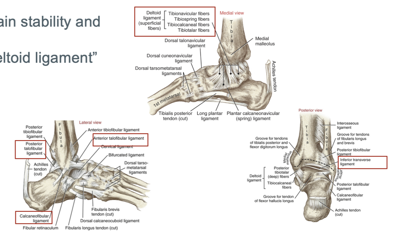
which motions stretch the deltoid ligament’s tibiotalar fibers?
talocrural joint eversion, dorsiflexion with associated posterior slide of the talus within the mortise
which full movements stretch the deltoid ligament’s tibionavicular fibers?
talocrural joint eversion, abduction, and plantarflexion with associated anterior slide of the talus, and talonavicular eversion and abduction
which full motions stretch the deltoid ligament’s tibiocalcaneal fibers?
talocrural and subtalar joint eversion
which full motions stretch the anterior talofibular ligament?
talocrural joint inversion, adduction, and plantarflexion with anterior slide of talus
which full motions stretch the calcaneofibular ligament?
talocrural joint inversion, dorsiflexion with associated posterior slide of the talus, and subtalar joint inversion
which motions stretch the posterior talofibular ligament?
talocrural joint abduciton, inversion, and dorsiflexion with associated posterior slide of the talus
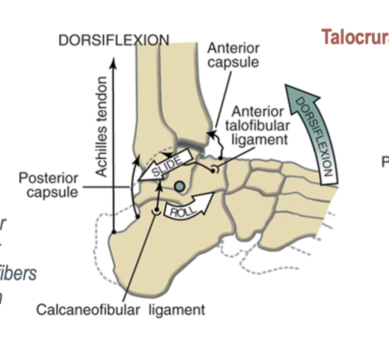
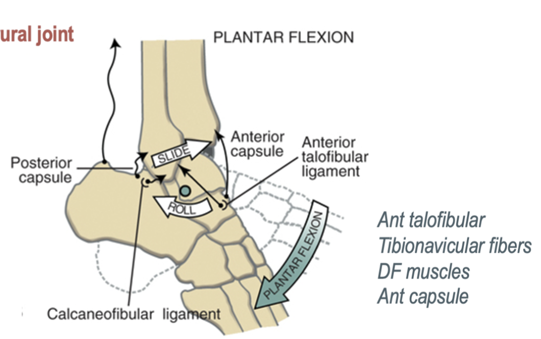
how many degrees of freedom are at the talocrural joint?
One degree of freedom, allowing for dorsiflexion and plantarflexion movements.
what are the two main motion of the talocrural joint?
dorsiflexion (combined with slight eversion and abduction to make pronation) and plantarflexion (combined with slight inversion and adduction to make supination)
what is the arthrokinematics of dorsiflexion of the talocrural joint?
the talus rolls anterior and slides posterior
what is the arthrokinematics of plantar flexion of the talocrural joint?
the talus rolls posterior and slides anterior
describe the arthrokinematics of dorsiflexion of the talocrural joint when the foot is fixed?
anterior roll and anterior slide of the tibia and fibula relative to the talus
describe the position of the talocrural joint during gait?
at heel contact (normally the initial contact), PF until the foot is flat, once the foot is flat, the leg rotates forward (dorisflexion) over grounded foot until after heel off, and at push off the joint is in full DF (closed packed and stable) position, ready to accept body weight compression forces
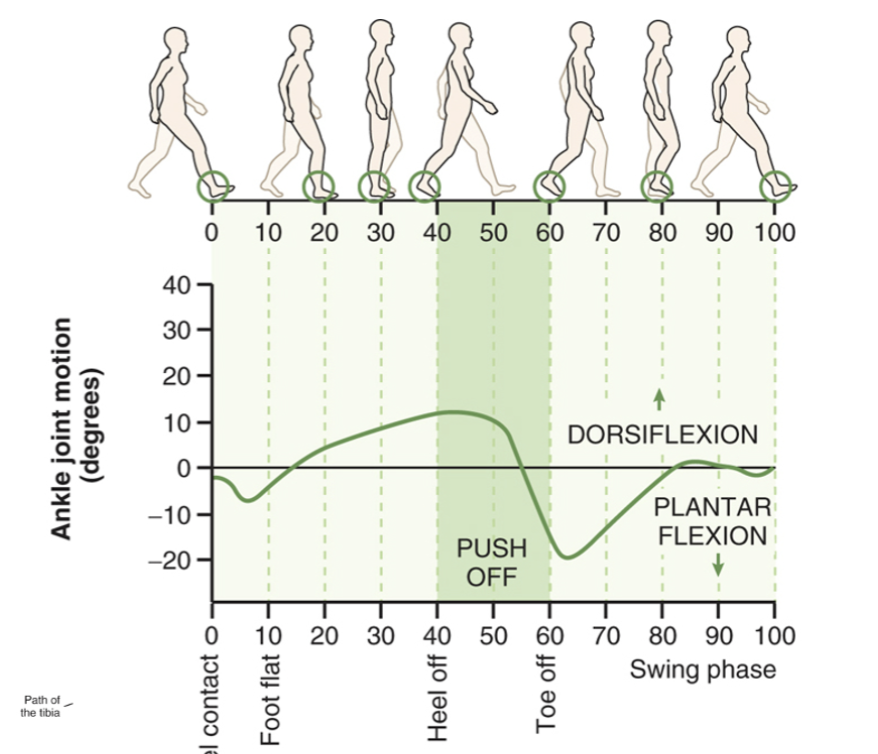
during which phase of the gait cycle is dorsiflexion greatest at the talocrural joint?
stance phase (specifically during the beginning of push off phase when the foot is still in contact with the ground)
what factors contribute to the stability of the talocrural joint during full dorsiflexion?
elongation of the collateral ligaments and plantarflexor muscles, as well as “widening” of the talus in the mortise since the talus is wider anteriorly
which bones make up the subtalar joint?
3 articulations between the posterior, middle and anterior facets of the calceneus and talus
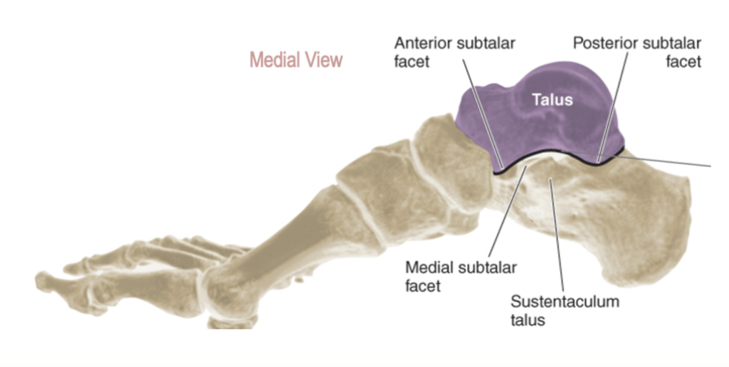
what ligaments offer the primary source of stabilization at the subtalar joint?
calcaneofibular, the tibiocalcaneal fibers of the deltoid ligament, and the interosseus (talocalcaneal) ligament, and the cervical ligaments
which motion does the calcaneofibular ligament resist at the subtalar joint?
excessive inversion
which motion does the tibiocalcaneal fibers of the deltoid ligament resist at the subtalar joint?
excessive eversion
which motion does the interosseus and the cervical ligaments limit in the subtalar joint?
limits the stress of all motions, especially inversion
what are the two main motions of the subtalar joint?
pronation (with major components of eversion and abduction, slight dorsiflexion is clinically ignored) and supination (with major components inversion and adduction, slight plantarflexion is clinically ignored)
where is the axis of rotation in the subtalar joint?
pierces lateral-posterior heel and passes through subtalar joint in anterior, medial, and superior directions
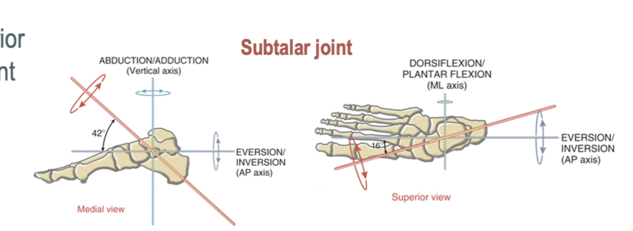
how many degrees of freedom are at the subtalar joint ?
1 DOF, for pronation/supination
describe the gait kinematics of the subtalar joint during the early to mid stance
during initial contact, see talocrural PF and slight subtalar supination (calcaneous inversion), immediately followed by talocrural DF and subtalar pronation (calcaneous eversion) (the calcaneous in contact with the ground pushes the talus medially and subtalar pronation causes the tibia/fibula and femur internal rotation)
describe the gait kinematics of the subtalar joint during the mid to late stance
at 15-20% of the entire stance limb (femur, tibia, and talus) reverses from internal to external rotation, and during mid to late phase see the pronated (everted) subtalar joint move toward supination (inversion), and the forefoot is pronated to remain in contact with the ground
what is another term for the transverse tarsal joint?
midtarsal or chopart’s joint
what regions do the transverse tarsal joint connect?
connects the rearfoot with the midfoot
what are the two articulations of the transverse tarsal joint?
the talonavicular and calcaneocuboid joints
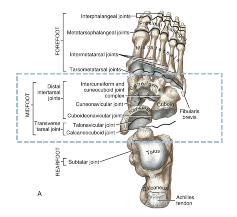
which joints make up the distal intertarsal joints?
the cuboideonavicular, intercuneiform and cuneocuboid joint complex, and the cuneonavicular

which is the most versatile joint of the foot?
the transverse tarsal joint, allows the weight bearing foot to adapt to a variety of surface contours
describe the talonavicular joint
is the transverse tarsal joints medial compartment, forms the articulation between the convex head of the talus and the concavity of the navicular’s proximal side and spring ligmament
what kind of joint is the talonavicular joint?
ball and socket joint, which provides substantial mobility to the foot’s medial (longitudinal) column
what kinds of motions does the talonavicular joint allow for between the midfoot and forefoot?
twists (inverts and everts), and bends (flexes and extends) the midfoot and forefoot relative to rearfoot
which of the following best describes the primary arthrokinematics at the talonavicular joint during inversion and eversion?
spinning motion between the concave, posterior side of the navicular bone and the head of the talus
which ligaments provide stability at the talonavicular joint?
the talonavicular ligaments- consisting of the spring, dorsal talonavicular, the tibionavicular, and tibiosping fibers of the deltoid ligament, and the bifurcated (calcaneonavicular fibers)
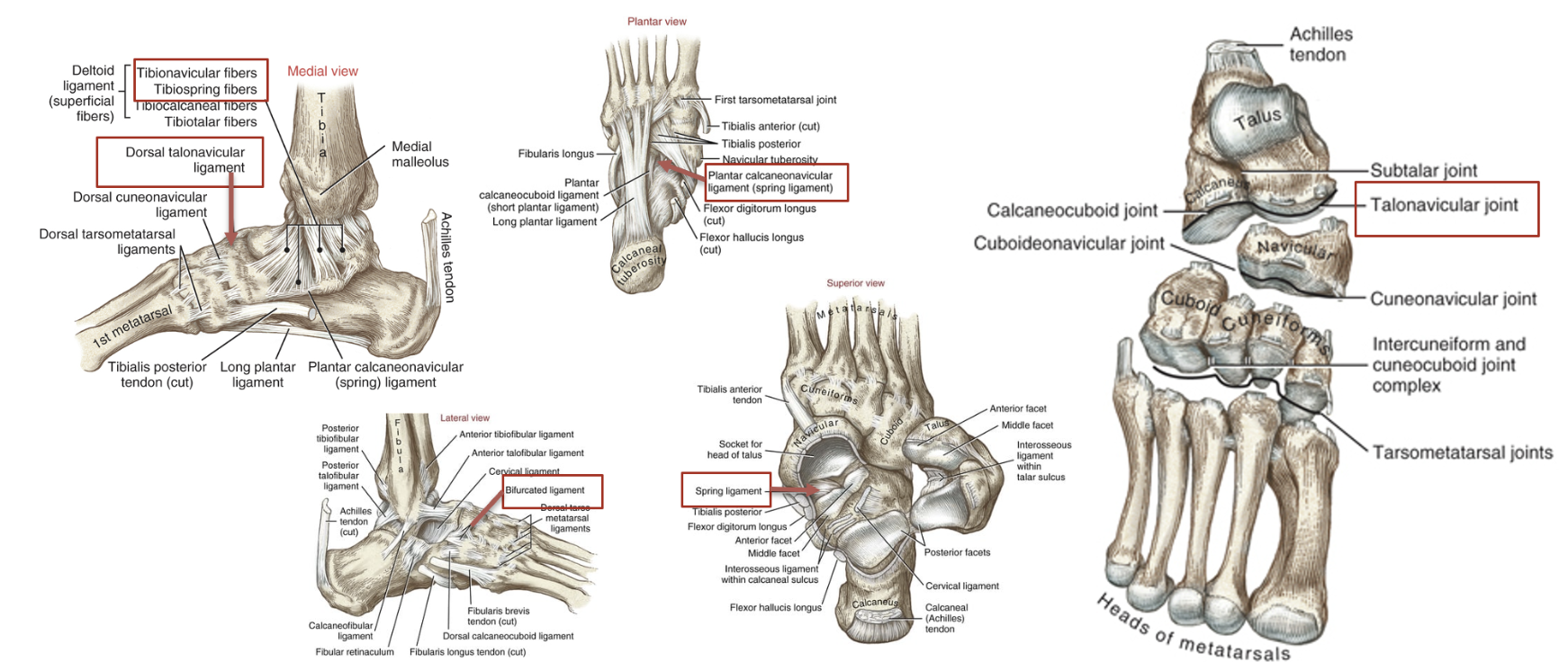
describe the calcaneocuboid joint
makes up the lateral compartment of the transverse tarsal joint, is the articulation between the calcaneous and the proximal surface of cuboid- each articular surface provides a concave and convex surface which forms an interlocking edge resistant to sliding
how much motion does the calcaneocuboid joint have in comparison to the talonavicular joint?
less motion, acts instead to provide stability to the lateral column of the foot
which ligaments provide stability to the calcaneocuboid joint?
dorsal calcaneocuboid, bifurcated, and long and short plantar ligaments
how many axes of rotation are at the transverse tarsal joints?
2- have a longitudinal axis allowing for pronation/supination primarily as a result of eversion/inversion, and an oblique axis allowing for pronation that is a result of abduction and DF and supination that is a result of adduction and PF
how much ROM is available at the transverse tarsal joint?
20-25 degrees of inversion, and 10-15 degrees of eversion
why are the multiple axis of rotation significant at the transverse tarsal joints?
allows for adaptation to a variety of surface contours, also allows for different amplitude and direction of movement during weight bearing and non-weight bearing activities, transverse tarsal joints also work with the subtalar joint to control pronation and supination of the entire foot, and the kinematics are influenced by position of the subtalar joint
what movement of the transverse tarsal joints occurs with full subtalar supination and when would it occur?
the lateral midfoot drops, the talonavicular and the calcaneocuboid joints become twisted (relative eversion), this occurs in late stance and adds rigidity to the foot to support large loads during push off of gait cycle
what movement of the transverse tarsal joint occurs with full subtalar pronation and when would it occur?
the talonavicular and calcaneocuboid joints become untwisted (relative inversion), this occurs in the first 35% of the gait cycle and adds flexibility
which is the foot’s primary load bearing and shock absorbing structure?
the medial longitudinal arch, which is made of the calcaneous, the talus, the navicular, the cuneiforms, and the three medial metatarsals, as well as nonmuscular structures like the plantar fascia, the sping ligament, the first tarsometatarsal joint
what mechanism stretches the fibers of the deep fascia of the medial longitudinal ligament, adding tension to the arch during the push off phase?
toe extension
what is the keystone of the medial longitudinal arch?
the talonavicular joint
how is body weight and load distributed through the medial longitudinal arch?
body weight is accepted and dissipated mainly by elongation of the plantar fascia, and load is distributed anterior and posteriorly throughout this arch to heel and the foot ball (metatarsal head region), the body weight pushes the talus inferiorly, and slightly lowers the arch and stretches the connective tissue (there is also some active muscle support that acts as a secondary line of support that is more active with dynamic activities)
how much is the medial longitudinal arch lowered during standing?
approx 7 mm
describe pes planus
aka flatfoot, characterized by chronically dropped or abnormally low arch, see excessive laxity and overstretching of the connective tissue (plantar fascia, the spring ligament, and the tibialis posterior tendon), compromises the ability to dissipate loads of the foot and have more intrinsic/extrinsic muscle force required while standing
what does the excessive subtalar pronation of pes planus cause?
valgus rearfoot (calcaneus everted away from the midline), and forefoot goes into excessive abduction
describe pes cavus
a condition where the foot has an abnormally high arch, leading to an inability to absorb shock effectively, is associated with excessive rearfoot varus (inversion) and excessive forefoot valgus (eversion) to keep the medial forefoot firmly in contact with the ground
what is the function of the distal intertarsal joints?
assist the transverse tarsal joint in pronating and supinating the midfoot (small motions), provides stability across the midfoot by forming the transverse arch, and depresses during weight bearing to allow the metatarsal heads to contact the ground
what is the transverse arch formed by?
intercuneiform and cuneocuboid complex, containing 3 joints: cuneonavicular, the cuboideonavicular, and the intercuneiform/cuneocuboid complex (so the distal intertarsal joints)
which articulations make up the tarsometatarsal joints?
articulation between the bases of the metatarsals and distal surfaces of three cuneiforms and cuboid
which of the TMT (tarsometatarsal joints) allow minimal movements, and provide stability for push off?
the 2nd and 3rd TMT joints, the 2nd ray is reinforced by the 2nd TMT joint
which combined motions of the first ray allow the forefoot to better conform to contour of surfaces?
combined PF and eversion of the TMT joint of the first ray and combined DF with slight inversion
describe the joint kinematics of the first TMT joint
plantarflexion occurs with slight eversion (PE), dorsiflexion occur with slight inversion (DI), and assists medial longitudinal arch in accepting and sharing loads incurred while walking
which ligaments connect the four lateral metatarsal joints?
the plantar, dorsal, and interosseus ligaments (the three points of contact form small synovial joints)
what feature of the first ray allows for increased relative movement?
the bases of the first and second metatarsals do not form a true joint, though interconnected by ligaments, so this lack of articulation allows for more movement
which ligament connects the distal ends of all five metatarsals?
the deep transverse metatarsal ligaments
what does slight motion at the intermetatarsal joints allow for?
augments the flexibility at the tarsometatarsal joints
what forms the metatarsophalangeal joints?
the convex head of the metatarsals and the concave proximal end of the proximal phalanx
how many degrees of freedom are available at the metatarsalphalangeal joints?
2 degrees of freedom, extension (dorsiflexion) and flexion (plantarflexion) and abduction and adduction
how many degrees of motion are available at the metatarsophalangeal joints?
great toe has about 85 degrees of extension, toes have about 65 degrees of passive extension and 30-40 degrees of flexion
describe the motion at the interphalangeal joints
mobility to limited primarily to flexion and extension, with flexion exceeding extension, and motion greater in the proximal joints, extension is limited primarily by passive tension in the toe flexor muscles and plantar ligaments
how does the midfoot and forefoot become stable during the end of the stance phase to accept stress associated with push off?
activation of intrinsic and extrinsic muscles, and rising of the medial longitudinal arch (about 6 mm)
what is the “windlass effect”?
the primary mechanism to raise the medial longitudinal arch in a normal foot, occurs by contraction of the extrinsic plantar flexors to lift the calcaneous, transferring BW forward over metatarsal heads, resulting extension of the MTP joints stretches the plantar fascia within the medial longitudinal arch, and increased tension from stretch raises the arch and strengthens the midfoot and forefoot (contraction of the intrinsic muscles provides additional reinforcement to the arch)
what is the role of the foot and ankle muscles?
controls specific actions of the underlying joints, provide stability, thrust, and shock absorptions necessary for locomotion, and the actions are evident by noting where the tendons cross the axes of rotation at the talocrural and subtalar joints
what are the extrinsic muscles of the anterior compartment?
the extensor hallucis longus which can perform joint dorsiflexion and great toe extension with negligible subtalar joint inversion, and tibialis anterior, which can also dorsiflex the subtalar joint, and invert the subtalar joint, invert and adduct the talonavicular joint, and supports the medial longitudinal ligament when needed, as well as the extensor digitorum longus and fibularis tertius, which evert the foot (and can dorsiflex the foot)
based on the analysis of the inversion torque potential at the subtalar joint, what is true regarding the TA and the EHL?
the TA has a greater inversion torque potential that the extensor hallucis longus due to its longer moment arm
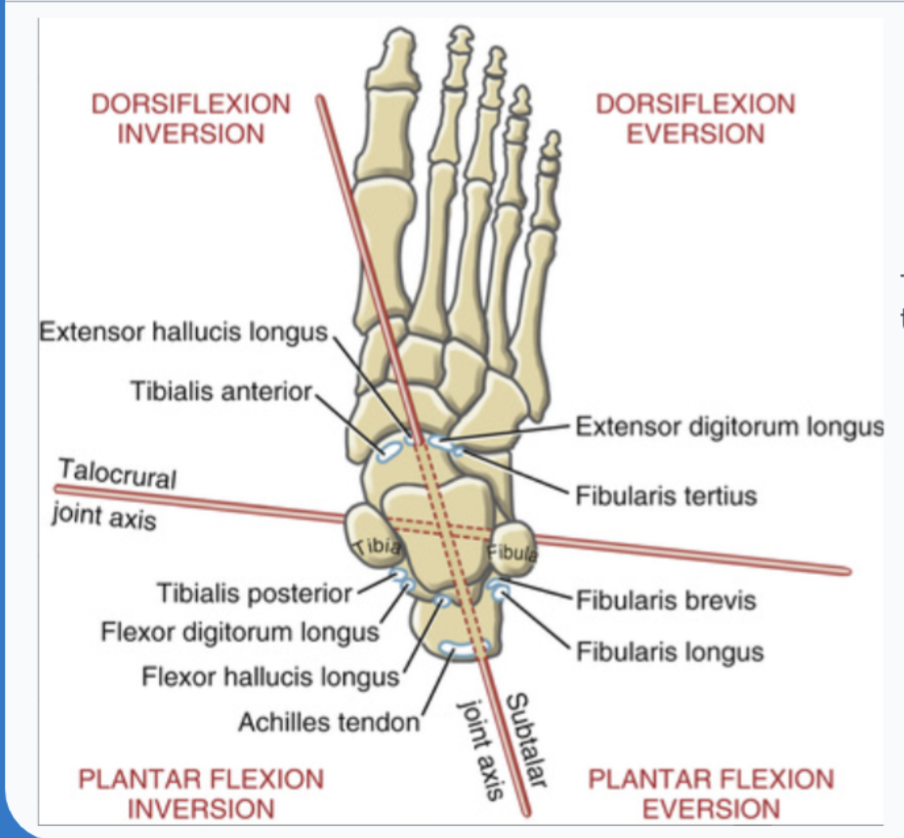
what is the function of the extrinsic muscles of the anterior compartment during the early stance phase of gait?
during early stance, eccentrically active to control the rate of plantar flexion (for soft landing of the foot), TA helps to decelerate lowering medial longitudinal arch and helps control prontation (eversion) of the rearfoot
what is the function of the extrinsic muscles of the anterior compartment during the swing phase of gait?
concentric activation to dorsiflex the ankle and extend the toes in order to ensure that the foot clears the ground
what is the function of the EDL and FT during gait?
they must counteract the inversion and adduction influence of the TA with their eversion and abduction influence
which extrinsic muscles form the lateral compartment?
the fibularis longus and brevis, which are the main source of stability to the lateral side of the ankle, are evertors and plantarflexors of the talocrural and abductors of subtalar and transverse tarsal joints
what is the function of the FL?
generates the forefoot eversion torque, base of the first ray everts and depresses slightly during pronation of the unloaded foot, also stabilizes the first tarsometatarsal joint against potent medial pull of TA
function of the fibularis longus in gait?
there is small concentric activation after heel contact to control pronating rearfoot, helps to fixate the first ray securely to the ground
what is the function of the lateral compartment during gait?
they are active through much of the stance phase, the highest level of activation is throughout mid stance and push off, and helps neutralize strong inversion (supination) bias of tibialis posterior, and forms a functional sling that supports the transverse and medial longitudinal arches, and during push off help to control supinating subtalar joint, assist with plantar flexion, and help transfer BW from lateral to medial side of the forefoot and toward the opposite foot
extrinsic muscles of the posterior compartment and their functions?
plantaris, gastrocneumis, and soleus which are plantarflexors, and the deep group of the FDL, the FHL, and the TP which are additionally supinators (inverters) (though the triceps surae also have slight inversion torque)
what are the characteristics of tibialis posterior?
likely produces greatest supination torque across subtalar and transverse tarsal joints, extensive distal attachments, especially to navicular, provide effective inversion “twist” of midfoot
what is the path of the tendon of the FHL from its origin to its insertion?
it originates from the distal two thirds of fibula and courses between the medial and lateral tubercles of the talus before inserting at the base of the distal phalanx of the great toe
what are the intrinsic muscles of the foot in layer 1?
flexor digitorum brevis, abductor hallucis, abductor digiti minimi
what are the intrinsic muscles of the foot in layer 2?
quadratus plantae, and lumbricals
what are the intrinsic muscles of the foot in layer 3?
adductor hallucis, flexor hallucis brevis, and flexor digiti minimi
what are the intrinsic muscles of the foot in layer 4?
dorsal interossei (four) and plantar interossei (three)