Perception WK 3 - Structure of Visual System
1/24
There's no tags or description
Looks like no tags are added yet.
Name | Mastery | Learn | Test | Matching | Spaced | Call with Kai |
|---|
No analytics yet
Send a link to your students to track their progress
25 Terms
Properties of light
Wavelength
Intensity (number of photons)
Spatial distributions of light is characterised by…
Sources of light
Reflectors of light
Optic Array
Structued observation of changing patterns of light in an environment, allowing us to infer depth, distance, layout etc
Structure of the visual system
Eye → Retina → Optic Chiasm → LGN → Striate Cortex → Extrastriate Cortex
Function of the visual system
Converts a structured pattern of light taken in from the retina into a perception of a 3D world
Encoding principles
Principle of least committment - Don’t throw away things you might need later
Principle of least redundancy - Do things as efficiently as possible
Principle of graceful degradation - If the system breaks, it should still be usable
Gross anatomy of retina (outer layer to inner layer; optic nerve)
(RCHBAG)
Rods and Cones (photoreceptors) - transmit signals
Horizontal Cells - modulate signal processing
Bipolar Cells - transmit signals
Amacrine Cells - modulate signal processing
Ganglion Cells - transmit signals
Rods and cones → Bipolar Cells → Ganglion cells → Optic nerve
Photoreceptor: Rods
100+ million
Provide black and white night vision
Photoreceptor: Cones
6+ million
Provide high-acuity colour vision in bright light
(Visual) Receptive field
Area of the retina that, when stimulated, influences the firing rate of neurons
Phototransduction
When photoreceptors convert light into electricity
Light is taken in by visual pigment molecules
The subsequent chemical changes results in electrical signals
Spectral sensitivities of photoreceptors
L, M and S (Long, Short and Medium) cones are responsible for colour vision
S cones are responsible for blue light
M cones are responsible for green light
L cones are responsible for red light
Blue/Green/Red (upwards)
Determining different colours
Different levels of each cone photoreceptor determines the colour
Can be fooled by METAMERS (making things look like a particular colour when it isn’t)
Visual electrophysiology
Experiments studying the activity of an individual neuron
Tiny electrode placed close to or inside a visual neuron
(Paralysed and anaesthetised) animals were shown visual stimuli
Electrical activity of the neuron is recorded and investigated
Action potentials on Oscilloscopes
We can see action potentials on Oscilloscope screens
This can look like a ‘spike’ if compressed
We measure the amount of action potentials/spikes given in a particular time
Centre/surround structure of ganglion cells (Kuffler, 1953)
Experiment on horse shoe crabs - areas outside the centre had lower responses
Retinal ganglion cells have an excitatory centre and an inhibitory surround (on-center cell)
Off-center cells have inhibitory centres and excitatory surrounds
When light shines in on the centre, this excitates the cell, whereas light on the surround inhibits the cell
This allows us to enhance edges and contrast by highlighting changes in light
Retinal function
Photoreceptors → Bipolar cells → Retinal Ganglion cells
Photoreceptors process light and turn it into an action potential
This is then synapsed into bipolar cells which segregate this into on (light-activated) or off (dark-activated) pathways which is sent to the ganglion cells
The ganglion cells axons form the optic nerve and sends signals to the brain
There are 1 million axons in the optic nerve → the 100+ million rods must compress this somehow
Horizontal and amacrine cells help to shape retinal ganglion cells’ receptive fields
Retinal ganglion cells
Respond with a series of nerve impulses (spike train)
More stimulation → Faster firing rate (spikes per second)
These cells react to contrast on the centre/surround rather than light
This means it responds more to high contrast colours or images (images that are “high information”)
Respond to blue/yellow, red/green and black/white
Structure of Laterate Geniculate Nucleus (LGN)
Size of a peanut, on both sides of the brain (bilateral nucleus)
Has 6 layers:
Layers 1, 4 and 6 process contrateral (opposite side as eye) visual information
Layers 2, 3 and 4 process ipsilateral (same side as eye) visual information
Retinotopic organisation - precise mapping of visual input from the retina onto the visual field (V1), where neurons located near eachother activate neighbouring neurons, creating a map, like a flipped picture
“The Club Sandwich” refers to the laminated structure of the LGN (with a red line as a toothpick)
Contains parvocellular, koniocellular and magnocellular layers
Function of Laterate Geniculate Nucleus (LGN)
Similar centre/surround structure to ganglion cells
Little modification of response, however many regulate information from retina to cortex:
Modificatory inputs from thalamus and brainstem
Feedback pathways from visual cortex
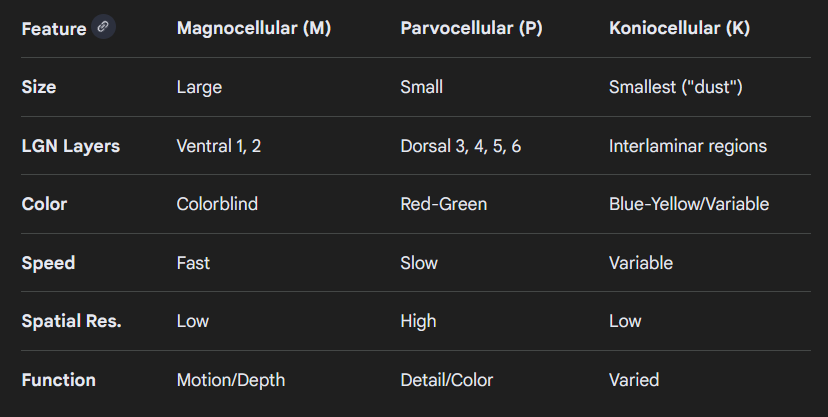
Parvocellular, magnocellular and koniocellular layers
These layers are specialised retinal ganglion cells forming parallel pathways for visual processing
Parvocellular (retinal P cells) - Dorsal layers (3, 4, 5, and 6), small receptive field, fine detail, red/green vision, → “what” an object is
Magnocellular layers (retinal M cells) - Ventral layers (1 and 2), large receptive field, motion detection, colourblind → “where” an object is
Koniocellular (b/y cells) - Interlaminar regions, smallest, blue/yellow vision
Encoding along the optic nerve
The optic nerve contains 3 channels via Retinal Ganglion Cells. These channels contain a centre-surround structure that computes colour contrast rather than absolute colour
Achromatic channel (Black and white → L + M)
“Red-Green” channel (L vs M)
“Blue-Yellow” channel ((L + M) vs S)
Structure of Striate Cortex (V1)
Striate = stripey
Connected to 1.5 million axons from LGN
Contains over 250 million neurons
6 layers (but not like LGN)
Function of Striate Cortex (V1)
Function is still hotly debated as highly complex
No conscious access
More refined than the LGN → deals with orientation, edge detecton, motion, contrast and eye-of-origin
No longer seen as the “seat” of visual perception → perception is more spaced out in brain
Essentially blind without it
Contains receptive fields → Simple cells, Complex cells and End-stopped cells (hypercomplex)
Simple cells
Neurons responsible for detecting orientation → orientation tuning
Responds optimally to bars or specific edges within narrow, spatially mapped receptive fields