biol 475 bones
1/101
Earn XP
Description and Tags
use fishboneviz to help
Name | Mastery | Learn | Test | Matching | Spaced | Call with Kai |
|---|
No analytics yet
Send a link to your students to track their progress
102 Terms
parasphenoid
A bone located on the floor of the skull in many vertebrates, it supports the brain and serves as an attachment point for muscles.
pterosphenoid
A bone in the skull that articulates with the parasphenoid and has a role in supporting the brain and muscle attachment, particularly in some vertebrates.
intercalar
A small bone located in the skull, often situated between the pterosphenoid and other cranial bones, contributing to the structure and integrity of the cranial vault.
vomer
A bone that forms part of the nasal septum, separating the left and right nasal cavities in vertebrates.
ethmoid
A complex bone in the skull that contributes to the structure of the nasal cavity and forms part of the eye socket, playing a key role in supporting the olfactory system.
pre-frontal
A bone located at the front part of the skull, involved in forming the forehead and contributing to the eye sockets and nasal cavity.
frontal
A bone located in the upper part of the head in fish, contributing to the formation of the forehead and being involved in the structure of the eye socket.
sphenotic
A small bone located in the skull of vertebrates, it is situated between the frontal and ethmoid bones, contributing to the eye socket and supporting the adjacent structures.
parietal
A bone located on the lateral sides of the skull in fish, contributing to the formation of the upper sides of the cranium and protecting the brain.
epiotic
A small bone located at the dorsal part of the skull in fish, situated behind the ear region, and contributes to the formation of the cranial roof.
supraoccipital
A median endochondral bone that forms the posterior-most portion of the cranial roof.
pterotic
A bone of the otic series that forms the outer-posterior corner of the neurocranium. It houses part of the lateral semicircular canal of the inner ear and often bears a process for muscle attachment.
exoccipital
Paired endochondral bones located at the posterior aspect of the skull. They form the lateral and dorsal margins of the foramen magnum and provide articulation points for the first vertebra of the spinal column.
basioccipital
It is a median, unpaired bone that forms the posterior base of the skull, situated between the two exoccipital bones. This bone plays a significant role in supporting the skull and connecting it to the vertebral column.
prootic
an essential endochondral bone in the fish braincase, located in the anterior-ventral portion of the otic (ear) region
dentary/meckels
this cartilage is covered and replaced by dermal bones like the dentary (outer jaw) and articular, forming the jaw joint. The cartilage itself acts as a developmental rod, with its posterior end ossifying into the articular bone.
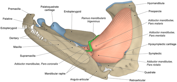
premaxilla/supra maxilla
key dermal bones forming the upper jaw apparatus in bony fishes (teleosts). They play a crucial role in the structure of the mouth, feeding mechanisms, and prey capture.
angular
structural element located at the posterior (rear) part of the lower jaw, or mandible. It articulates with the quadrate bone to form the hinge joint that allows fish to open their mouths and bite. It is part of the dermal bone series in the skull.
articular
paired component of the lower jaw, forming the posterior, moveable joint with the quadrate bone of the suspensorium
symplectic
small, rod-shaped endochondral bone in the jaw suspension (suspensorium) of many bony fishes (teleosts), connecting the hyomandibular bone above to the quadrate bone below. It acts as a key structural element, supporting the lower jaw and facilitating jaw movement, particularly in opening and closing.
branchiostegals
thin, curved, dermal bones located on the ventral part of a bony fish's head, posterior to the gill covers (operculum). They support the gill membranes, acting as a valve to manage water flow during ventilation
ceratohyal
a paired, endochondral, dumbbell-shaped bone located in the middle region of the hyoid arch, acting as a critical support for the branchiostegal rays and gills
epihyal
a, paired, endochondral, "small bone" located in the fish hyoid arch, situated directly behind the main ceratohyal bone. It plays a critical role in the hyoid system for feeding, anchoring branchiostegal rays that support the gill membranes, and connects to the interhyal bone.
interhyal
a small, rod-like element connecting the dorsal and ventral components of the hyoid arch. It serves as a crucial, often cartilaginous, articulation point between the posterior end of the epihyal and the junction of the hyomandibula and symplectic, aiding in jaw and gill support.
hypohyal
a paired, ventral bony element in the fish hyoid arch that connects the ceratohyal to the basihyal, playing a crucial role in jaw support, extension, and respiration
hyomandibula
critical endochondral bone in the fish skull, acting as a suspension bridge between the braincase and the jaws/operculum. It is a boomerang-shaped, dorsal-most component of the hyoid arch that facilitates jaw movement (hyostyly), allowing for suction feeding and breathing
urohyal
single, median, and unpaired bone in teleostean fishes, located ventrally in the head within the branchial and hyoid arches. Forming from the ossification of the sternohyoideus muscle tendon, it is essential for mouth opening and closing mechanics
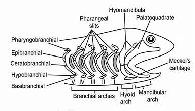
hypobranchial
paired endochondral bones located in the ventral (lower) portion of the gill arches, connecting the ceratobranchials to the median basibranchials. essential to gill arch skeleton and supports gills and allow movement
ceratobranchial
longest, paired endochondral elements within a fish's gill arches, located ventrally between the hypobranchials and epibranchials. Typically numbering five pairs, these bones support gill rakers and filaments, and the fifth pair often acts as tooth-bearing pharyngeal jaws in bony fishes.
basibranchial
median, endochondral elements located on the floor of the mouth/throat behind the tongue, forming part of the branchial (gill) arch skeleton in fishes
epibranchial
main upper, dorsal, endochondral elements of the gill arches in fish, typically forming the upper section of the gill basket
pharyngobranchial
the most dorsal (upper) paired bony elements in the gill arch system of fishes, located at the roof of the throat
interopecle
located between the preopercle and the branchiostegals. It is part of the opercular series, which supports and protects the gill chamber
subopercle
flat, thin dermal bone located at the bottom-rear margin of the gill cover (operculum) in bony fish. It is one of four, the others being the preopercle, opercle, and interopercle, that together form the protective gill flap. This bone often has a rectangular shape and helps protect the gills
opercle
hard, bony flap in bony fish that covers and protects the gills, located on both sides of the head. It plays a crucial role in respiration by pumping water over the gills, even when the fish is stationary
preopercle
boomerang or L-shaped, paired dermal bone located on the cheek region of bony fish. As the most anterior bone of the opercular series (gill cover), it supports the gill flap, houses a sensory canal, and often features serrations or spines used for identification and defense.
quadrate
critical bone in the fish skull that forms the upper part of the jaw joint, hinging with the lower jaw's anguloarticular bone to enable biting. Positioned in the suspensorium, this bone suspends the jaw and is essential for feeding
ectopterygoid
paired dermal bone located in the roof of the mouth (palate) of bony fish, connecting the pterygoid bone with the quadrate in the upper jaw/palatoquadrate arch. It serves as a structural component in the fish skull and can sometimes bear small teeth
metapterygoid
an endochondral bone in the skull of teleostean fishes, forming part of the suspensorium or palatoquadrate arch
mesopterygoid
paired dermal bone located in the roof of the mouth of bony fishes. It is a thin, laminar bone that forms part of the palatoquadrate arch, articulating with the palatine (anteriorly) and metapterygoid (posteriorly)
cleithram
large bone that extends upwards from the base of the pectoral fin and anchors to the cranium above the gills, forming the posterior edge of the gill chamber
postcleithram
dermal bone located behind the cleithrum, which is part of the shoulder girdle in bony fishes. plays a role in supporting the pectoral fin and is involved in the overall structure of the fish's skeletal system.
supracleithram
paired dermal bone found in the secondary pectoral girdle of bony fish. It is located above the cleithrum and below the posttemporal bone of the skull. This bone plays a role in the structural support of the pectoral fin and contributes to the overall skeletal framework of the fish
posttemporal
a paired, often forked, dermal bone that connects the pectoral girdle (shoulder) to the back of the neurocranium (skull), specifically anchoring to the epiotic or pterotic bones. It sits behind the skull and above the supracleithrum, playing a key role in suspending the shoulder girdle
scapula
paired bone that supports the pectoral fins. It articulates with other bones like the coracoid and cleithrum, playing a crucial role in fin movement and stability
coracoid
paired endochondral bone located at the base of the pectoral fin. It plays a crucial role in the skeletal structure, providing support and attachment for the pectoral girdle
supratemporal
small bone located at the back of the skull. It is positioned in front of and slightly outside the posttemporal bone. This bone plays a role in the structure and support of the skull
basipterygia/lepidotrichia
basal bone or cartilage that supports one of the paired fins of a fish, particularly the pelvic fins
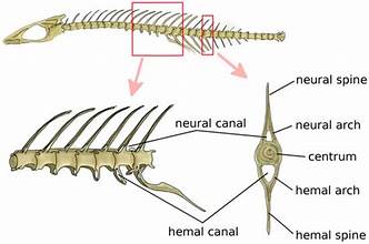
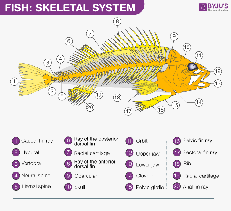
vertebrae centrum
centrum of a vertebra in fish is the central cylindrical part of each vertebra. It serves as a crucial structural element that provides support along the length of the vertebral column.
neural arch
bony structure that forms part of the vertebrae. It is located dorsally (on the upper side) and serves to protect the spinal cord
neural spine
bony projection that extends upward from the neural arch of the vertebrae. This structure serves to protect the spinal cord and provides attachment points for muscles
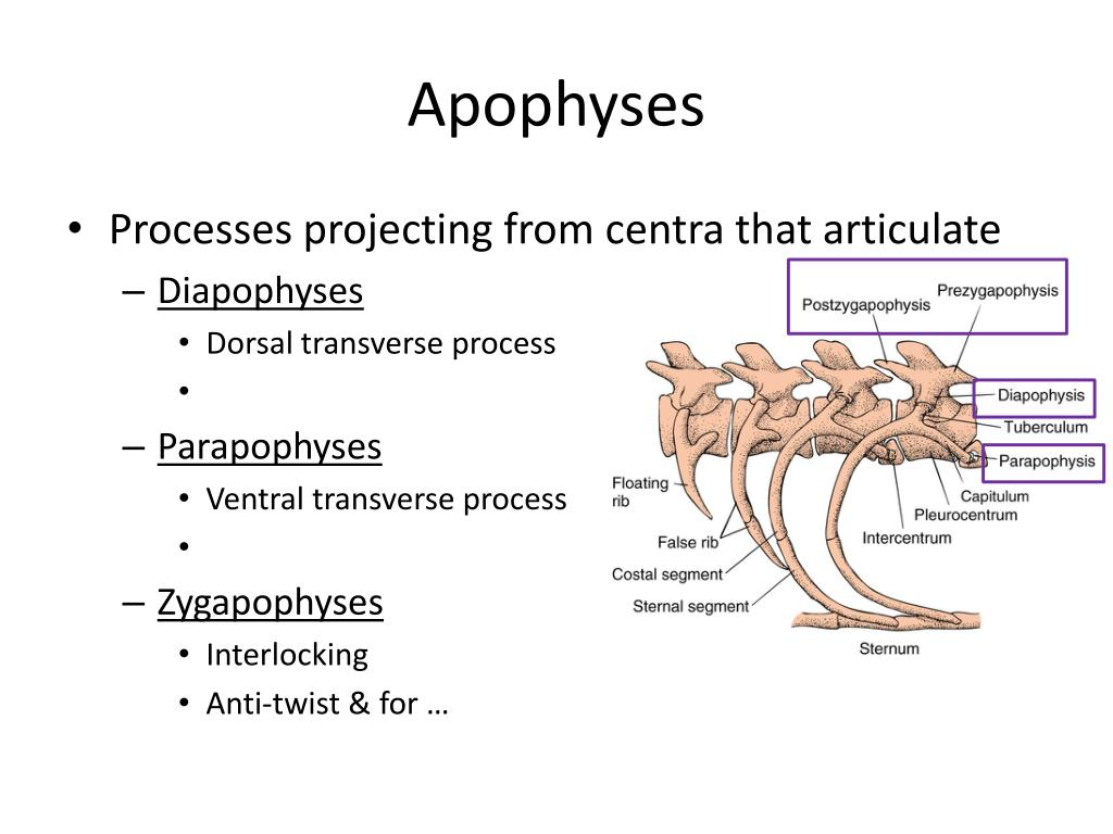
parapophyses
transverse processes that project from the centrum of vertebrae
hemal arch
small, V-shaped bone located on the ventral side of the caudal (tail) vertebrae. The primary function of the hemal arch is to enclose the hemal canal, which allows for the passage of caudal blood vessels, specifically the caudal artery and vein.
hemal spine
V-shaped bone located at the anterior end of the ventral surface of the caudal vertebrae.
ribs
bony or cartilaginous structures that are part of the skeletal system, located in the trunk region and associated with the vertebrae. They serve to protect internal organs and provide structural support.
intermuscular
slender linear bones embedded in muscle, which ossify from tendons through a process of intramembranous ossification
pterygoiphores
skeletal elements in fish that support the fins. They are typically found at the base of dorsal and anal fins, providing articulation and stabilization for the fin rays
epineurals
attach to the upper surface of neural spines and play a role in the musculoskeletal system of teleostean fishes.
spines
spine, or vertebral column, is a crucial component of fish anatomy, providing support and protection for the spinal cord
rays
supportive structures in the fins of fish. These rays can be categorized into two types: soft rays and hard rays. Soft rays are typically branched, segmented, and flexible, while hard rays are unbranched, segmented, and provide greater rigidity.
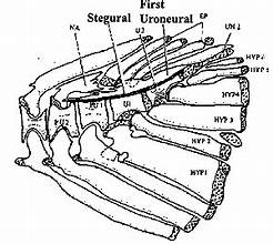
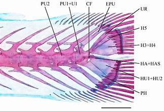
epurals
found in the tails of teleost fish. They are modified neural spines that contribute to the structure of the hypochordal skeleton, playing a crucial role in supporting the caudal fin
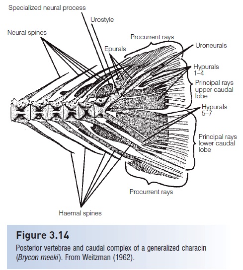
hypurals
bony structures in fish that support the caudal fin. They are primarily formed from the expanded and fused hemal spines of the last few vertebrae in most teleost fishes.
parahypurapophyses
part of the vertebral column and are associated with the support of the pelvic fins.
uroneural
paired, elongated endochondral bones found in the caudal skeleton of certain fish. They project from the lateral surfaces of the urostyle, which is part of the vertebral column. These bones play a role in the structural support of the tail
parahypural
bone element in the caudal fin skeleton of fish. It is part of the hypural complex and serves as an attachment point for muscles that control the caudal fin's movement
anterior (rostial)
front
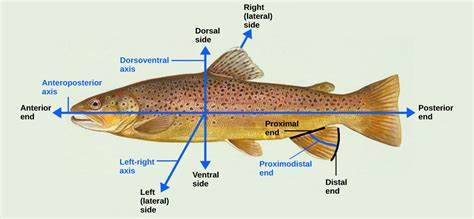
posterior (caudal)
back
Dorsal
up
ventral
down
dorsal fin (spines and rays)
unpaired fin located on the back
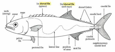
pectoral fins
paired appendages located on the sides of a fish's body, typically positioned just behind the gill
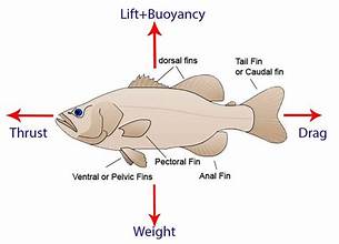
pelvic fins
paired fins located on the ventral (belly) surface of fish
anal fins
located on the ventral side, typically behind the anus
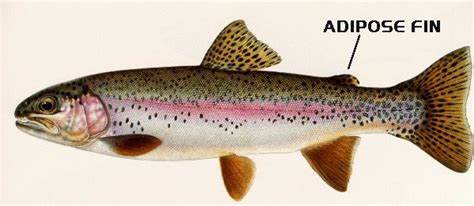
adipose fin
small, fleshy fin found on the backs of certain fish species. It is located between the larger dorsal fin and the caudal (tail) fin
caudal fin
tail fin
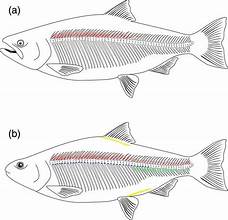
lateral line
lateral line organ, is a system of sensory organs
axial region
central part of their body, includes vertebral column, skill, and ribs
jaw region
anatomical structures that supports feeding and prey capture
ethmoid region
area of the skull that includes bones associated with nasal cavity and orbits
optic region
area of the brain sponsible for visual
otic region
area associated with auditory system includes structures like inner ear
cranial region
skull of fish to protect brain and vital sensory organs
suspensorial/hyoid region
structure that connects with skull to that jaw, mechanics of feeding and jaw movement
branchiostegal/hyoid
series of bony structures that supports gill membranes
opercular region
area surrounding the operculum which is a bony flap that covers and protects the gills
clavicular region
associated with the pectoral girdle, specifically where the clavicle or its equivalent structures are located, supports pectoral fins
caudal region
tail section, posterior part of the fish. helps with propulsion, steering, balance
appendicular region
skeletal structures that is associated with locomotion, like fins (pectoral and pelvic) and unpaired fins (dorsal, anal, and caudal)
branchial arches region
paired structures in fish that supports gills, located behind throat (pharyngeal cavity)
levator arcus palatini
a fan-shaped muscle found in bony fishes (teleosts) that plays a critical role in feeding and respiration. It is part of the facial musculature located behind the eye, responsible for expanding the buccal (mouth) cavity by lifting or abducting the suspensorium (the bony structure supporting the jaw)
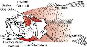
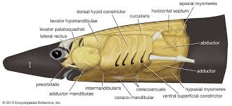
dilator operculi
cranial muscle in fish responsible for abducting—or opening—the operculum (gill cover). It is a critical component of the respiratory and feeding mechanisms in teleost fishes, helping to expand the buccal and opercular cavities to create suction.
adductor mandibuli
complex used by fish to close their jaws, playing a critical role in feeding and, in many cases, respiration. It is located on the lateral surface of the head (cheek area) and originates on the suspensorium (specifically the quadrate process or palatoquadrate) and inserts on the lower jaw (Meckel's cartilage or angulo-articular)
intermandibularis
thin sheet of cranial muscle in fish located on the floor of the mouth between the two halves of the lower jaw (mandibles)
adductor arcus palatini
responsible for pulling the palatal arch (suspensorium) medially, which decreases the volume of the mouth cavity during respiration and feeding
adductor operculi
paired hyoid muscle found in fish, primarily responsible for the adduction (closing/inward movement) of the operculum, which is the bony flap covering the gills.
levator operculi
key cranial muscle in bony fishes responsible for lifting or elevating the operculum (the bony gill cover). This action is critical for both respiration and feeding mechanisms, specifically for initiating mouth opening and creating suction
protractor hyoidei
paired muscle in teleost (bony) fishes that connects the lower jaw (dentary bone) to the hyoid arch (anterior ceratohyal). It plays a critical role in the fish's feeding and respiratory mechanism by controlling the movement of the hyoid apparatus and the lower jaw.
epaxials
muscles located on the dorsal (upper) half of the body, specifically situated above the horizontal septum that separates them from the ventral (lower) hypaxial muscles. hypaxial muscles, they act as the primary motors for locomotion, contracting to create the side-to-side, S-shaped, lateral undulations of the body. They are essential for suction feeding, where they act to dorsally rotate (lift) the neurocranium (head) to expand the mouth cavity.
hypaxials
block of skeletal muscle located on the ventral (lower) side of the body, specifically below the horizontal septum (a connective tissue sheet separating the upper and lower halves of the fish). They represent the lower portion of the fish's trunk musculature and are responsible for ventral movement, body wall tension, and powering suction feeding
lateralis superficialis
thin layer of muscle that lies directly on the surface of the deeper, main muscle mass (profundus) along the lateral line, extending from the head to the tail. slows swimming activities