Brain and cranial nerves
1/39
There's no tags or description
Looks like no tags are added yet.
Name | Mastery | Learn | Test | Matching | Spaced |
|---|
No study sessions yet.
40 Terms
What is the brain called
Cerebrum right or left cerebral hemisphere
How is the brain divided?
5 lobes
Frontal- Executive function and higher intelligence
Partial lobe-
Temporal lobe-
Occipital lobe-
Insular lobe-
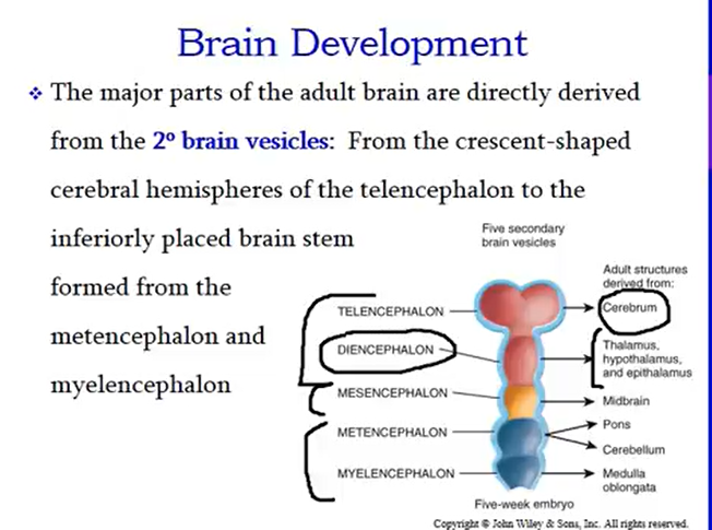
Stages of brain development in embryo
Primary
3 weeks brain vesicles called forebrain, midbrain and hindbrain.|
Prosencephalon Forebrain
Mesencephalon Midbrain
Rhombencephalon Hindbrain
Secondary
Forebrain divide into 2
hindbrain turns into two
What adults structures are developed from the 5 vesical of embryo.
Study cerebral hemisphere
What are cranial meninges
Continues meninges that mirror structure and function.
Dura mater
Spidery arachnoid mater
Thin delicate pia mater
What is a dura mater?
Cranial dura mater has two layers- an external periosteal layer and internal meningeal layer. The spinal dura only has one.
The important Dural extensions are the falx cerebri, falx cerebelli and the tentorium cerebelli.
Explain blood supply to brain.
brain receive 20% of body’s blood supply and consume 20% of the 02 in glucose
Anteriorly, the internal carotoid arteries supply blood to the brain; postierior supply is via vertebral arteries
internal Juglar veins are venous return
What forms blood brain barrier?
Neuroglial cells form blood brain barrier that isolate the parenchyma of the brain from many substances in the blood that would not notmally be able to
Cerebral spinal fluid is present in meninges.
Its is a clear fluid that circulates through internal cavities in the brain (ventricles)
and spinal cord (the central canal) and follows over and around the brain and cord in the subarachnoid space. The brain floats in it
CSF is a shock absorber, protects the brain and cord.
Also helps transport nutrients and waste between blood and nervous tissue.
CSF produces byÉ
Ependymal cells in the choroids plexus (network of blood capillaries that line ventricles)
What is the path of CSF?
Lateral ventricals-interventricular foramina-third ventricle- cerebral aqueduct- fourth ventricle- midian aperture and the lateral apertures - SAS Sub arachnoid space
What reabsorbs CSF
Arachnoid granulation
Where does the medulla start and estend to?
Inferior border of the pons and extends to the foramen magnum. Contains all ascending and decending tracts extending between the spinal cord and cerebum. Medulla contains nuclei that regulate vital body functions.
Describe the strictures of the medulla oblongata
Two externa bulges called pyramids formed by largest motor tracts.
Axons from left pyramid that cross over to the right and axons on the right cross over to the left.
The left hemisphere of the brain controls the right side of muscles while the right control the left side.
What does medulla oblongata do?
The cardio vascular center
Respiuratoy rythmicity center
Vomiting, coughing, and sneezing centers
The nuclei associateed with 5 of 12 cranial nerves originates in the medulla.
Part of the 4th ventrical extends to medulla.
If damaged causes death
Where is pons located?
above medulla and anterior to cerebellum. It acts as aq bridge connecting spinal cord with the brain and parts of the brain with other
What does pons do?
Pontine respiratory group
5-8 |
Cranial emerges from nerve 5 directly from poms
678 emerge from the space between pons and medulla.
Where’s mid brain located?
mid brain extends from pons to the diencephalon
Cerebral aqueduct passes through midbrain connecting 3rd ventricle above with the 4th ventricles below.
Describe the structure of the mid brain
Cerebral peduncles
Peduncles contain axons of corticospinal, corticobulbar and corticopontine tracts. These conduct nerve impulses from motor areas in the cerebral cortex to the spinal cord, medulla and pons.
What are cpllicoli?
Superior for visual reflexes and inferior for auditory reflexes
What is substancia nigra? What does it do?
Dark pigmented area
Located in mid brain that releases a neuro transmitter called dopamine. The red nucleus helps control voluntary movements of the limbs.
What is reticular formation?
Collection of different neurons that pass through mid brain. Bundle of myelinated axons (High speed conduction axons). Reticular formation. Keeps us awake and alert.
Where is cerebellum located?
located inferior to the cerebum and posterior to the brain stem.
Seperated from cerebum by transverse fissure (in which the falx cerebelli is located)
Cerebellar hemisphere?
Vermis “wings” or lobes. Responsible for posture, equilibrium and balance.
Where is diencephalon located? and function.
The thalamus functions as a relay station for all sensory impulses to the cerebral cortex. Pain, Temp and pressure are all relayed to the thalamus en route to the higher centers of the cerebral cortex.
While not precisely localized here. All peripheral sensations in the cerebral cortex. All of these peripheral sensations are processed in the thalamus in conjunction with their attendant memories and the emotions.
Epithalamus is suprior and posterior to the thalamus.
consists of the pineal gland (melatonin) and habenular nuclei (emotional response to orders)
More melatonin is secreted in darkness then light. thought to promote sleepiness and help regulate our biological clocks.
What is hypothalamus?
Controls homeostatic functions
Controls ANS
Cordinates between NS and endocrine
Controls temp
Regulates hunger/thirst
Assists with the internal circadian clock by regulating biological activivty
The cerebrum
The cerebral cortex is the “seat of our intelligence” Neurons in the cortex that we are able to read, write, speak, remember and plan our life.
The cerebrum consists of an outer cerebral cortex, an internal region of cerebral white matter, and gray matter nuclei deep within white matter.
What is cerebral cortex
Superficial outermost of brain. Folds are called “Gyri”, the deepest folds are fissues’s; the shallower grooves between folds are sulci.
Longitudial fissue seperates the cerebrum into right and left cerebral hemispheres. The central sulcus further divides the anterior frontal lobe from the posteriorly situated parietal lobe.
The precentral gyrus and postcentral gyrus of those two lobes
Explain the function and location of each highlighted region.
Primary sensory area is located where?
The somatosensory area recieves impulses from the lips and fingertips then from thorax and hip.
Distorted somatic sensory map is known as sensory homunculus.
Primary motor area
located un the precentral gyrus of the frontal lobe controls voluntary contractions of specific muscles or groups of muscles.
The motor cortex also has homunculus map, with more cortical area devoted to muscles involved in skilled complex delicate movements
Where are each located?
Visual- occipital lobe
Gustatory area- Somatosensory area
Auditory area-In the superior part of temporal lobe
Olfactory area- Inferomedial temporal lobe
What is white matter
Consists of myelinated axons in three types of tracts.
Association tracts contain axons that conduct nerve impulses between gyri in the same hemisphere
Commissural tracts conduct nerve impulses between corresponding gyri from one hemisphere to another.
Projection tracts convey impulses to lower parts of the CNS (Thalamus, brain stem, spinal cord)
Corpus callosum
Thick band of axons that connects corresponding areas of two hemispheres
Through the corpus callosum, the left motor cortex (which controls the right side) is linked to right motor cortex (which controls the left sdie)
Basal nuclei
Control subconscious contractions of skelital muscles. Examples include automatic arm swings while walking and true laughter in response to a joke.
Hemispheric lateralization
Divison of labor. Each hemisphere divides the funcvtions between both. They do different functions.
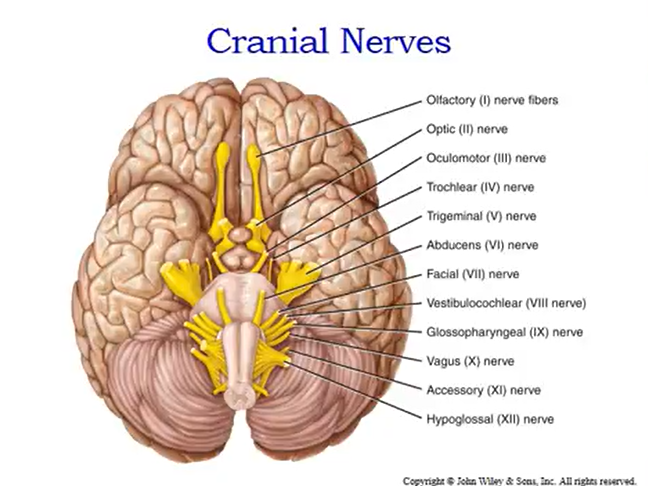
Identify cranial nerves. What do they control? Are they sensory or motor nerve?