PNS: CMA
1/29
Earn XP
Description and Tags
Write the correct answer to each question.
Name | Mastery | Learn | Test | Matching | Spaced | Call with Kai |
|---|
No analytics yet
Send a link to your students to track their progress
30 Terms
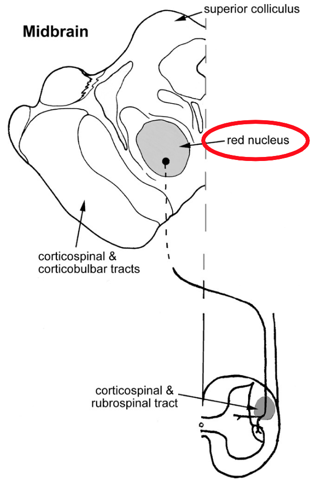
What is the function of the red nucleus?
The red nucleus helps coordinate movement by facilitating flexor activity, especially in the upper limbs, and acts as a motor relay between the cerebellum and spinal cord.

The reticulospinal and vestibulospinal tracts organize…
reflexes and postural adjustments.

Do all rubrospinal tracts cross the midline and end up in the intermediate zones?
No, not all rubrospinal tracts cross the midline; some may remain ipsilateral.
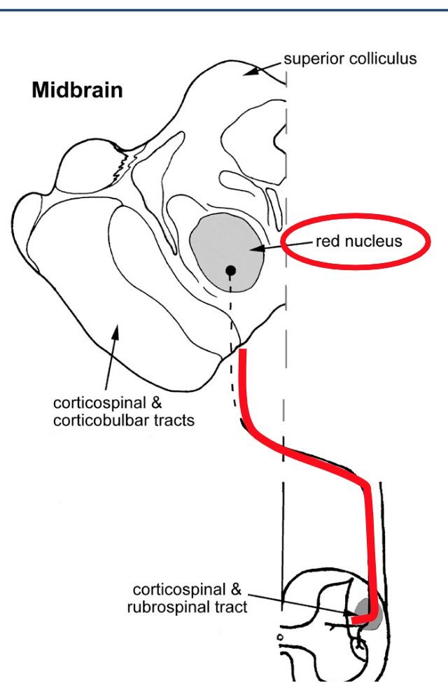
Where are the corticospinal and rubrospinal tracts located?
The corticospinal and rubrospinal tracts are located in the lateral within the ventral.
Give an example of synergy.
Holding a ball with all five of your fingers together is an example of synergy. It demonstrates how muscles and joints work together to perform a coordinated movement.
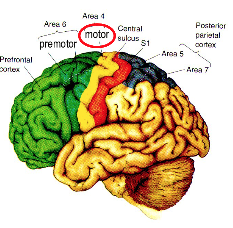
The motor cortex is located where?
In the postcentral gyrus.

What is somatotopic organization?
The mapping of bodies is upside down, and the sizing is not really proportionate.

Where do the axons of the motor cortex head to?
Axons of the motor cortex go through the brainstem. From there, it crosses to the opposite side of the body and continues down the spinal cord to synapse with motor neurons.

Where do 20% of the axons of the motor cortex head to?
They go directly to the motor neurons in the spinal cord without crossing.

Where do 80% of the axons of the motor cortex go to?
80% goes directly to the interneurons in the spinal cord that facilitate communication between motor neurons and the brain.

How could a single motor nucleus (muscle) be represented in columns at many loci?
This is because different parts of the zone exhibit the same movement. For example, the index finger flexion is represented multiple times in the zone.
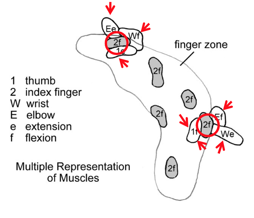
How is synergy represented in each muscle column from this image?
Synergy is represented by the activation of multiple muscle columns that work together to produce a coordinated movement, allowing for complex muscle actions.
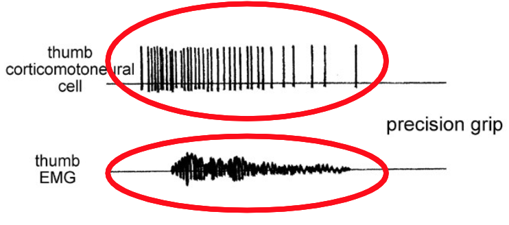
What can you tell about the activity in the thumb cell from this motor map?
You perform a precision grip, and the thumb is contracting. This means that the motor cortex is active.
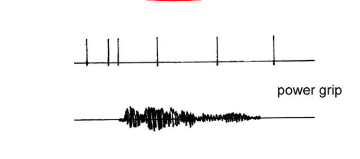
What can you tell about the activity in the thumb cell from this motor map?
When you perform a power grip, this particular neuron up in the motor cortex is not active during that specific movement. Instead, the neuron is primarily active during precision grips, indicating its role in fine motor control.
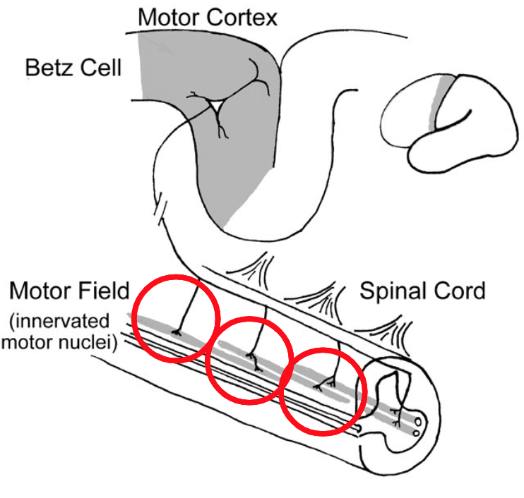
What is a motor field?
A motor field is the specific area of the motor cortex that governs the movement of a particular muscle or group of muscles, influencing the execution of voluntary movements.
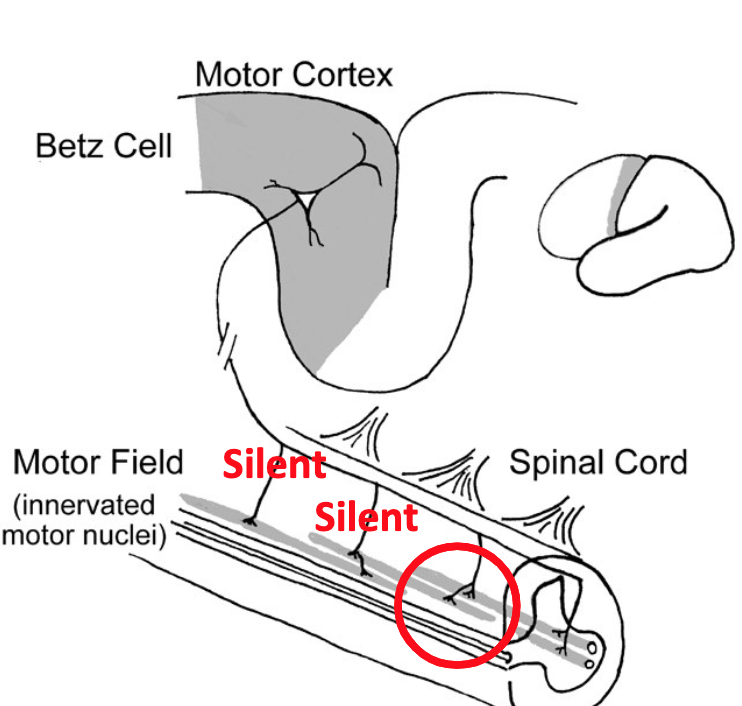
When synapses of a motor field are silent…
It means that they aren’t being used. This indicates that the corresponding motor behaviours associated with that motor field are not being executed.
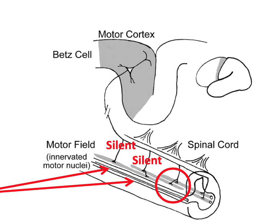
The motor field provides potential for plasticity. What happens to the motor field if you lose a muscle in an injury?
The corticomotor neurons will be useless. Plasticity extent depends on the motor field; it allows you to learn new skills. The motor field may reorganize, allowing adjacent areas of the cortex to take over control of the movements previously governed by the lost muscle, potentially leading to re-establishment of function.
Mechanoreceptors input come from…
the skin, muscles, and joints, providing sensory information about touch and pressure before going to the motor cortex.
Proprioceptive inputs direct from…
the thalamus, expect for olfaction.
What is the function of the premotor cortex?
The premotor cortex is involved in sensory inputs, especially visual and auditory information, to plan and coordinate movements.
What are sensorimotor cues?
Environmental sounds, such as a door knob, push button, etc.
What is a visuomotor response?
You’re told, “When the light turns green, press the button.” After hearing the instruction but before the light turns green, motor neurons show preparatory activity. When the light turns green, execution-related activity occurs, and muscles contract
What is preparatory activity? Give an example.
You select the synergies, but once the movement starts, it’s silenced. It’s now the motor cortex’s job to produce all of those movements.
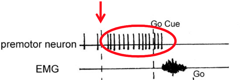
Describe what’s happening in this graph in terms of preparatory activity.
First, a “warning” cue appears, telling the brain to get ready. Once preparatory activity begins, the premotor neuron starts firing repeatedly after the warning cue. When the “go” cue appears, the EMG activates, telling the muscles to contract.
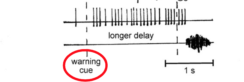
What does it mean when there is a “longer delay period” in preparatory firing?
It indicates that the motor system is taking more time to organize and select appropriate motor synergies before execution, potentially reflecting increased uncertainty or complexity of the upcoming movement. EMG is flat, so muscles are not activated.
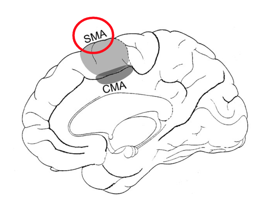
Where is the supplementary motor area located?
The supplementary motor area (SMA) is located on the medial surface of the frontal lobe, just anterior to the primary motor cortex and superior to the cingulate gyrus.

What does the supplementary motor area control?
It controls bilateral coordination of limbs when different motions are done on each side. For example, it assists in tasks requiring both hands to perform different movements, such as playing a musical instrument. Activities of both sides must be adjusted to one another.

Where is the cingulate motor area located?
It’s located within the cingulate sulcus.
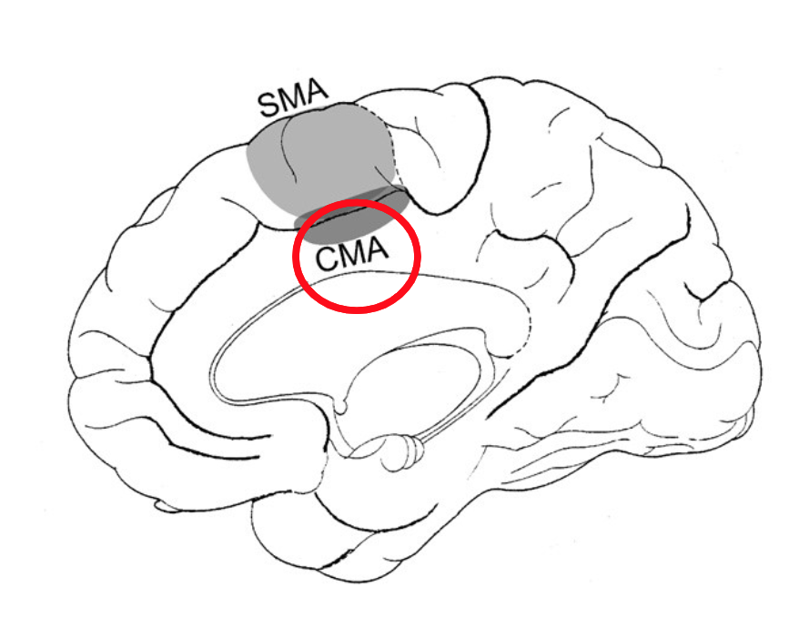
What is the role of the CMA?
It helps integrate motor control with emotional responses and decision-making.

How could you tell the difference between a fake smile and a real smile? Between the SMA and the CMA, which motor area drives what?
The genuine smile will be driven by the cingulate motor area, whereas the fake smile will be driven by the supplementary motor area. The cingulate motor area drives true emotional expressions, while the supplementary motor area controls voluntary, practiced actions like fake smiles.