Lab 14 - Isolation and Identification of Streptococcus and Enterococcus
1/45
There's no tags or description
Looks like no tags are added yet.
Name | Mastery | Learn | Test | Matching | Spaced | Call with Kai |
|---|
No analytics yet
Send a link to your students to track their progress
46 Terms
Why is it hard to tell apart Streptococcus and Enterococcus?
It is difficult to distinguish them because both are gram-positive cocci that appear in pairs or chains and share similar morphology and biochemical traits, requiring additional tests for identification.
What is the shape and arrangement of Streptococcus and Enterococcus?
They are gram-positive cocci that occur in pairs or chains of varying lengths.
Can catalase test distinguish Streptococcus and Enterococcus?
No, both genera are catalase-negative and do not produce bubbles when hydrogen peroxide is added.
What are the two major properties used to classify Streptococci clinically?
Streptococci are classified based on hemolytic properties on blood agar and serologic grouping (Lancefield classification).
What is the most commonly used agar to isolate Streptococci?
Blood agar is the most commonly used medium.
What is blood agar made of?
It consists of tryptic soy agar (TSA) enriched with intact sheep red blood cells.
What type of medium is blood agar?
Blood agar is an enriched and differential medium that supports fastidious organisms and differentiates bacteria based on hemolysis.
What type of organisms grow on blood agar?
Fastidious organisms, including Streptococci and many other pathogens, grow on blood agar.
What is hemolysis?
Hemolysis is the lysis of red blood cells surrounding bacterial colonies on blood agar.
What causes hemolysis?
Hemolysis is caused by bacterial exotoxins called hemolysins.
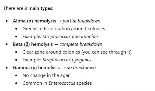
What is beta hemolysis?
Beta hemolysis is complete destruction of red blood cells, producing a clear zone around colonies.
How do you identify beta hemolysis on blood agar?
It appears as a clear, transparent zone surrounding bacterial colonies.
What is alpha hemolysis?
Alpha hemolysis is partial hemolysis of red blood cells, producing a greenish discoloration around colonies.
How do you identify alpha hemolysis on blood agar?
It appears as a green or brownish zone around colonies due to incomplete RBC destruction.
What is gamma hemolysis?
Gamma hemolysis indicates no hemolysis, meaning no change in the agar surrounding colonies.
How do you distinguish alpha vs beta hemolysis?
Alpha hemolysis shows a greenish partial clearing, while beta hemolysis shows a completely clear zone.
What is the Lancefield classification system?
It classifies Streptococci into groups based on carbohydrate antigens in their cell walls.
Which Lancefield groups infect humans?
Groups A, B, C, D, F, and G infect humans and are typically beta-hemolytic.
What is GAS?
GAS stands for Group A Streptococcus, which is Streptococcus pyogenes.
What diseases does Streptococcus pyogenes cause?
It causes strep throat, pneumonia, cellulitis, impetigo, scarlet fever, rheumatic fever, necrotizing fasciitis, and more.
What are the growth characteristics of GAS on blood agar?
It forms small white/gray colonies with beta hemolysis.
What antibiotic is GAS sensitive to?
GAS is sensitive to bacitracin.
How is bacitracin sensitivity determined?
A zone of inhibition around a Taxo A disc indicates sensitivity.
What is GBS?
GBS is Group B Streptococcus, or Streptococcus agalactiae.
Where is Streptococcus agalactiae normally found?
It is found in the gastrointestinal and genitourinary tracts.
What is the clinical significance of GBS?
It causes neonatal infections like pneumonia, meningitis, and sepsis, and opportunistic infections in adults.
Where is Streptococcus pneumoniae found?
It is part of normal microbiota in the nasopharynx.
What diseases does Streptococcus pneumoniae cause?
It causes pneumonia, otitis media, bacteremia, and meningitis.
What is the most common cause of community-acquired pneumonia?
Streptococcus pneumoniae.
What are its blood agar characteristics?
It forms small, shiny colonies with alpha hemolysis.
What antibiotic is S. pneumoniae sensitive to?
It is sensitive to optochin.
How is optochin sensitivity determined?
A zone of inhibition around a Taxo P disc indicates sensitivity.
What are Enterococci and where are they found?
Enterococci are gram-positive cocci found as normal microbiota in the intestinal tract.
Why are Enterococci clinically important?
They cause opportunistic infections and are highly antibiotic resistant.
What Lancefield group do Enterococci belong to?
Group D.
What is the most common Enterococcus species?
Enterococcus faecalis.
What infections are Enterococcus faecalis associated with?
UTIs, wound infections, abdominal infections, and hospital-acquired infections.
Do Enterococci produce hemolysins?
No, they are typically gamma hemolytic (no hemolysis).
How are gram-positive cocci identified using catalase and blood agar?
Catalase-negative organisms with specific hemolysis patterns on blood agar are identified as Streptococci or Enterococci.
What medium is used to isolate Enterococci?
Bile esculin azide agar.
What makes bile esculin azide agar selective and differential?
Bile and azide inhibit other bacteria (selective), and esculin hydrolysis causes a color change (differential).
What bacteria are inhibited on this medium?
Bile inhibits most gram-positive bacteria, and sodium azide inhibits gram-negative bacteria.
What is the color of the medium before inoculation?
Light brown.
Why is esculin added?
It allows detection of esculin hydrolysis by Enterococci.
What happens when Enterococci grow on this medium?
The agar turns black due to esculetin reacting with iron salts.
How do you identify Enterococcus on bile esculin agar?
Growth with blackening of the medium indicates a positive result.