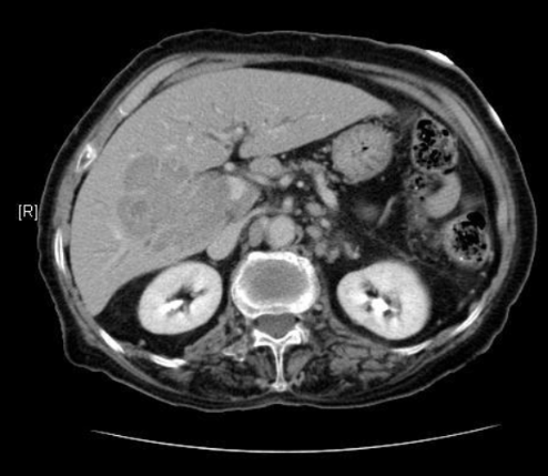
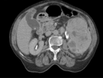
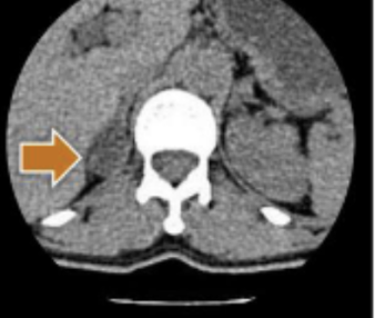
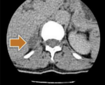
CT abdomen and pelvis
1/98
There's no tags or description
Looks like no tags are added yet.
Name | Mastery | Learn | Test | Matching | Spaced |
|---|
No study sessions yet.
99 Terms
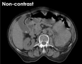
CT abdomen and pelvis protocol is a general survey of
GI tract and accessory organs
indications for CT abdomen and pelvis
trauma and general concerns (nausea, vomiting)
CT abdomen and pelvis slice parameters
5.0 mm with matching interval
CT abdomen and pelvis gantry tilt
none
CT abdomen and pelvis contrast
100 ml IV contrast 3 ml/sec
CT abdomen and pelvis scan delay
6o second in portal venous phase
CT abdomen and pelvis for non trauma contrast
600 ml of oral water diluted 1 hour before scanning
CT abdomen and pelvis algorithm and window
standard: 400 WW and 40 WL
CT abdomen and pelvis slices for post processing
thin
CT abdomen and pelvis delayed images through __ for trauma patients
kidneys and bladder
CT Appendicitis indications
appendicitis, RLQ pain, elevated WBC
CT Appendicitis scan type
helical
CT Appendicitis slice parameters
3.75 mm with matching interval
CT Appendicitis gantry tilt
none
CT Appendicitis contrast IV contrast
100 ml, 3 ml/sec
CT Appendicitis scan delay
60 second in portal venous phase
CT Appendicitis oral contrast
600 ml, 40-60 minutes prior to scanning
CT Appendicitis rectal contrast
1000 ml of water with diluted contrast
CT Appendicitis algorithm
standard 400 WW and 40 WL
CT Appendicitis ___ slices should be used for post processing
thin slices
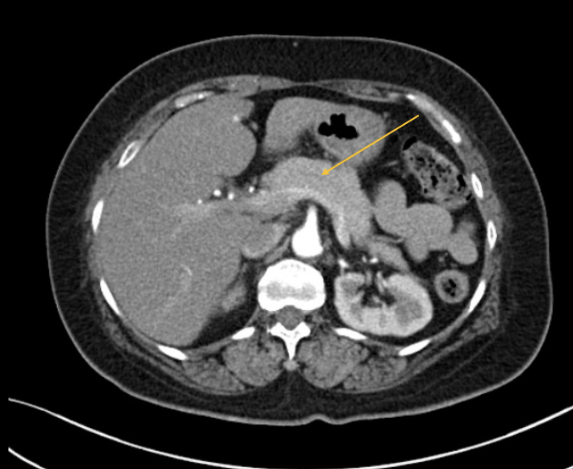
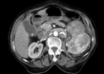
CT Liver evaluates liver for
cancer
CT Liver includes scan and re-scans in multiple phases of contrast enhancement to
differentiate between primary liver cancer, metastatic cancer, benign tumors
CT Liver slice parameters
2.5 mm with matching interval
CT Liver contrast
600 ml oral contrast 40-60 minutes before scanning
CT Liver scan 1 contrast
no IV contrast
CT Liver scan 2 contrast
30 second delay arterial phase
CT Liver scan 3
60 second scan delay portal venois phase
CT Liver scan 4
5+ minute delay, delayed phase
CT Liver IV contrast
100 ml, 3 ml/sec
CT Liver algorithm
standard 400 WW and 40 WL
CT Liver non contrast scan is baseline view of liver can be compared to
contrast enhanced phase
CT Liver arterial phase (30 sec) used for
primary liver cancers (hepatocellular carcinoma) b/c they typically have arterial blood supply
CT Liver portal venous phase (60 sec) used for
metastatic liver cancers b/c they have portal venous blood supply
CT Liver delayed phase (5 minutes) used for
hemangiomas (benign) fill with contrast
CT Pancreas additional oral contrast given to help differentiate
duodenum from pancreatic head
T/F CT Pancreas delayed phase not necessary
true
CT Pancreas scan parameters
2.5 mm with matching interval
CT Pancreas oral contrast
600 ml, 40-60 minutes prior to scanning
CT Pancreas scan 1
no contrast
CT Pancreas scan 2
30 second scan delay (arterial phase)
CT Pancreas scan 3
scan delay (portal venous)
CT Pancreas IV contrast
100 ml at 3 ml/sec
CT Pancreas algorithm
standard 400 WW and 40 WL
CT Enterography indications
crohn’s or small bowel
CT Enterography scan parameters
2.5 mm with matching interval
CT Enterography IV contrast
100+ ml at 4+ ml/sec
CT Enterography contrast delay
60 seconds
CT Enterography oral contrast
1500-2000 ml over 40-60 minutes to distend bowels
CT Enterography algorithm
standard 400 WW and 40 WL
CT Enterography administered at same time as water to stimulate emptying of the stomach to help water move into the bowels
Metoclopramide (Reglan)
CT Enterography temporarily stops peristalsis to decrease bowel motion and enhance visibility)
glucagon
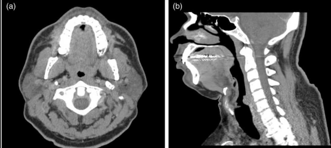
CT soft tissue neck indications
mass, swelling, abscess, cancer
CT soft tissue neck slice parameters
2.5 mm with matching intervals
CT soft tissue neck contrast
100 ml IV at 3 ml/sec
CT soft tissue neck algorithm
standard 400 WW and 40 WL
CT soft tissue neck may include algorithm for
visible portions of lung. Include orbital roof through carina
CT stone protocol used to
identify stones in urinary tract
CT stone protocol stones more visible
without contrast
how is CT stone protocol different from an abdoment and pelvis?
-no contrast
-thinner slices
-only kidneys through bladder included in scan
CT stone protocol indications
renal stone, flank pain, hematuria, dysuria
CT stone protocol slice parameters
3.75 mm with matching interval
CT stone protocol algorithm
standard 400 WW and 40 WL
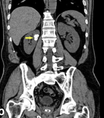
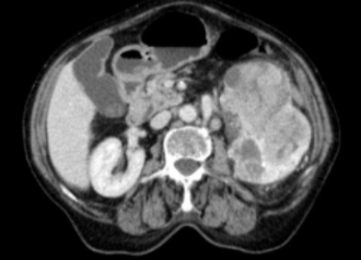
CT Kidneys indications
renal mass or abnormality
CT Kidneys slice parameters
3.75 mm with matching interval
CT Kidneys contrast
600 ml oral, 40-60 minutes before scan
CT Kidneys scan 1
no contrast
CT Kidneys scan 2
30 second delay (corticomedullary phase)
CT Kidneys scan 3
90 second delay (nephrographic phase)
CT Kidneys scan 4
4 minutes (excretory phase)
CT Kidneys IV contrast
100 ml, 3 ml/sec
CT Kidneys algorithm
standard 400 WW and 40 WL
CT Kidneys non contrast establish
baseline for comparison
CT Kidneys corticomedullary phase (30 sec) evaluates
renal arteries and renal cortex (receives arterial blood supply)
CT Kidneys nephrographic phase (90 sec) evaluate
nephrons
CT Kidneys excretory phase (4 mins) evaluate
renal pelvis and collecting system of kidney
CT adrenals uses scans in multiple phases to differentiate
benign adrenal tumors from adrenal cancer
CT adrenals ____ is a presentation of an adrenal tumor
abnormal hormone levels
CT adrenals adrenal glands produce what hormones for males
androgrens
CT adrenals adrenal glands produce what hormones for females
estrogens
CT adrenals indication
adrenal tumor
CT adrenals slice parameters
3.75 mm with matching interval
CT adrenals IV contrast
100 ml IV 3 ml/sec
CT adrenals scan 1
no contrast

CT adrenals scan 2
60 second delay (portal venous)
CT adrenals scan 3
15 minutes (delayed phase)
CT adrenals algorithm
standard 400 WW and 40 WL
CT adrenals commonly reduce
DFOV to 18 cm to only include adrenal glands
CT adrenals usually includes
coronal reformations
CT Urogram is for __ of urinary system
functionality and anatomy
CT Urogram scan entire collecting system during
excretory phase
CT Urogram may be used as pre-planning for
ureteral stent placement
CT Urogram may also be used to diagnose
functional and anatomical causes of recurrent stones
CT Urogram indications
hematuria, renal stone, pain
CT Urogram slice parameters
3.75 mm with matching intervals
CT Urogram oral contrast
1000 ml, 30 minutes prior to scanning
CT Urogram scan 1
no contrast
CT Urogram scan 2
4+ minute delay (excretory phase)
CT Urogram IV contrast
100 ml, 3 ml/sec
CT Urogram algorithm
standard 400 WW and 40 WL