Week 11 - Radiographic Diagnosis Part 2 (Abnormalities and Osseous Diseases)
1/70
There's no tags or description
Looks like no tags are added yet.
Name | Mastery | Learn | Test | Matching | Spaced | Call with Kai |
|---|
No analytics yet
Send a link to your students to track their progress
71 Terms
What are the two major structural types of bone?
Cortical bone and trabecular (cancellous) bone
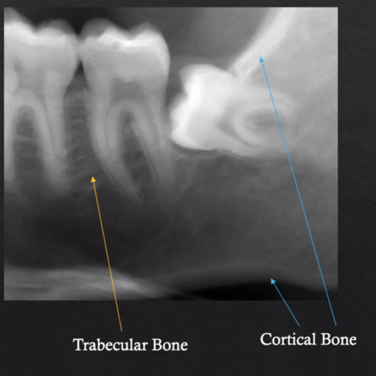
What is cortical bone?
Dense outer bone layer that provides structural strength
What is trabecular bone?
Spongy inner bone composed of trabecular networks and marrow spaces
What minerals make up most of bone's mineralized component?
Calcium and phosphate
What are osteoblasts?
Bone forming cells that synthesize new bone
What are osteoclasts?
Bone resorbing cells that break down bone
What are osteocytes?
Mature bone cells that regulate bone remodeling
What do hematopoietic stem cells in bone marrow produce?
Red blood cells, white blood cells, and platelets
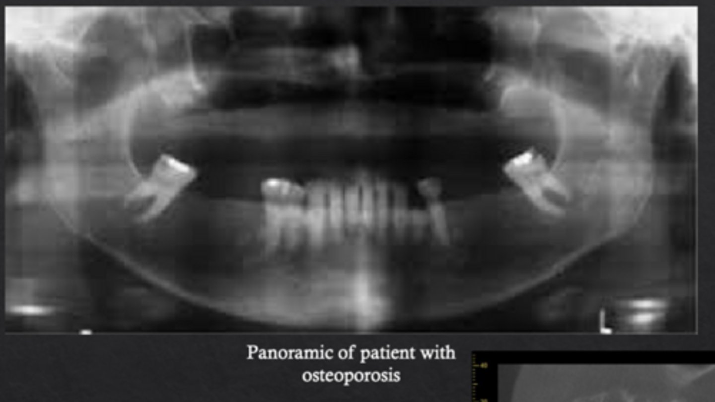
What is osteoporosis?
Systemic skeletal disease characterized by low bone mineral density and deterioration of bone microarchitecture
What is osteopenia?
Intermediate stage between normal bone and osteoporosis
What radiographic changes are associated with osteoporosis?
Loss of trabecular architecture and thinning of mandibular cortical bone
What is idiopathic osteosclerosis?
"Dense bone island" an area where normal trabecular bone is replaced with dense cortical bone
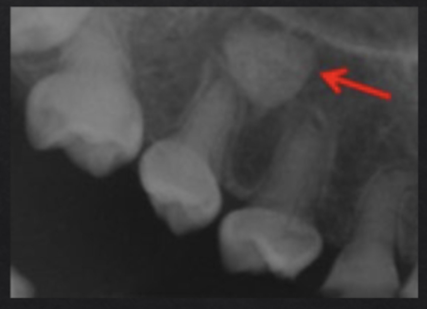
How does idiopathic osteosclerosis appear radiographically?
Region of dense radiopaque bone, blends with surrounding bone
Does idiopathic osteosclerosis typically affect adjacent structures?
No
What are exostoses or tori?
Outgrowths of cortical bone
How do tori appear radiographically?
Where are tori most commonly located?
Mid-palate (torus palatinus) and lingual mandible (mandibular tori)
Do tori usually require treatment?
No, unless they interfere with prosthetics
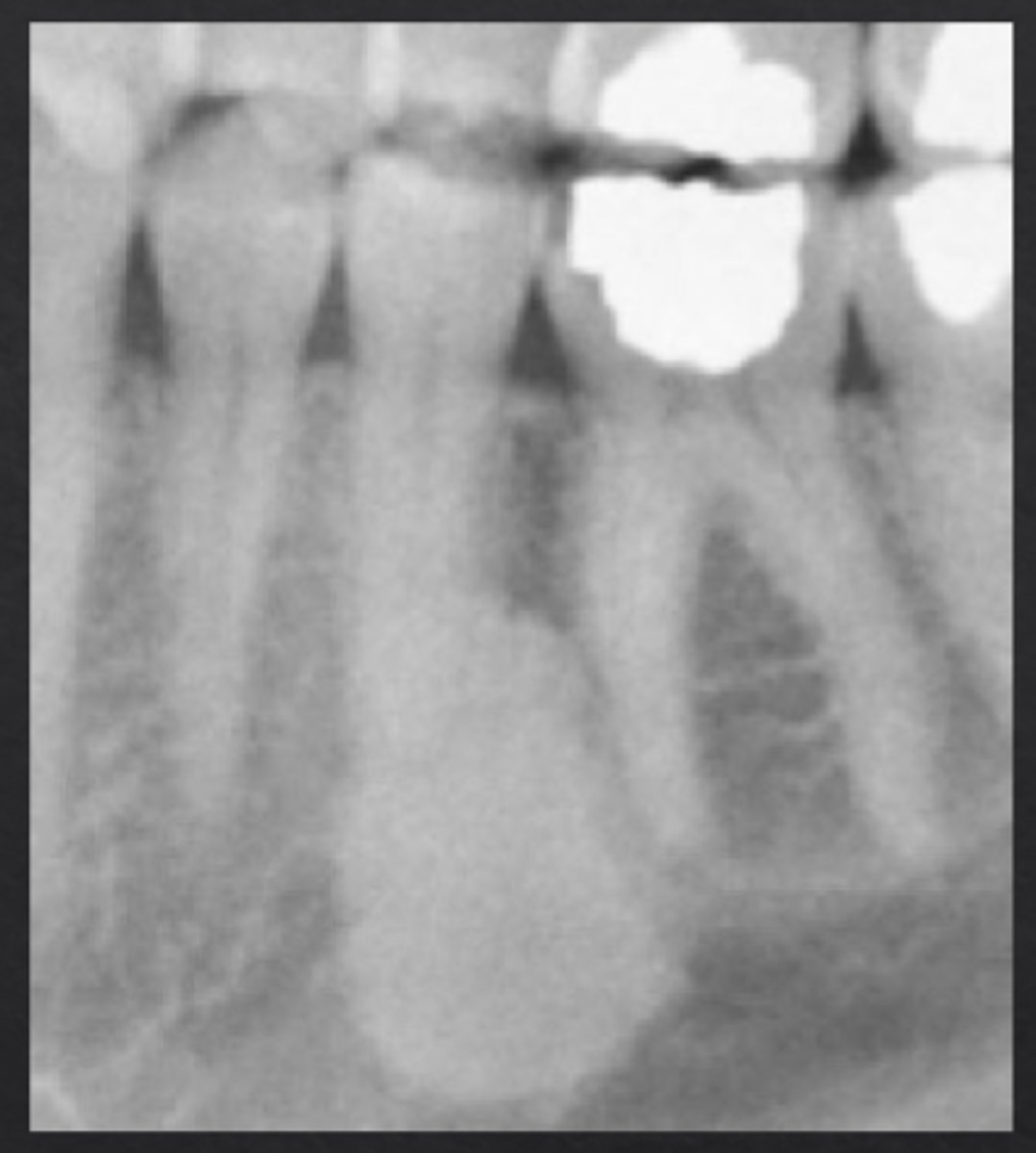
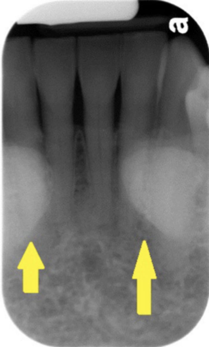
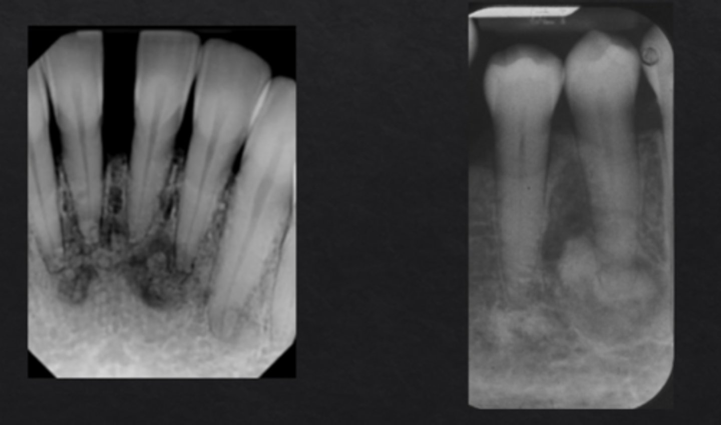
What is cemento-osseous dysplasia?
Localized bone dysplasia that becomes more calcified over time
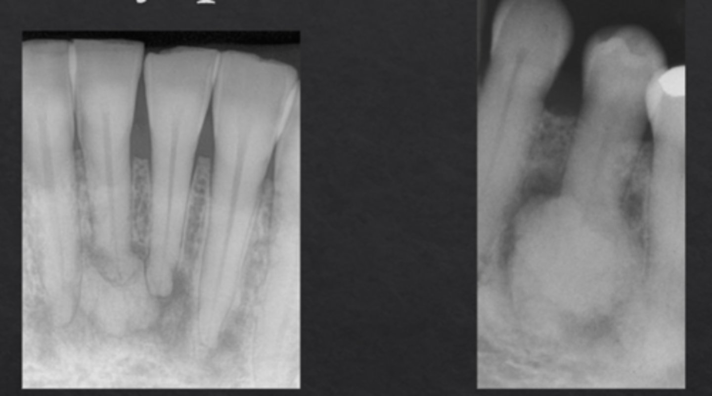
Cemento-Osseous Dysplasia: _____ if located in one sextant, _____ if located in more than one sextant
Focal, florid
What demographic most often develops cemento-osseous dysplasia?
Middle-aged females, especially African or East Asian ethnicity
How does cemento-osseous dysplasia appear radiographically?
Preapical radiolucency with central calcifications, will become more calcified over time (may lose radiolucent rim)
What is fibrous dysplasia?
Genetic condition where normal bone is replaced by fibrous connective tissue and immature bone
What gene mutation is associated with fibrous dysplasia?
GNAS gene mutation
What radiographic appearance is associated with fibrous dysplasia?
"Orange peel" bone, mixed density, poorly defined
Does fibrous dysplasia typically cross the skeletal midline?
No
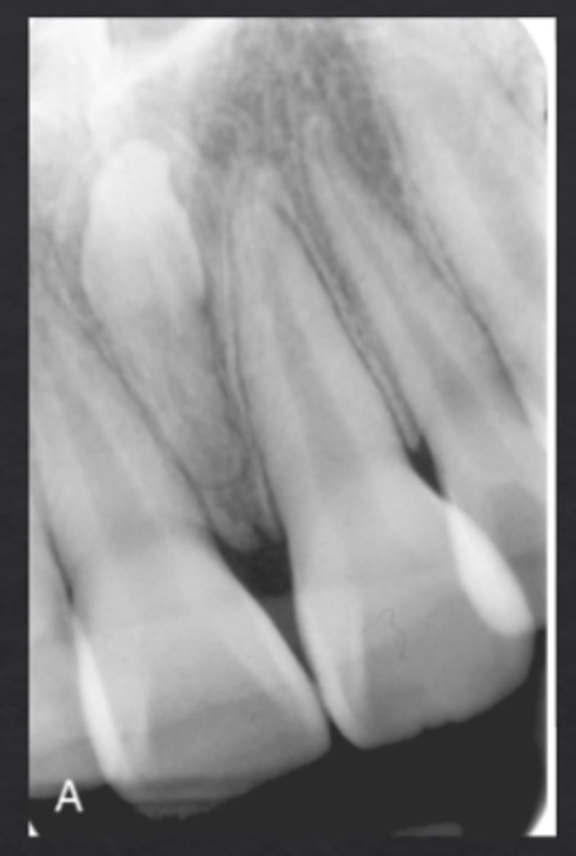
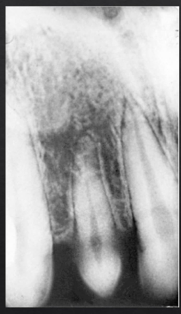
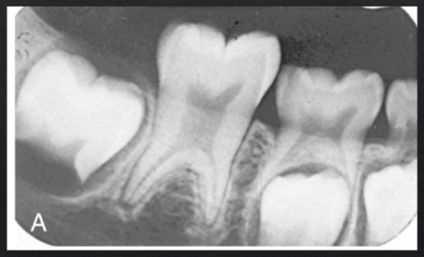
What is dens invaginatus (dens in dente)?
Infolding of enamel into the tooth, forming "tooth within a tooth"
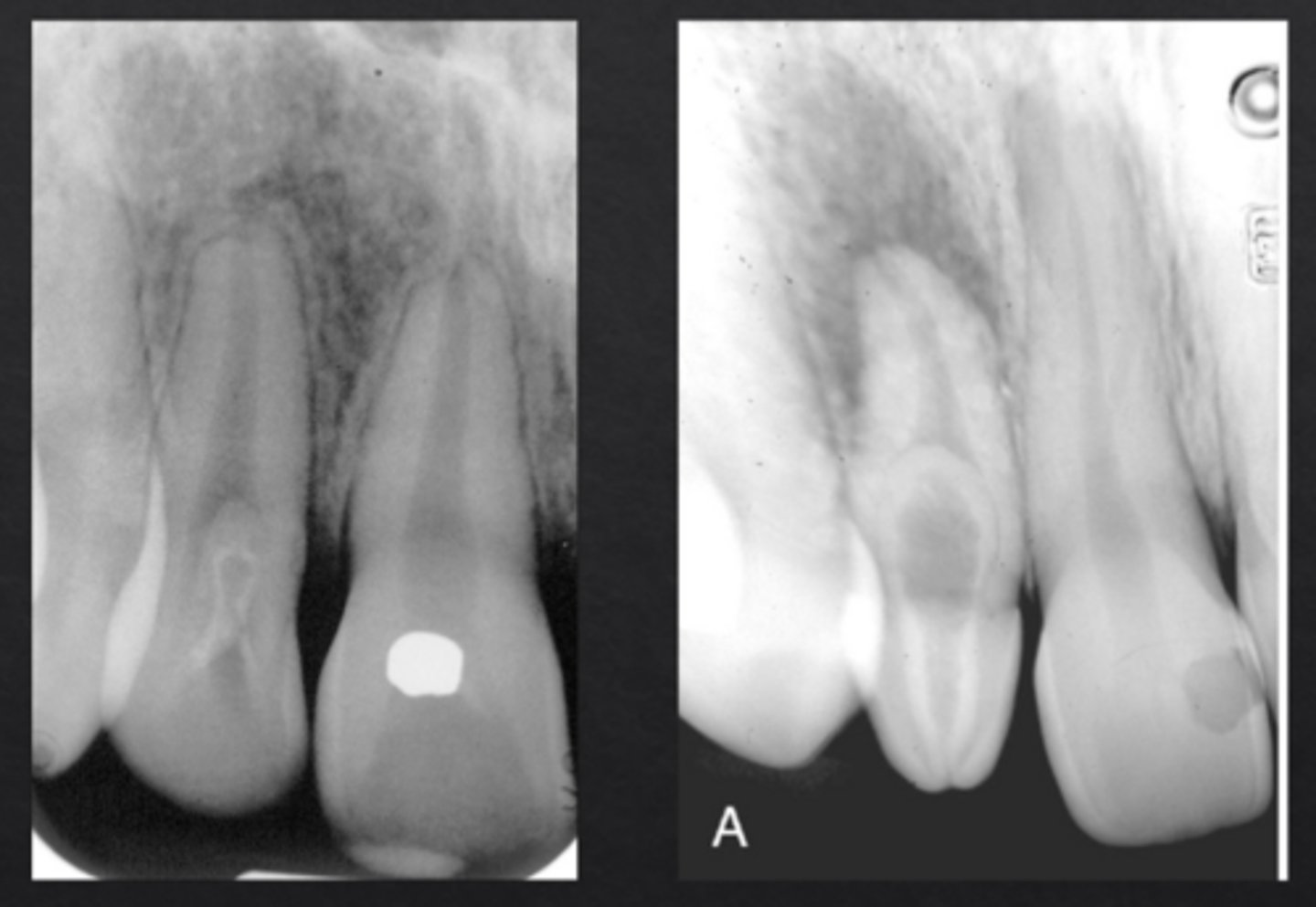
Which tooth is most commonly affected by dens invaginatus?
Maxillary lateral incisors
What is dens evaginatus?
Enamel grows outward forming an extra cusp
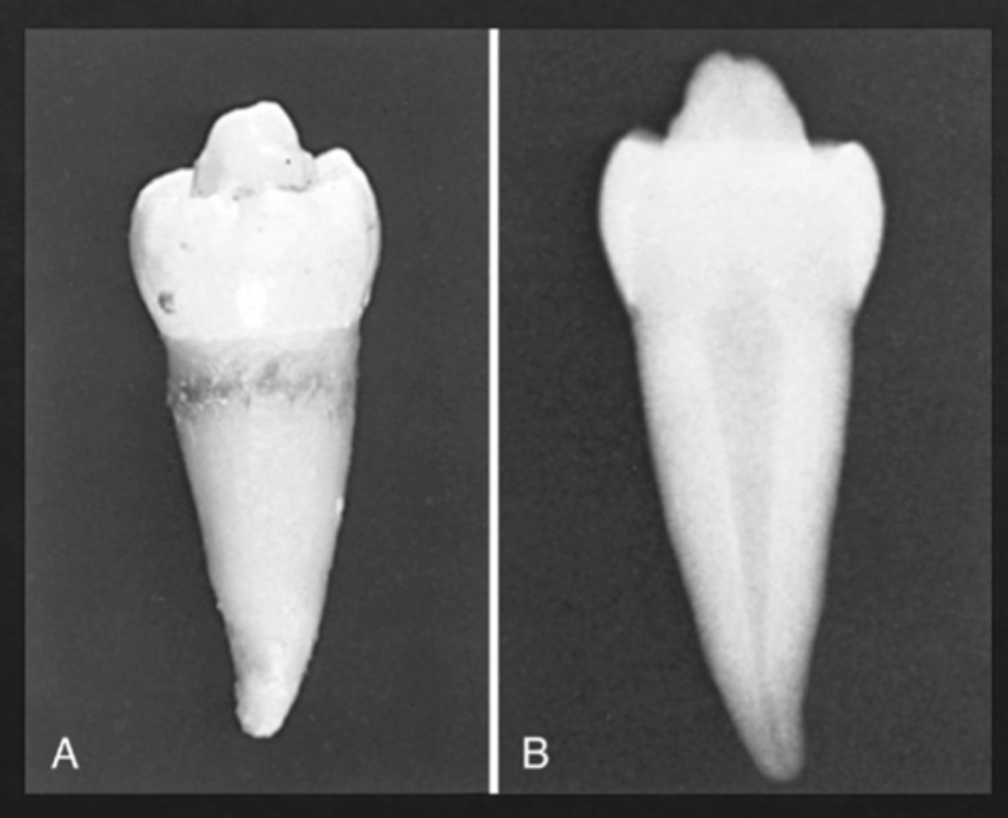
Which teeth are most commonly affected by dens evaginatus?
Premolars and lateral incisors
What are supernumerary teeth?
Extra teeth beyond the normal dentition
What is a mesiodens?
Supernumerary tooth between maxillary central incisors
What is a peridens?
Supernumerary tooth in premolar region
What is a distodens?
Supernumerary tooth in molar region
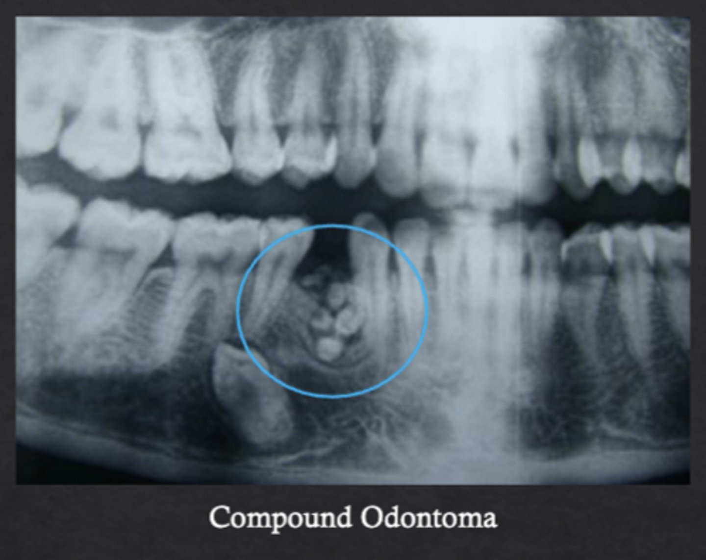
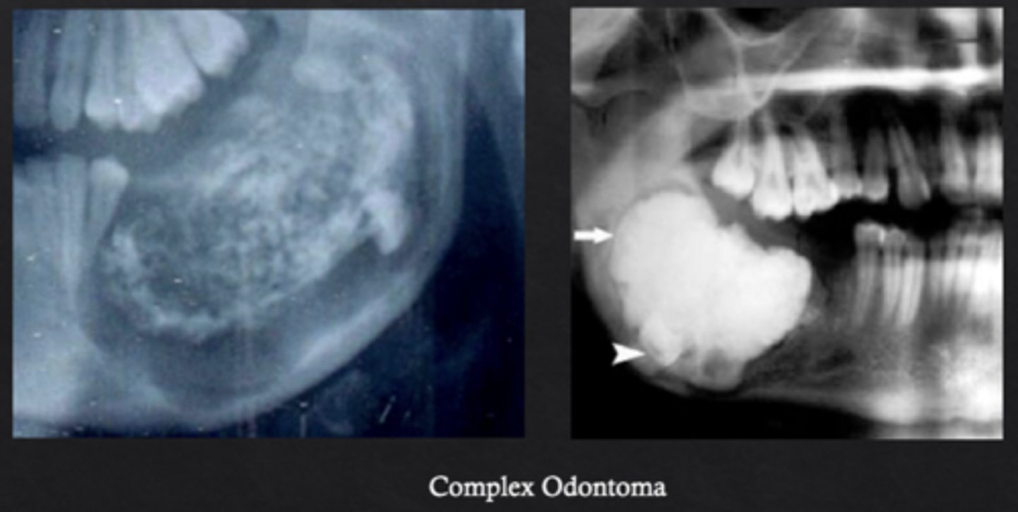
What is an odontoma?
Benign odontogenic tumor composed of enamel, dentin, cementum, and pulp
What is a compound odontoma?
Forms multiple small tooth-like structures with recognizable layers of dental antomy
What is a complex odontoma?
Disorganized mass of dental tissues without a tooth-like form
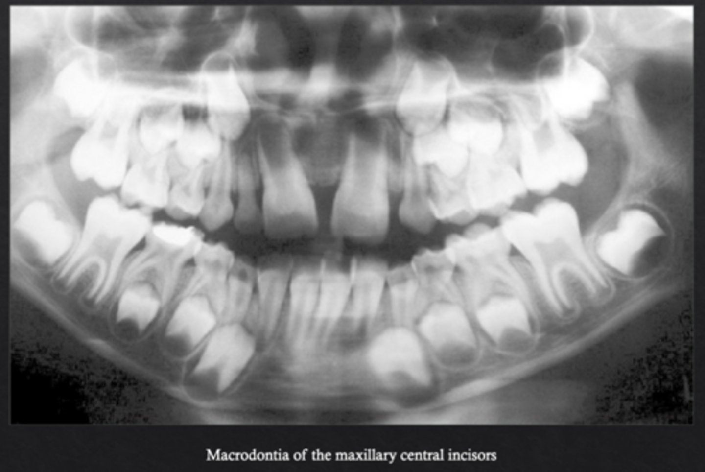
What is macrodontia?
Teeth are larger than normal *may be syndrome associated
What is microdontia?
Teeth are smaller than normal
Which tooth most commonly exhibits microdontia?
Maxillary lateral incisor (peg lateral)
What is fusion?
Two tooth buds fuse to form one tooth
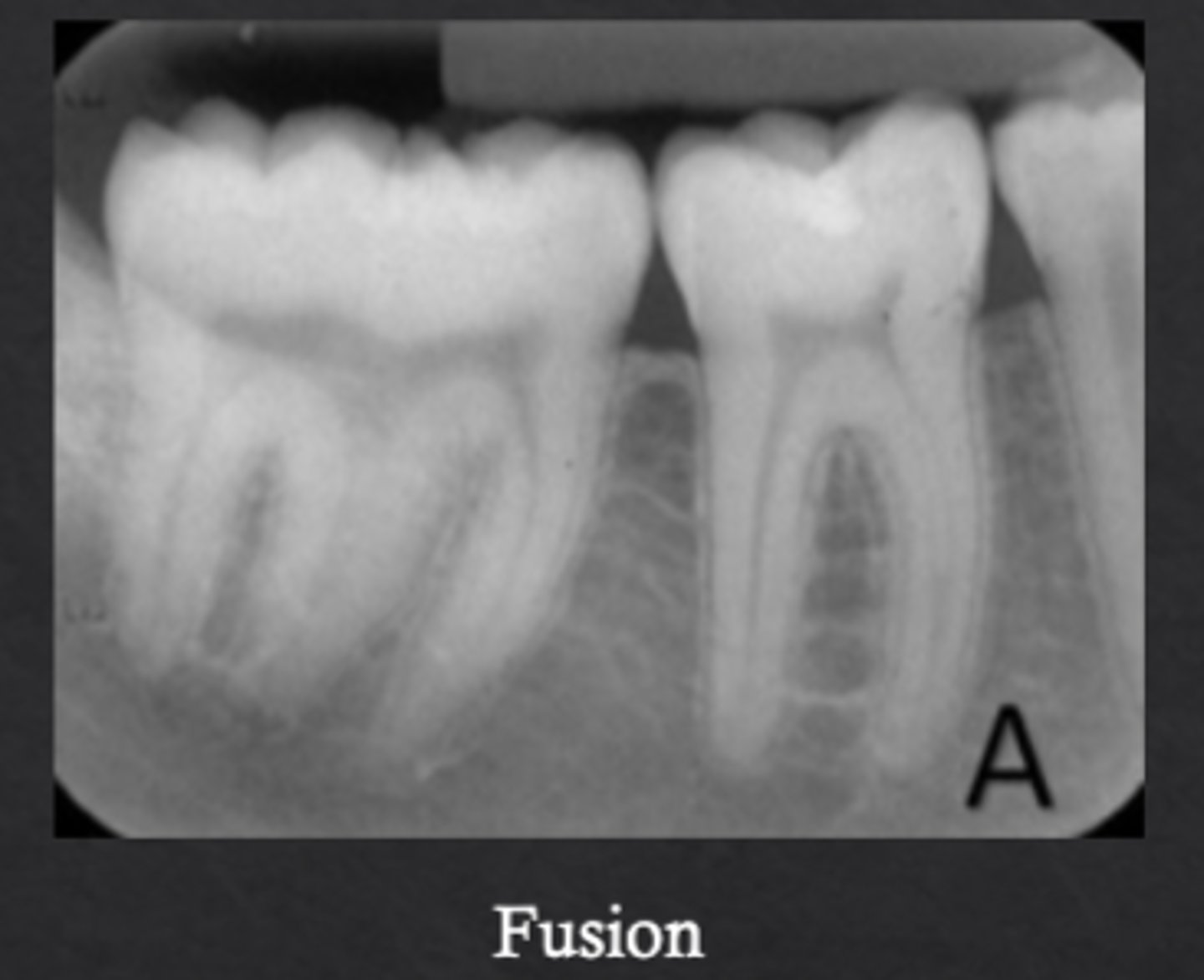
How many teeth are present in fusion cases?
One fewer than normal
What is gemination?
One tooth bud attempts to divide into two crowns
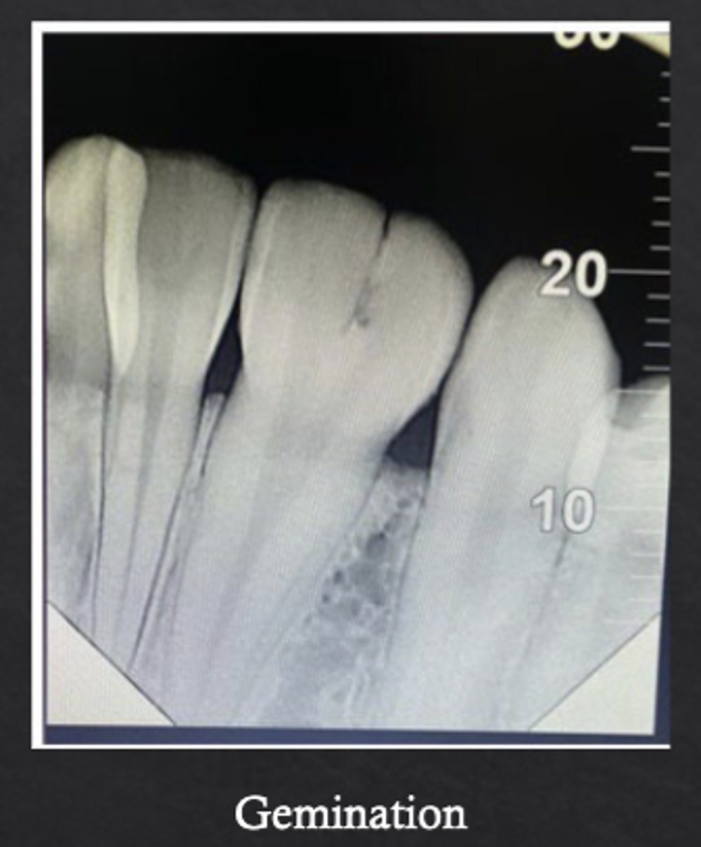
How many teeth are present in gemination cases?
Normal number of teeth
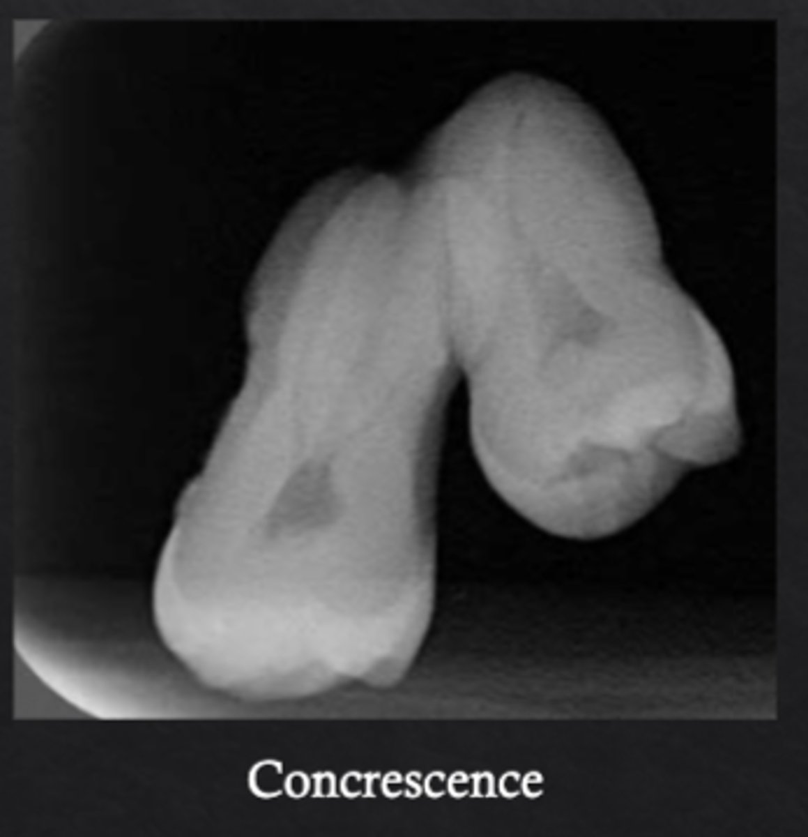
What is concrescence?
Fusion of the cementum of two adjacent teeth
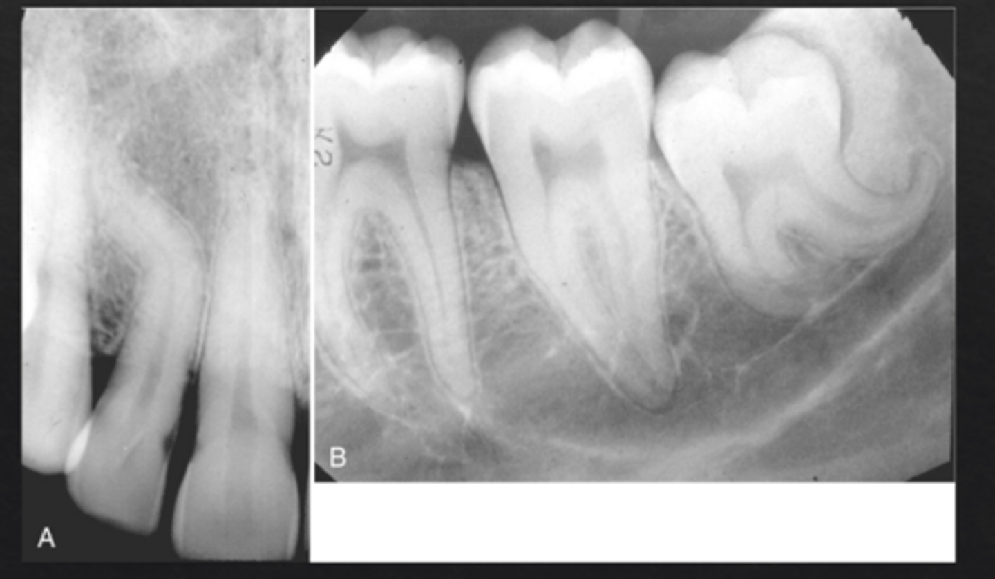
What is taurodontia?
Tooth with elongated pulp chamber and shortened roots, normal crown presentation
Which teeth most commonly show taurodontia?
Molars
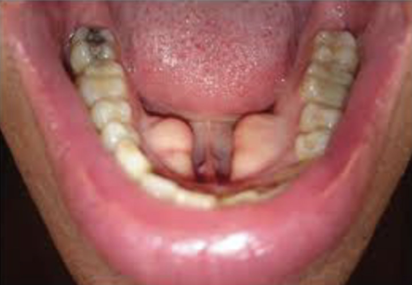
What is an enamel pearl?
Small globule of enamel on the root surface or CEJ
Where are enamel pearls most often found?
Furcation of molars
Why are enamel pearls clinically important?
they prevent normal periodontal attachment and may cause perio bone loss
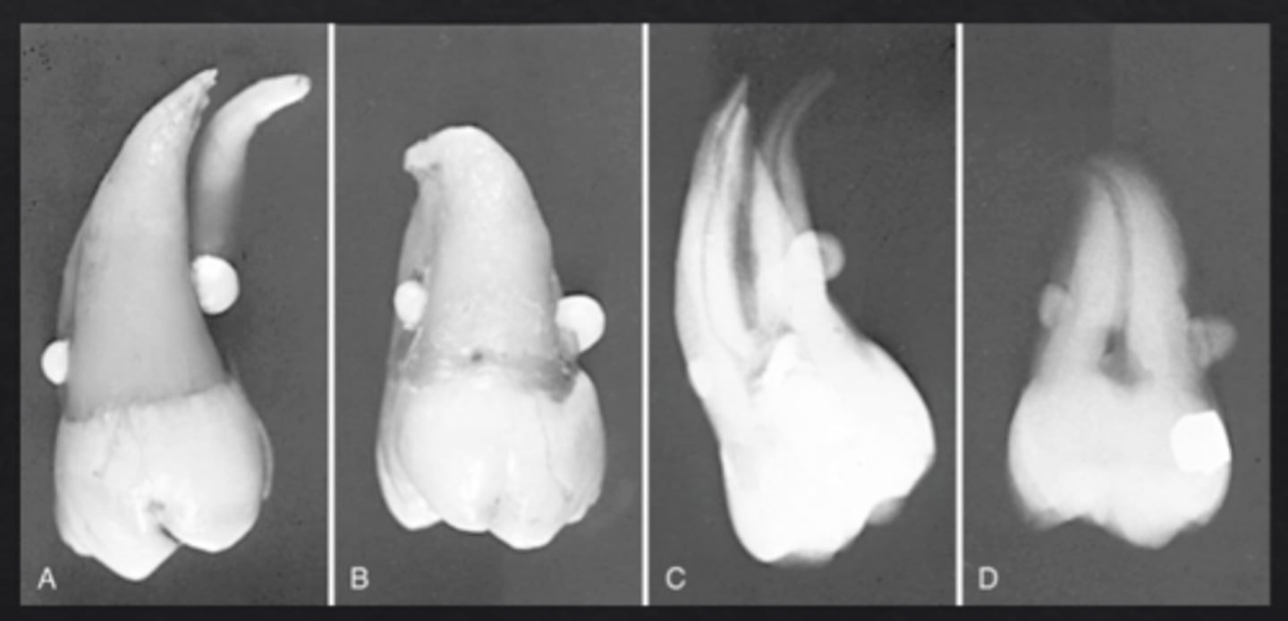
What is dilaceration?
Sharp bend or curvature of a tooth root
Why is dilaceration clinically important?
Can complicate extractions and RCT
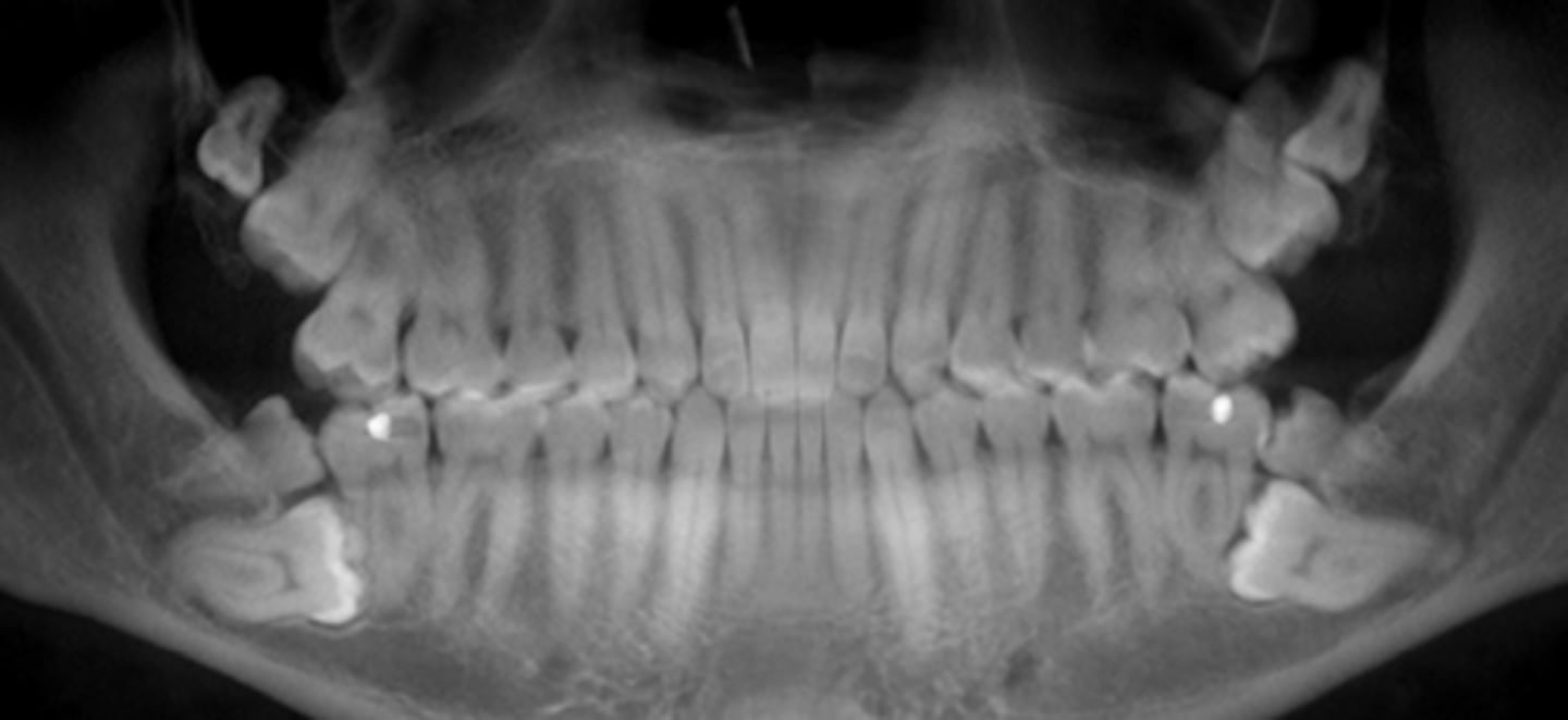
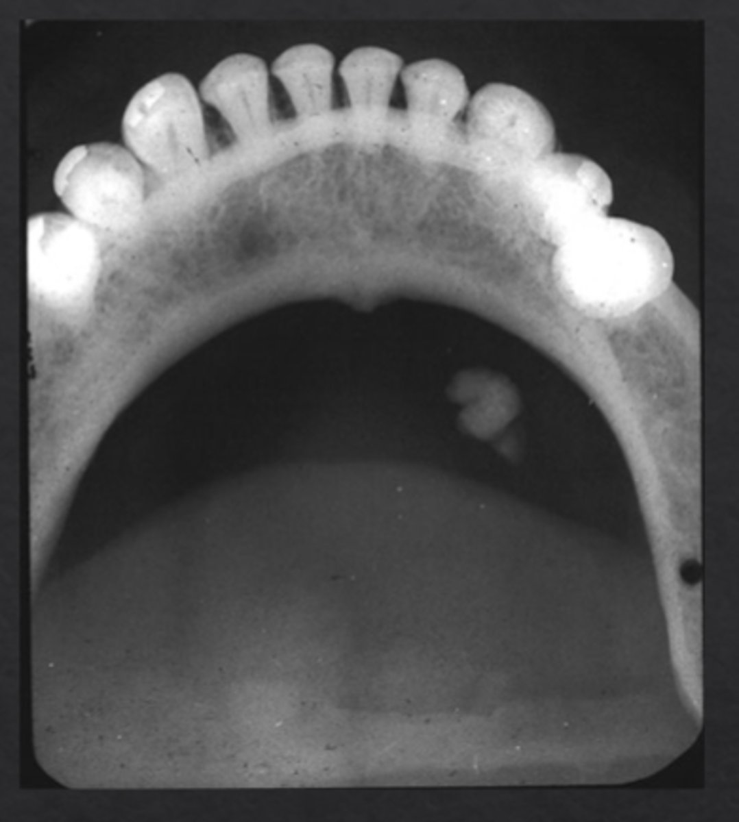
What are tonsiloliths?
Tonsil Stones: calcified masses of debris in tonsillar crypts
How do tonsiloliths appear radiographically?
Multiple small radiopacities superimposed over mandibular ramus, can be uni or bilateral
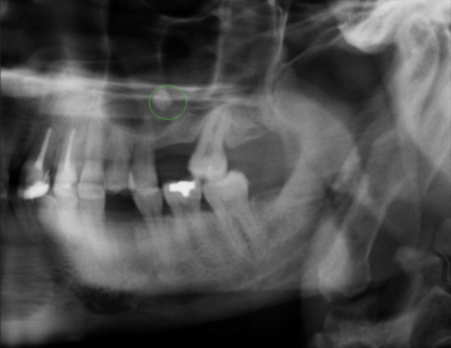
What are sialoliths?
Salivary Stones: calcified stones within salivary ducts
Which salivary gland most commonly develops sialoliths?
Submandibular gland (>80%)
What are antroliths?
Sinus Stones: calcified masses within paranasal sinuses
Where are antroliths most commonly found?
Maxillary sinus
What are carotid artery calcifications?
Calcified atherosclerotic plaques in carotid artery
Where do carotid artery calcifications appear radiographically?
Near C3-C4 vertebrae
*will not be tested to ID on final
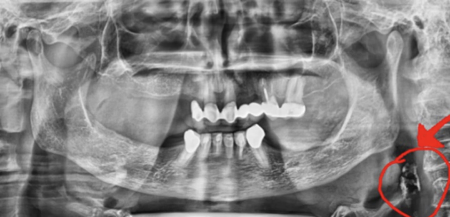
What should be done if you detect a carotid calcifications?
Refer pt to cardiology
What is stylohyoid ligament calcification?
Calcification of the ligament between the styloid process/hyoid bone, often due to aging
How does stylohyoid ligament calcification appear radiographically?
Linear radiopacity extending from skull base toward hyoid
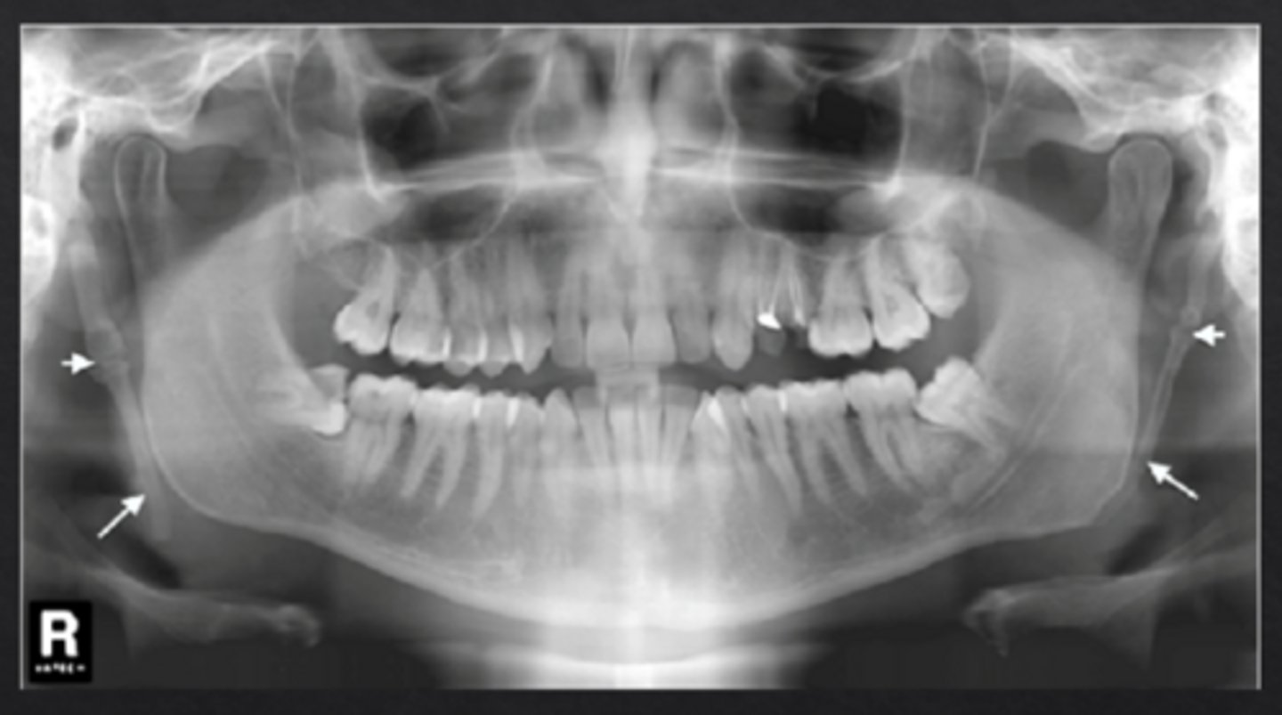
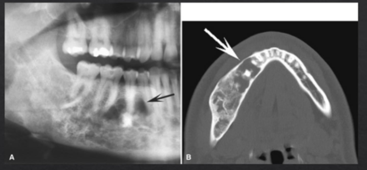
What is Mönckeberg's arteriosclerosis?
Calcification of the medial layer of muscular arteries
What systemic diseases are associated with Mönckeberg's arteriosclerosis?
Diabetes and chronic kidney disease/end stage renal
How does Mönckeberg's arteriosclerosis appear radiographically?
Worm-like tubular radiopacities near mandibular ramus, *often bilateral
What is a cyst?
Epithelial lined sac filled with fluid
What is a neoplasm?
Abnormal mass of tissue caused by uncontrolled cell proliferation
How to differentiate benign vs malignant neoplasms?
Malignant neoplasms can metastasize
What is metastasis?
Spread of tumor cells from primary site to other tissues (can be local or distant)
Describe the BLESSED approach
How to describe unknown lesions:
B borders
L location
E internal Enitities
S size
S shape
E effect on adjacent structures
D density